Abstract
Background
Oral squamous cell carcinoma is a global threat and accounts for approximately 90% of malignant oral lesions. The emergence of oral carcinoma is linked to precancerous lesions, which act as precursors of the disease. Matrix metalloproteinases appear to play a significant role in the pathogenesis of both precancerous conditions and oral malignancies due to their participation in remodeling of the extracellular matrix.
Methodology
This is an analytical study conducted at Dow University of Health Sciences, Pakistan. Unstimulated saliva samples were collected from healthy, oral submucous fibrosis and oral squamous cell carcinoma patients. The level of MMP-12 was estimated using enzyme-linked immunosorbent assay. One-way Analysis of variance was run to determine if MMP-12 levels differ between the three groups, which was preceded by post hoc Tuckey test. MMP-12 cut off values were determined using Receiver operating characteristic curve.
Results
A significant difference in salivary MMP-12 expression was observed in OSF and OSCC (p < 0.001). The expression of salivary MMP-12 was higher in OSF and OSCC patients as compared to the healthy group (p < 0.001). The mean MMP-12 expression in OSCC appeared higher than in OSF cases (p < 0.05). MMP-12 value of 4.05 ng/ml and 4.20 ng/ml is predictive of OSF and OSCC respectively, with 100% sensitivity and specificity (p < 0.001).
Conclusion
Increased expression of MMP-12 appears as the healthy patient advances to OSF and OSCC. The study results also demonstrate higher MMP-12 expression in OSCC patients as compared to OSF. Therefore, the estimation of salivary MMP-12 serves as a valuable non-invasive early diagnostic tool in diagnosing oral submucous fibrosis and oral squamous cell carcinoma.
Keywords: Matrix metalloproteinase, MMP-12, Oral squamous cell carcinoma, Oral submucous fibrosis, Salivary biomarker, ELISA
Background
Oral cancer poses a significant health threat worldwide with noteworthy incidence and mortality in developing countries [1]. Despite an increase in the knowledge of disease prevention and treatment, an increasing number of cases every year is quite evident. Oral cavity cancers appear to be among the most prevalent cancers globally and occur in nearly one-fifth of all cancers in males and one-tenth of all cancers in females globally [2].
The emergence of oral carcinoma is linked to precancerous lesions, which act as precursors of the disease [3]. These precancerous lesions include erythroplakia, leukoplakia, and oral submucous fibrosis (OSF), with oral submucous fibrous having high potential for malignant transformation among people residing in south and southeast Asia [4]. The rate of transformation of oral submucous fibrosis to oral squamous cell carcinoma (OSCC) is roughly around 2.3–7.6% [5]. The risk is increased in patients consuming heavy tobacco and alcohol [6]. Early and correct diagnosis of highly suspicious precancerous lesions helps in timely treatment and prevents transformation into malignancy. Tumor responds well to treatment modalities in the early stage as compared to the late stage. This is evident by the outcome that approximately 80% of patients have an expected 5- years of survival [7].
Histological examination is the gold standard but, to some extent, not practicable because of the nature and therapeutic response of the tumor [8]. It has a major drawback of being an invasive method as well as painful and time-consuming [9, 10]. Therefore, the focus is being directed towards non-invasive methods for the detection of oral squamous cell carcinoma. Changes in human genetics can be identified in patient's body fluid like saliva, cerebrospinal fluid, blood serum, and urine. These body fluids reflect an alteration in proteins and nucleic acid and therefore can be considered effective biomarkers for detection of oral squamous cell carcinoma at an early stage [11].
Until now, several oral salivary biomarkers have been studied for different premalignant lesions like oral lichen planus [12], leukoplakia [13], OSF [14], but not enough data is available for a biomarker that could aid in primary OSCC detection at the non-detectable stage or precursor stage [15]. Identifying a biomarker may serve threefold benefits; detection of the tumor at an early stage, serving as a prognostic marker, and a therapeutic target [15]. Matrix metalloproteinase (MMPs) are considered one of those potential biomarkers that can theoretically fulfil all these purposes. It has been suggested that estimation of MMPs levels in various tissue fluids serve as an accessible, non-time-consuming, and non-invasive tool for diagnosis of primary disease along with subsequent prognostic monitoring [15].
Several studies observed MMPs expression in oral cancer and demonstrated enhanced expression of different MMPs and advancement of the disease as compared to controls. Generally, increased expression of MMP-1, MMP-2, MMP-3, MMP-7, MMP-9, MMP-13, and MMP-14 are associated with cancer advancement, which relates to poor cell differentiation, tumor invasion, distant metastasis, and poor prognosis [15]. The protein concentrations of MMP-1, MMP-2, MMP-3, and MMP-9 also have been seen to be greater in OSCC tumor tissue in contrast to control tissue [16].
The aim and objective of this study is to estimate and compare the levels of salivary MMP-12 in patients presenting with oral submucous fibrosis and patients presenting with oral squamous cell carcinoma.
Methodology
Study design and setting
It is a descriptive & analytical cross-sectional study. The study was conducted on 90 randomly selected participants presenting to Oral and maxillofacial surgery departments of Dow University of health sciences, Karachi, after obtaining permission from Institutional Review Board (IRB- 1322/DUHS/Approval/2019/71).
Sample size
The sample size of 6 subjects per group was calculated using PASS version 15 software, based on two sample T test, allowing unequal variance with 99% Confidence of interval and 99% power of test, SD of MMP-12 in healthy subjects (0) and in OSCC (0.365 0.113) [17]. Keeping in view low subjects in each group, we increased the sample size to 90 subjects (30 per group).
Study participants
There were three groups of patients contributing to the study. Each group consisted of an equal number of participants (30 participants), selected randomly based on inclusion and exclusion criteria. Group 1 represented healthy individuals who demonstrated an absence of clinical and radiographic manifestation of precancerous condition and oral carcinoma and answering 'no' to dryness- related questions. Group 2 consisted of patients presenting with clinical evidence of oral submucous fibrosis, and group 3 represented patients presenting to the OPD with biopsy-proven cases of oral squamous cell carcinoma regardless of any grade.
Participants for the study were enrolled after performing a thorough oral examination, including assessment of oral mucosa, intra- oral bony areas, teeth, and periodontal tissues. Patients presenting with uncontrolled systemic diseases (uncontrolled diabetes/ hypertension), periodontal disease, salivary gland pathology, or autoimmune diseases such as SLE, rheumatoid arthritis, or Sjogren's syndrome were excluded from the study. We also excluded patients reporting acute or chronic use of drugs known to induce oral dryness, including antidepressants, antipsychotics, and antihistamines.
Study questionnaire
The previously validated OSF and OSCC patients' questionnaire is department-designed (OMFS department, Dow University of Health Sciences). It has been pretested on several patients for the past few years for maintaining the record. The first part of the questionnaire contained questions related to socio-demographic data. The second part of the questionnaire contained questions about disease status.
The Performa for healthy participants was filled out to facilitate whether the participant qualifies the inclusion criteria. The healthy participants included students, OPD assistants, friends, and family members. The questionnaire consists of five parts: the patient's demographic features and personal information, medical history, tobacco and alcohol habits, oral hygiene practice, and other information related to oral dryness [18].
Informed consent in accordance with Helsinki's declaration was sought from all participants, and assurance of confidentiality about personal data was provided.
Saliva collection & quantitative assessment by ELISA
For saliva sample collection, patients were seated in a clean and comfortable environment with the dental chair in an upright position. They were advised not to communicate during the procedure and not to forcefully spit or cough up mucus but let the saliva drool in 15 ml Falcon tubes once collected on the floor of the mouth. Around 2-5 ml of unstimulated saliva was gathered in a falcon tube by passive drooling method, which is considered an assuring alternative for minimizing potential error sources [18]. The effect of possible environmental factors (tobacco chewing, betel nut chewing, smokeless tobacco consumption, smoking) was controlled at the analysis phase through Analysis of covariance (ANCOVA).
Collected saliva was centrifugated at 6000×g at 4 °C for 10 min, and the supernatant obtained was carefully transferred equally into 3 Eppendorf tubes (microcentrifuge tubes- 1 ml) via pipette Juster. After disinfection and labelling with patient's name and hospital registration number, microcentrifuge tubes were stored in the freezer at − 80 °C until further ELISA investigation. ELISA was performed according to the manufacturer's instruction manual, and the colored product was read immediately at 450 nm wavelength. The excess sample was washed from the plate. Each sample was analyzed in duplicate for statistical analysis. MMP12 concentration in saliva was calculated with the help of standard curve using a known concentration of standard MMP12.
Statistical analysis
Readings were obtained by using ELISA reader software. Data was analyzed using SPSS 23.0 version (SPSS, Inc., Chicago, IL). Pearson correlation test was applied to analyze the correlation between salivary MMP-12 level and age. Comparison of means among genders was tested by applying independent sample t-test. The statistical Analysis was run with a significance level of p < 0.05. One-way Analysis of variance (ANOVA) was run to determine if MMP-12 levels differ between healthy, OSF and OSCC groups, preceded by post hoc Tuckey test. The cut- off values of MMP-12 were determined by performing Receiver Operating Characteristic (ROC) curve analysis.
Results
The socio-demographic data of all subjects included in the study is presented in Table 1. This data includes age, gender, marital status, employment status and oral habits of all participants.
Table 1.
Sociodemographic data of patients
| Category | Healthy n (%) |
OSF n (%) |
OSCC n (%) |
|---|---|---|---|
| Age | |||
| (Mean S.D) | 28.87 6.81 | 33.27 12.43 | 47.13 13.38 |
| Gender | |||
|
Male Female |
5 (16.6%) 25 (83.3%) |
25 (83.3%) 5 (16.6%) |
20 (66.6%) 10 (33.3%) |
| Marital status | |||
| Unmarried | 8 (26.6%) | 10 (33.3%) | 1 (3.3%) |
| Employment Status | |||
| Married | 22 (73.3%) | 20 (66.6%) | 29 (96.6%) |
|
Employed Unemployed |
23 (76.6%) 7 (23.3%) |
19 (63.3%) 11 (36.6%) |
20 (66.6%) 10 (33.3%) |
| Oral habits | |||
|
Betel nut Tobacco smoking Smokeless tobacco None |
7 (23.3%) 4 (13.3%) 0 (0%) 19 (63.3%) |
17 (56.6%) 6 (20%) 7 (23.3%) 0 (0%) |
10 (33.3%) 7 (23.3%) 4 (13.3%) 9 (30%) |
| Total | 30 | 30 | 30 |
A statistical test was applied to evaluate the correlation between age and MMP-12 expression among healthy, OSF and OSCC group. A positively moderate correlation was observed between age and MMP-12 expression among the three groups (r = 0.35, p = 0.001). It demonstrates that as age increases, MMP-12 levels also increase.
A test was run to compare mean MMP-12 expression between gender. A highly statistically significant mean difference in MMP-12 expression was observed between gender (p < 0.001). Higher mean MMP-12 expression was found in Males (M = 12.5 ng/ml) compared to Females (M = 5.59 ng/ml). (Table 2).
Table 2.
Mean comparison of Matrix metalloproteinases- 12 expressions with genders
| Gender | N = 90 | Mean (S.D) | M.D (p value ó) |
|---|---|---|---|
| Male | 50 | 12.5 (5.84) | 6.91 (< 0.001) |
| Female | 40 | 5.59 (6.72) |
ó- p value computed using Independent t- test
M.D- Mean difference, S.D- Standard Deviation
Oral habits of healthy, OSF and OSCC group were recorded. A statistical test was applied to compare the means of MMP-12 levels among participants with various oral habits. A statistically significant difference in means was observed (p < 0.05) among participants with distinct oral habits (Table 3).
Table 3.
Descriptive ANOVA- MMP-12 expression & oral habits
| Category | N = 90 | Mean (SD) | MSE (p value ò) |
|---|---|---|---|
| Betel nut | 34 | 10.5b (5.8) | 44.46 (0.003*) |
| Tobacco smoking | 17 | 10.5 (6.9) | |
| Smokeless tobacco | 11 | 13.8a (4.2) | |
| Non-significant oral habit | 28 | 5.6a,b (8.0) |
Ò—p value computed using One-way ANOVA
*- Significant at 0.05
MSE- Mean square error
a on comparison p < 0.01
b on comparison p < 0.05
The above table (Table 3) demonstrates a significant difference in mean between smokeless tobacco consumers and those with non-significant oral habits. MMP-12 levels among smokeless tobacco consumers appear higher as compared to participants with non-significant oral habits. A statistically significant difference in mean is also evident in betel nut consumers and participants with non-significant oral habits. Higher MMP-12 levels appear in betel nut consumers as compared to individuals with no significant oral habits.
The difference in mean MMP-12 level among healthy, OSF and OSCC group was analyzed. The result demonstrated a statistically significant difference in salivary MMP-12 means among the groups (p < 0.001) (Table 4).
Table 4.
Descriptive ANOVA- MMP-12 expression (ng/ml)
| Category | N = 90 | Mean (SD) | MSE (p value ò) |
|---|---|---|---|
| Healthy | 30 | 0.82 (0.45) | 12.48 (< 0.001) |
| OSF | 30 | 12.53 (3.2) | |
| OSCC | 30 | 14.92 (5.1) |
| Comparison | Mean difference | p value ø | |
|---|---|---|---|
| OSF vs Healthy | 11.70 | < 0.001 | |
| OSCC vs Healthy | 14.09 | < 0.001 | |
| OSCC vs OSF | 2.39 | 0.028 |
Ò- p value computed using One-way ANOVA
MSE- Mean square error, SD- Standard Deviation
ø- p value computed using Post hoc Tuckey test
Healthy participants demonstrated significantly different salivary MMP12 expression compared to patients belonging to Oral Submucous Fibrosis (p < 0.001) & Oral Squamous Cell Carcinoma (p < 0.001) group, respectively. Table 4 depicts the mean comparison of MMP-12 expression among the healthy, OSF, and OSCC groups at a statistically significant mean level (p = 0.028).
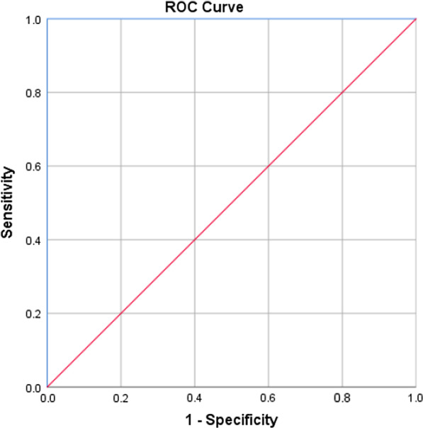
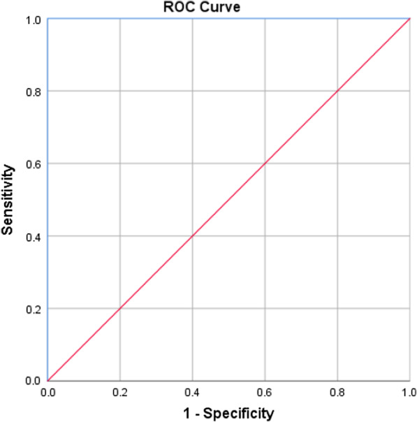
MMP-12 value of ≥ 4.06 ng/ml is predictive of Oral submucous fibrosis and MMP- 12 value of ≥ 4.21 ng/ml is predictive of Oral squamous cell carcinoma, with 100% sensitivity and 100% specificity (p < 0.001) (Table 5). The area under curve observed for OSF and OSCC is 100% (Figs. 1, 2).
Table 5.
MMP12 Cut-off for OSF & OSCC using receiving operative curve (ROC)
| Category | MMP12 Cut-Off |
Sensitivity | Specificity | AUC (p value) |
|---|---|---|---|---|
| OSF | ≥ 4.06 | 100% | 100% | 100% (< 0.001) |
| OSCC | ≥ 4.21 | 100% | 100% | 100% (< 0.001) |
AUC: Area Under the Curve
Fig. 1.

Receiver operating characteristics (ROC) curve analysis of the predictive value of MMP-12 for OSF
Fig. 2.

Receiver operating characteristics (ROC) curve analyses for predictive value of MMP-12 for OSCC
Discussion
The level of MMP-12 appears to be highly expressed in a wide range of cancers, including colorectal, gastric, skin nasopharyngeal, and lung cancer [19–24]. A study was carried out to identify the expression of LIFR, PIK3R1, and MMP- 12 in gall bladder carcinoma. This study validated MMP-12 as a significant prognostic biomarker in this rare and aggressive tumor [25]. Another study was conducted to analyze the expression of MMP-12 level in patients presenting with laryngeal squamous cell carcinoma. There was a poorer degree of tumor differentiation as the expression of MMP-12 went higher [26]. Elevated levels of MMP-12 were also correlated with pathological stage and metastasis of lung adenocarcinoma. Hence, targeting MMP-12 for the treatment of lung adenocarcinoma seemed promising. In addition to this, high expression of MMP-12 was observed in esophageal squamous cell carcinoma compared to normal epithelial cells [27]. Due to its functional properties and its role in tissue destructive disease, MMP 12 can be used as a biomarker for various oral diseases [28]. It plays a significant part in tumorigenesis and progression. This includes tumor growth, migration, invasion and tumor metastasis [15, 29, 30]. MMP-12 is recommended as a diagnostic biomarker for OSCC due to its significant sensitivity and specificity [15].
The current study consisted of participants with a minimum age of 18 years and maximum age of 78 years with a mean age of 36.42 13.60 years. Statistical Analysis was applied to observe the relationship between salivary MMP-12 expression and participants' age. The results demonstrated a statistically significant direct relationship between the two variables, which explains that MMP-12 expression in saliva increases as age increases. In contrast to our findings, lower salivary MMP-12 levels were reported in a study in individuals aged 40–60 years compared to individuals under 40 years [28].
A higher proportion (56%) of research participants were males, whereas 44% were females. A statistically significant difference was observed in salivary MMP-12 expression among genders, implying a higher mean expression of salivary MMP-12 in males than females. However, no difference in salivary MMP-12 was observed between males and females in a study focusing on MMP-12 levels in patients with periodontal inflammation [28].
The study also illustrates the oral habits of participants. Majority of the participants were betel nut consumers, followed by patients reporting tobacco smoking as their oral habit. A comparison was made between distinct oral habits and salivary MMP-12 expression. A significant difference was observed between MMP-12 expression of individuals with distinct oral habits. Smokeless tobacco consumers demonstrated higher MMP-12 expression compared to other oral habits. However, individuals who reported tobacco and betel nut as their oral habit demonstrated lower MMP-12 expression comparatively. In contrast to the results mentioned, another study evidences a non-significant difference in MMP-12 expression among patients with different oral habits, including smoking, alcohol, and betel nut chewing [31].
The current study results demonstrated a statistically significant difference in salivary MMP-12 expression in OSF and OSCC group compared to healthy participants. OSF and OSCC group showed higher salivary MMP-12 expression, and lower expression was observed in healthy group. The results coincide with a previous study that indicated a high association of salivary protease spectrum with oral health status [17]. It demonstrated increased protease levels in OSCC patients as compared to patients presenting with other oral diseases. MMP- 12 was detected only in the saliva of patients with OSCC along with other MMPs, such as MMP-1, MMP-2, MMP-3, MMP-10, and MMP-13. In addition to this, MMP-1, MMP-2, MMP-10, and MMP-12 were also observed to be significantly increased in patients with OSCC compared to healthy patients and patients with benign oral masses (OBM) and mild chronic periodontitis (CPD). The concentration of salivary MMP-12 in OSCC patients demonstrated in this study is around 1300 pg ml−1, which is comparatively more than healthy (700 pg ml−1), benign oral mass (900 pg ml−1), and chronic periodontal disease patients (900 pg ml−1) [17].
A cohort study carried out in Sweden on 436 participants aimed to investigate salivary MMP-12 levels to various aspects of oral health. The influence of non-disease covariates on MMP-12 levels was also assessed. The results revealed an association between MMP-12 levels and the percentage of gingival pockets 4 mm. The study concluded that MMP-12 reflects various aspect of periodontal disease and the levels are contrarily affected by the presence of tumor [28]. The results corroborate the results of our study in which MMP-12 levels are affected in the presence of tumor.
A study was conducted to compare the MMP12 level in patients presenting with OSCC and verrucous carcinoma (VC) in tissue samples. The study results showed that VCs were devoid of epithelial MMP-12 expressions compared to SCC [32]. Another study was undertaken to estimate serum MMPs levels in OSCC patients compared to healthy participants. Serum level of MMP-12 was notably arisen in OSCC patients as compared to healthy participants [15].
MMP-12 expression found elevated in patients with chronic periodontitis with identification of CD68+ CD14+ CD64+ cells [28]. Also, the expression of MMP 12 in tissue samples appear high in patients with extracapsular spread compared to those without extracapsular spread [24]. Hence, it may be a useful predictive marker for extracapsular spread (ECS) in head and neck tumors [24].
In recent years, the prevalence of OSF has increased from 8.3/105 to 16.2/105 [33, 34]. The rate of malignant transformation to oral cancer is 9.13%, and there is a 29.26 times higher risk in OSF patients than non-OSF patients [35, 36]. In Pakistan, OSF is contemplated as a public health concern as oral malignancies are one of the most common malignancies reported [37]. Also, oral habits like tobacco smoking and consumption of betel nut and smokeless tobacco are major risk factors of OSF and are common in Pakistan. Studying the role of several markers present in saliva will help in devising a non-invasive investigation for OSF diagnosis, which will ultimately result in early diagnosis of OSF and prevent it from advancing to OSCC, if treated promptly. Since OSF is an oral potentially malignant disorder and is common in our part of the world, we have studied the expression of MMP-12 in OSF patients along with OSCC patients.
Statistically, a significant difference in salivary MMP-12 level was observed in OSF patients in comparison with healthy participants. OSF patients demonstrated higher MMP-12 levels. However, in comparison with salivary MMP-12 in OSCC, salivary MMP-12 in OSF demonstrated lower expression explaining the increase in MMP-12 expression as an oral potentially malignant disorder (OSF) drifts towards malignancy (OSCC). Since the significant difference in salivary MMP-12 levels is observed between OSF and OSCC, this investigation may serve as a beneficial non-invasive tool for differentiating OSCC from specifically last stage OSF in which patients present with nil mouth opening and the surgeon suspects a lesion intraorally.
Conclusion
The current study indicates salivary MMP-12 expression in patients presenting with oral submucous fibrosis and biopsy-proven oral squamous cell carcinoma. We observed a statistically significant difference in salivary MMP-12 expression in OSF and OSCC group as compared to healthy patients, with higher expression in OSF and OSCC patients. The study results also demonstrate higher expression of salivary MMP-12 in OSCC as compared to OSF. Therefore, the estimation of salivary MMP-12 may serve as a useful non-invasive early diagnostic tool in diagnosing oral submucous fibrosis and oral squamous cell carcinoma.
Acknowledgements
We would like to thank all the participants who cooperated with us, without whom this study would not have been possible.
Abbreviations
- OSCC
Oral squamous cell carcinoma
- OSF
Oral submucous fibrosis
- MMP
Matrix metalloproteinases
- ELISA
Enzyme-linked immunosorbent assay
- ANOVA
One-way analysis of variance
- OBM
Oral benign mass
- CPD
Mild chronic periodontitis
- VC
Verrucous carcinoma
- ROC
Receiver operating characteristics
- SD
Standard deviation
- MD
Mean difference
- MSE
Mean square error
Authors' contributions
ZS, the principal investigator, made a substantial contribution to the conception, design of the work, data collection, sample processing, and led the writing. AS and UZ supervised the project and contributed to data collection, sample processing interpretation & critical evaluation. SA, MM and AK contributed to the sample collection, processing and drafting. WA contributed in data analysis and interpretation. All authors contributed to the interpretation of data, read and approved the final manuscript and agreed to be both personally accountable for their contributions and ensure that questions related to the accuracy or integrity of any part of the work are appropriately investigated, resolved and the resolution documented in the literature. All authors read and approved the final manuscript.
Funding
The current study did not receive any funding.
Availability of data and materials
The datasets used and analyzed during the current study are available from principal investigator (corresponding author) on reasonable request.
Declarations
Ethical approval and consent to participate
The study was approved by Dow University of health sciences' Institutional Review Board (IRB- 1322/DUHS/Approval/2019/71). Informed consent to participate was taken from all participants according to Helsinki's declaration.
Consent for publication
The consent for publication was taken from all participants.
Competing interests
The authors declare that they have no competing interests.
Footnotes
Publisher's Note
Springer Nature remains neutral with regard to jurisdictional claims in published maps and institutional affiliations.
References
- 1.Yang Y, Zhou M, Zeng X, Wang C. The burden of oral cancer in China, 1990–2017: an analysis for the Global Burden of Disease, Injuries, and Risk Factors Study 2017. BMC Oral Health. 2021;21(1):1–11. doi: 10.1186/s12903-020-01362-6. [DOI] [PMC free article] [PubMed] [Google Scholar]
- 2.Saleem Z, Abbas SA, Nadeem F, Majeed MM. The habits and reasons of delayed presentation of patients with oral cancer at a tertiary care hospital of a third world country. Pakistan J Public Health. 2018;8(3):165–169. doi: 10.32413/pjph.v8i3.97. [DOI] [Google Scholar]
- 3.Goyal D, Goyal P, Singh H, Verma C. An update on precancerous lesions of oral cavity. Int J Med Dent Sci. 2013;2:70–75. [Google Scholar]
- 4.Ekanayaka RP, Tilakaratne WM. Oral submucous fibrosis: review on mechanisms of malignant transformation. Oral Surg Oral Med Oral Pathol Oral Radiol. 2016;122(2):192–199. doi: 10.1016/j.oooo.2015.12.018. [DOI] [PubMed] [Google Scholar]
- 5.Selvam NP, Dayanand AA. Lycopene in the management of oral submucous fibrosis. Asian J Pharm Clin Res. 2013;6(3):58–61. [Google Scholar]
- 6.Lingen MW, Abt E, Agrawal N, Chaturvedi AK, Cohen E, D’Souza G, Gurenlian J, Kalmar JR, Kerr AR, Lambert PM. Evidence-based clinical practice guideline for the evaluation of potentially malignant disorders in the oral cavity: a report of the American Dental Association. J Am Dent Assoc. 2017;148(10):712–727. doi: 10.1016/j.adaj.2017.07.032. [DOI] [PubMed] [Google Scholar]
- 7.Wang Z, Jiang L, Huang C, Li Z, Chen L, Gou L, Chen P, Tong A, Tang M, Gao F. Comparative proteomics approach to screening of potential diagnostic and therapeutic targets for oral squamous cell carcinoma. Mol Cell Proteomics. 2008;7(9):1639–1650. doi: 10.1074/mcp.M700520-MCP200. [DOI] [PMC free article] [PubMed] [Google Scholar]
- 8.Khan MSR, Siddika F, Xu S, Liu XL, Shuang M, Liang HF. Diagnosing oral squamous cell carcinoma using salivary biomarkers. Bangabandhu Sheikh Mujib Med Univ J. 2018;11(1):1–10. doi: 10.3329/bsmmuj.v11i1.35399. [DOI] [Google Scholar]
- 9.Ishikawa S, Sugimoto M, Kitabatake K, Sugano A, Nakamura M, Kaneko M, Ota S, Hiwatari K, Enomoto A, Soga T. Identification of salivary metabolomic biomarkers for oral cancer screening. Sci Rep. 2016;6:31520. doi: 10.1038/srep31520. [DOI] [PMC free article] [PubMed] [Google Scholar]
- 10.Kaur J, Jacobs R, Huang Y, Salvo N, Politis C. Salivary biomarkers for oral cancer and pre-cancer screening: a review. Clin Oral Invest. 2018;22(2):633–640. doi: 10.1007/s00784-018-2337-x. [DOI] [PubMed] [Google Scholar]
- 11.Javaid MA, Ahmed AS, Durand R, Tran SD. Saliva as a diagnostic tool for oral and systemic diseases. J Oral Biol Craniofac Res. 2016;6(1):67–76. doi: 10.1016/j.jobcr.2015.08.006. [DOI] [PMC free article] [PubMed] [Google Scholar]
- 12.Santarelli A, Mascitti M, Rubini C, Bambini F, Zizzi A, Offidani A, Ganzetti G, Laino L, Cicciù M, Muzio LL. Active inflammatory biomarkers in oral lichen planus. Int J Immunopathol Pharmacol. 2015;28(4):562–568. doi: 10.1177/0394632015592101. [DOI] [PubMed] [Google Scholar]
- 13.von Zeidler SV, de Souza BT, Mendonça EF, Batista AC. E-cadherin as a potential biomarker of malignant transformation in oral leukoplakia: a retrospective cohort study. BMC Cancer. 2014;14(1):1–7. doi: 10.1186/1471-2407-14-1. [DOI] [PMC free article] [PubMed] [Google Scholar]
- 14.Bazarsad S, Zhang X, Kim KY, Illeperuma R, Jayasinghe RD, Tilakaratne WM, Kim J. Identification of a combined biomarker for malignant transformation in oral submucous fibrosis. J Oral Pathol Med. 2017;46(6):431–438. doi: 10.1111/jop.12483. [DOI] [PMC free article] [PubMed] [Google Scholar]
- 15.Choudhry N, Sarmad S, Gondal AJ. Estimation of serum matrix metalloproteinases among patients of oral squamous cell carcinoma. Pakistan J Med Sci. 2019;35(1). [DOI] [PMC free article] [PubMed]
- 16.Baker E, Leaper D, Hayter J, Dickenson A. The matrix metalloproteinase system in oral squamous cell carcinoma. Br J Oral Maxillofac Surg. 2006;44(6):482–486. doi: 10.1016/j.bjoms.2005.10.005. [DOI] [PubMed] [Google Scholar]
- 17.Feng Y, Li Q, Chen J, Yi P, Xu X, Fan Y, Cui B, Yu Y, Li X, Du Y. Salivary protease spectrum biomarkers of oral cancer. Int J Oral Sci. 2019;11(1):7. doi: 10.1038/s41368-018-0032-z. [DOI] [PMC free article] [PubMed] [Google Scholar]
- 18.Bhattarai KR, Kim H-R, Chae H-J. Compliance with saliva collection protocol in healthy volunteers: strategies for managing risk and errors. Int J Med Sci. 2018;15(8):823–831. doi: 10.7150/ijms.25146. [DOI] [PMC free article] [PubMed] [Google Scholar]
- 19.Zheng J, Chu D, Wang D, Zhu Y, Zhang X, Ji G, Zhao H, Wu G, Du J, Zhao Q. Matrix metalloproteinase-12 is associated with overall survival in Chinese patients with gastric cancer. J Surg Oncol. 2013;107(7):746–751. doi: 10.1002/jso.23302. [DOI] [PubMed] [Google Scholar]
- 20.Kahlert C, Pecqueux M, Halama N, Dienemann H, Muley T, Pfannschmidt J, Lasitschka F, Klupp F, Schmidt T, Rahbari N. Tumour-site-dependent expression profile of angiogenic factors in tumour-associated stroma of primary colorectal cancer and metastases. Br J Cancer. 2014;110(2):441. doi: 10.1038/bjc.2013.745. [DOI] [PMC free article] [PubMed] [Google Scholar]
- 21.Chung I-C, Chen L-C, Chung A-K, Chao M, Huang H-Y, Hsueh C, Tsang N-M, Chang K-P, Liang Y, Li H-P. Matrix metalloproteinase 12 is induced by heterogeneous nuclear ribonucleoprotein K and promotes migration and invasion in nasopharyngeal carcinoma. BMC Cancer. 2014;14(1):348. doi: 10.1186/1471-2407-14-348. [DOI] [PMC free article] [PubMed] [Google Scholar]
- 22.Lv F, Wang J, Wu Y, Chen H, Shen X. Knockdown of MMP12 inhibits the growth and invasion of lung adenocarcinoma cells. Int J Immunopathol Pharmacol. 2015;28(1):77–84. doi: 10.1177/0394632015572557. [DOI] [PubMed] [Google Scholar]
- 23.Liu L, Sun J, Li G, Gu B, Wang X, Chi H, Guo F. Association between MMP-12-82A/G polymorphism and cancer risk: a meta-analysis. Int J Clin Exp Med. 2015;8(8):11896. [PMC free article] [PubMed] [Google Scholar]
- 24.Kim JM, Kim HJ, Koo BS, Rha KS, Yoon Y-H. Expression of matrix metalloproteinase-12 is correlated with extracapsular spread of tumor from nodes with metastasis in head and neck squamous cell carcinoma. Eur Arch Otorhinolaryngol. 2013;270(3):1137–1142. doi: 10.1007/s00405-012-2161-x. [DOI] [PubMed] [Google Scholar]
- 25.Zhao X, Xu M, Cai Z, Yuan W, Cui W, Li MD. Identification of LIFR, PIK3R1, and MMP12 as novel prognostic signatures in gallbladder cancer using network-based module analysis. Front Oncol. 2019;9:325. doi: 10.3389/fonc.2019.00325. [DOI] [PMC free article] [PubMed] [Google Scholar]
- 26.El-Hennawi DE-DM, El Tabbakh MT, Sabek NAE-A, Abou-halawa AS, Fareed AM. High Mobility Group A2 (HMGA2) and Matrix Metalloproteinase 12 (MMP12) genes expression in Egyptian patients with laryngeal squamous cell carcinoma. Egypt J Ear Nose Throat Allied Sci. 2019;20(2):94–98. doi: 10.21608/ejentas.2019.6403.1056. [DOI] [Google Scholar]
- 27.Han F, Zhang S, Zhang L, Hao Q. The overexpression and predictive significance of MMP-12 in esophageal squamous cell carcinoma. Pathol-Res Pract. 2017;213(12):1519–1522. doi: 10.1016/j.prp.2017.09.023. [DOI] [PubMed] [Google Scholar]
- 28.Holmström SB, Lira-Junior R, Zwicker S, Majster M, Gustafsson A, Åkerman S, Klinge B, Svensson M, Boström EA. MMP-12 and S100s in saliva reflect different aspects of periodontal inflammation. Cytokine. 2019;113:155–161. doi: 10.1016/j.cyto.2018.06.036. [DOI] [PubMed] [Google Scholar]
- 29.Rahamthulla SU, Priya PV, Hussain SJ, Nasyam FA, Akifuddin S, Srinivas VS. Effectiveness of the supraomohyoid neck dissection in clinically N0 neck patients with squamous cell carcinoma of buccal mucosa and gingivobuccal sulcus. J Int Soc Prevent Commun Dent. 2015;5(2):131. doi: 10.4103/2231-0762.155740. [DOI] [PMC free article] [PubMed] [Google Scholar]
- 30.Mishev G, Deliverska E, Hlushchuk R, Velinov N, Aebersold D, Weinstein F, Djonov V. Prognostic value of matrix metalloproteinases in oral squamous cell carcinoma. Biotechnol Biotechnol Equip. 2014;28(6):1138–1149. doi: 10.1080/13102818.2014.967510. [DOI] [PMC free article] [PubMed] [Google Scholar]
- 31.Yen C-Y, Chen C-H, Chang C-H, Tseng H-F, Liu S-Y, Chuang L-Y, Wen C-H, Chang H-W. Matrix metalloproteinases (MMP) 1 and MMP10 but not MMP12 are potential oral cancer markers. Biomarkers. 2009;14(4):244–249. doi: 10.1080/13547500902829375. [DOI] [PubMed] [Google Scholar]
- 32.Impola U, Uitto V-J, Hietanen J, Hakkinen L, Zhang L, Larjava H, Isaka K, Saarialho-Kere U. Differential expression of matrilysin-1 (MMP-7), 92 kD gelatinase (MMP-9), and metalloelastase (MMP-12) in oral verrucous and squamous cell cancer. J Pathol. 2004;202(1):14–22. doi: 10.1002/path.1479. [DOI] [PubMed] [Google Scholar]
- 33.Cai X, Yao Z, Liu G, Cui L, Li H, Huang J. Oral submucous fibrosis: a clinicopathological study of 674 cases in China. J Oral Pathol Med. 2019;48(4):321–325. doi: 10.1111/jop.12836. [DOI] [PMC free article] [PubMed] [Google Scholar]
- 34.Yang S-F, Wang Y-H, Su N-Y, Yu H-C, Wei C-Y, Yu C-H, Chang Y-C. Changes in prevalence of precancerous oral submucous fibrosis from 1996 to 2013 in Taiwan: a nationwide population-based retrospective study. J Formos Med Assoc. 2018;117(2):147–152. doi: 10.1016/j.jfma.2017.01.012. [DOI] [PubMed] [Google Scholar]
- 35.Yang PY, Chen YT, Wang YH, Su NY, Yu HC, Chang YC. Malignant transformation of oral submucous fibrosis in Taiwan: a nationwide population-based retrospective cohort study. J Oral Pathol Med. 2017;46(10):1040–1045. doi: 10.1111/jop.12570. [DOI] [PubMed] [Google Scholar]
- 36.Speight PM, Khurram SA, Kujan O. Oral potentially malignant disorders: risk of progression to malignancy. Oral Surg Oral Med Oral Pathol Oral Radiol. 2018;125(6):612–627. doi: 10.1016/j.oooo.2017.12.011. [DOI] [PubMed] [Google Scholar]
- 37.Shaikh AH, Ahmed S, Siddique S, Iqbal N, Hasan SMU, Zaidi SJA, Ali A. Oral submucous fibrosis. Professional Med Jl. 2019;26(02):275–281. [Google Scholar]
Associated Data
This section collects any data citations, data availability statements, or supplementary materials included in this article.
Data Availability Statement
The datasets used and analyzed during the current study are available from principal investigator (corresponding author) on reasonable request.


