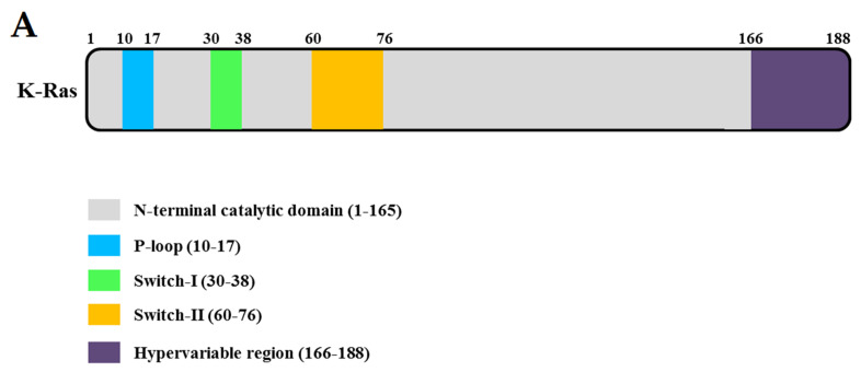
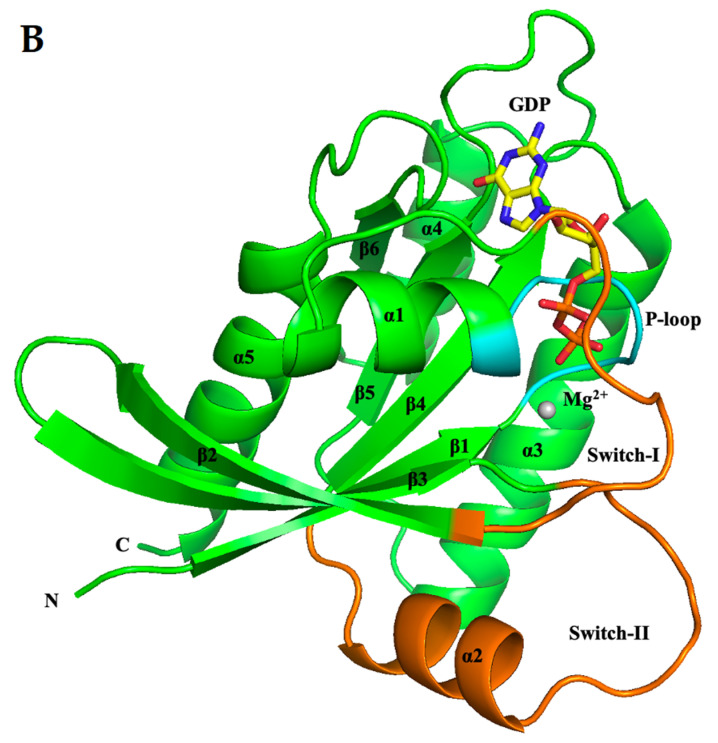
Figure 1.
Domain structures and the three-dimensional structure. (A) Domain structure of full-length KRAS. (B) Ribbon representation of the monomeric crystal structure of KRAS (PDB ID: 7C40) using the program PyMOL. The Mg2+ ion is indicated by a gray circle and guanosine diphosphate (GDP) by a yellow rod. The MgGDP molecule is color-coded as follows: C yellow, O red, N blue, P purple, and Mg2+ gray.


