Abstract
The dopamine D2/D3 receptor (D2R/D3R) agonists are used as therapeutics for Parkinson’s disease (PD) and other motor disorders. Selective targeting of D3R over D2R is attractive because of D3R’s restricted tissue distribution with potentially fewer side-effects and its putative neuroprotective effect. However, the high sequence homology between the D2R and D3R poses a challenge in the development of D3R selective agonists. To address the ligand selectivity, bitopic ligands were designed and synthesized previously based on a potent D3R-preferential agonist PF592,379 as the primary pharmacophore (PP). This PP was attached to various secondary pharmacophores (SPs) using chemically different linkers. Here, we characterize some of these novel bitopic ligands at both D3R and D2R using BRET-based functional assays. The bitopic ligands showed varying differences in potencies and efficacies. In addition, the chirality of the PP was key to conferring improved D3R potency, selectivity, and G protein signaling bias. In particular, compound AB04-88 exhibited significant D3R over D2R selectivity, and G protein bias at D3R. This bias was consistently observed at various time-points ranging from 8 to 46 min. Together, the structure-activity relationships derived from these functional studies reveal unique pharmacology at D3R and support further evaluation of functionally biased D3R agonists for their therapeutic potential.
Keywords: dopamine D3 receptor, dopamine D2 receptor, bitopic ligand, biased agonism, functional selectivity, subtype selectivity, subtype affinity, chirality
1. Introduction
Dopamine (DA) is a major neurotransmitter in the central nervous system responsible for various physiological functions such as motor control, cognition, reward, pain, and memory and learning [1]. Dopamine signaling is mediated by five G protein-coupled receptors (GPCRs) classified into two subgroups based on distinct sequence homologies and signal transduction activities [2]: D1-like receptors that primarily couple to Gs protein and enhance the activity of adenylyl cyclase, leading to an increase in intracellular cAMP production, and D2-like receptors that primarily couple to the Gi/Go class of G proteins and suppress the activity of adenylyl cyclase. The dopamine D2 and D3 receptors (D2R and D3R), both belonging to the D2-like receptor subfamily, represent the major targets for neuropsychiatric disorders such as schizophrenia, Parkinson’s disease (PD), and substance use disorders (SUDs) [3,4]. Between the two receptor subtypes, selective targeting of D3R would have lower potential for side effects because of its relatively restricted distribution in the ventral striatum compared to the expression of D2R both centrally and peripherally [5,6,7]. Additionally, D3R has also emerged as the target for L-DOPA induced dyskinesia (LID) in PD [8,9,10]. D3R is upregulated in animal models of PD, essentially in the same regions of the brain where D1R is expressed. As such, D3R-D1R heteromerization has been suggested to underlie the LID in PD [8,10].
While D3R antagonists or partial agonists have been studied for schizophrenia, substance use disorders, and L-DOPA induced dyskinesia in PD, D3R-preferential agonists, which is the focus of this study, have been studied for PD and other motor-associated disorders [3,11,12]. Particularly, D3R has emerged as a potential target for the treatment of PD due to their pharmacological similarity to the D2R, but without their added risk for the side-effects likely associated with the peripherally distributed D2Rs [5,6,13]. For example, D3R agonists have been shown to alleviate the symptoms of PD in a chemically induced mouse model of PD. Rescue of DA depletion in the striatum as well as DA neuronal death in the substantia nigra was posited as the mechanism of action of these agents. Importantly, these beneficial effects were not observed in the D3R knockout mice, thus validating the role of D3R in mediating these effects [14]. Thus, D3R agonists have been shown to not only improve PD symptoms, such as motor perturbation and cognitive deficits, but also appear to slow down the neurodegenerative process that underlies PD progression, in these animal models [13]. However, not all reports point to the beneficial roles of D3R, as the involvements of D3R in neuroinflammation and PD pathogenesis have been reported [4]. Despite tremendous efforts, the development of highly selective D3R agonists has remained a challenge, because of the nearly identical orthosteric binding site (OBS) and the 78% sequence identity in the transmembrane domain between the D2R and D3R [15,16,17]. This not only poses a challenge in discerning the individual contributions in conferring the therapeutic effects of D2R/D3R agonists, but also in understanding the receptor specific tissue distribution and signaling pathways such as dimerization, transactivation, biased signaling, and allosterism. Indeed, D3R signaling is very intricate that several ex vivo and in vitro studies demonstrate its existence as homomers and heteromers with either D1R or D2R [18,19,20]. The understanding of D3R specific signaling is further complicated by the distinct mechanisms of action and downstream functional profiles for homomers and heteromers [20,21]. These unique mechanisms of action may contribute to the therapeutic properties of antiparkinsonian agents as suggested by their high potency at D2R-D3R heteromers [22]. D3R agonists currently used as therapies or as research tools exhibit limited D3R selectivity of ~10 fold over D2R [2]. Thus, novel D3R agonists with high affinity, selectivity, over D2R and other homologous GPCRs (e.g., 5HT1A), are required to probe their unique pharmacological properties and to determine their therapeutic efficacy as well as side effect profiles.
One important concept in GPCR pharmacology is functional selectivity, whereby GPCR biased ligands selectively modulate canonical G protein dependent pathways or G protein independent pathways such as β-arrestin signaling [23,24]. Increasing evidence suggests that functional selectivity may provide increased efficacy, improved safety profiles, and overall therapeutic advantage [25]. Indeed, G protein biased agonism at D2R has been suggested to underlie antipsychotic efficacy [26,27,28]. At D3R, a series of agonists have been synthesized and identified such as SK609 and SK213 with higher potency for G protein dependent pathways and low tendency to recruit β-arrestin. Among this series, studies using the lead D3R selective G protein biased agonist revealed efficacy in improving PD symptoms in hemiparkinsonian rodent models [29,30], indicating a potential therapeutic utility of G protein functional bias at D3R. In case of β-arrestin bias, there is no evidence suggesting therapeutic utility of D3R specific β-arrestin bias; however, studies in several other GPCRs indicate the potential therapeutic utility of β-arrestin bias in psychiatric and neurological disorders including schizophrenia, PD and SUDs [31].
Although several G protein versus β-arrestin biased agonists for D3R have been successfully identified using cellular functional assays, many studies lack a biophysical approach that can directly demonstrate coupling of G protein versus β-arrestin [29,32,33]. Thus, there is a need for biophysical characterization of novel agonists with D3R specific functional selectivity before evaluating whether such agonists can provide therapeutic value in in vivo models and beyond.
Bitopic ligands are comprised of a primary pharmacophore (PP) that binds to the OBS—the endogenous ligand binding site, and a secondary pharmacophore (SP) that binds to a secondary binding pocket (SBP), connected by a chemically defined linker [34,35,36]. Previous studies reveal that the bitopic ligand strategy can provide improved receptor subtype selectivity, affinity, and functional selectivity [37,38,39]. Additionally, bitopic ligands can also confer unique receptor signaling an example of which is the bitopic ligand SB269,652 that behaves as an allosteric antagonist at D3Rs and D2Rs [40,41]. Indeed, we have successfully utilized a bitopic design strategy to synthesize potent, selective, and G protein biased full agonists at D2R [42]. Using a similar strategy, we recently synthesized bitopic D3R compounds with increased binding affinity and selectivity for D3R demonstrated in radioligand binding studies [43]. In this study, we utilize bioluminescence resonance energy transfer (BRET) based biophysical functional assays to probe structure activity relationships (SAR) in conferring D3R over D2R functional selectivity.
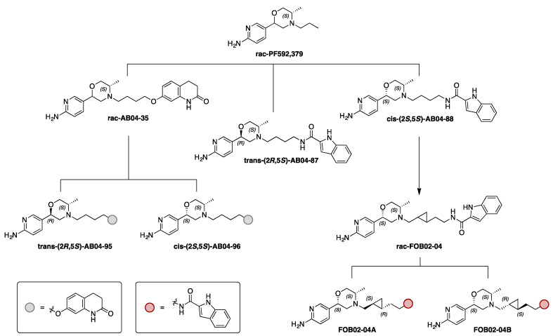
Specifically, we tested a series of congeneric bitopic compounds with their PP based on PF592,379 (Figure 1), an agonist at D3R developed by Pfizer for the treatment of sexual dysfunction and pain [43,44,45]. Among the series of compounds generated in Battiti et al. [43,46], we selected the ones that showed interesting SAR in radioligand binding assays for functional characterization [43]. In particular, we first selected the compounds that meets at least one of the two criteria: (i) a Ki < 32 nM in D3R binding (ii) a D3R over D2R selectivity > 22 fold. In order to comprehensively probe the role of chirality in conferring unique pharmacology, we further evaluated the corresponding racemic mixture or stereoisomers of the selected compounds in our BRET assays. First, a 3,4-dihydroquinoline-2(1H)-one SP, inspired by the antipsychotic D2R/D3R partial agonist Aripiprazole, was tethered to the PP with a butyl linker to generate rac-AB04-35, a mixture of two diastereoisomers. Based on the chirality at the PP morpholine ring, specifically in the 2-position, trans-(2R, 5S)-AB04-95 and cis-(2S, 5S)-AB04-96 were prepared via diastereospecific synthesis [43]. Second, a 2-indole amide SP was connected to the PP with the same butyl linker. In order to further assess the role of chirality in the PP, both trans-(2R, 5S)-AB04-87 and cis-(2S, 5S)-AB04-88 diastereoisomers were synthesized. Third, based on the observation of the optimal cis-(2S, 5S)-PP stereochemistry for D3R binding, the 2-indole amide SP was connected via a more rigid trans-cyclopropyl [47] containing linker to generate rac-FOB02-04 as a diastereoisomeric mixture of the trans cyclopropyl ring. Chiral resolution of the cyclopropyl linker gave two different isomers (1R, 2S)-FOBO2-04A and (1S, 2R)-FOB02-04B (Figure 1). Investigating pairs of diastereoisomers, we report functionally selective bitopic compounds that only show the bias characteristic in one stereoisomer, underscoring the importance of stereochemistry as a fundamental structural characteristic in D3R functional selectivity.
Figure 1.
Drug design for D3R agonists. For the bitopic ligand design, the primary pharmacophore (PP) scaffold used was inspired by PF592,379. Several bitopic ligands based on the PF592,379 moiety were synthesized [43], among which the ligands shown were tested for functional characterization.
2. Materials and Methods
2.1. Bioluminescence Resonance Energy Transfer (BRET) Studies
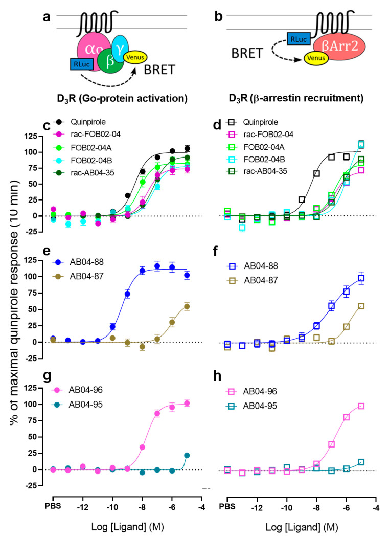
The BRET-based Go protein activation and β-arrestin recruitment assays were performed as described previously [34,42]. Go protein activation assay uses Renilla luciferase 8 (Rluc8; provided by Dr. S. Gambhir, Stanford University, Stanford, CA, USA)-fused GαoA and mVenus-fused Gγ2 as the BRET pair (Figure 2a). β-arrestin recruitment assay uses RLuc8-fused D3R or D2R and mVenus-fused β-arrestin2 as the BRET pair (Figure 2b). HEK293T cells were transiently transfected with the above constructs using polyethyleneimine (PEI) at a ratio of 2:1 (PEI:total DNA by weight). After ~48 h of transfection, cells were washed, harvested, and resuspended in PBS + 0.1% glucose + 200 µM Na bisulfite buffer. Then, 200,000 cells were transferred to each well of the 96-well plates (White Lumitrac 200, Greiner bio-one, Monroe, NC, USA) followed by addition of 5 µM coelenterazine H, a luciferase substrate for BRET. Test compounds, reference D2/D3 agonist-quinpirole (Tocris Bioscience, Minneapolis, MN, USA), and vehicle controls were then added by Nimbus liquid handling system (Hamilton, Reno, NV, USA) with its stamping protocol and cells were incubated at 37 °C for 10 min. BRET signal was then measured using a Pherastar FSX plate reader (BMG Labtech, Cary, NC, USA). For kinetic experiments, cells were incubated at 37 °C within the Pherastar FSX plate reader (BMG Labtech, Cary, NC, USA) with BRET signal measurements taken at various time-points ranging from 2–46 min. BRET ratio was calculated as the ratio of mVenus (530 nm) over RLuc8 (480 nm) emission.
Figure 2.
Pharmacological comparison of D3R bitopic ligands in both G protein activation and β-arrestin recruitment at 10 min. (a). Scheme for the bioluminescence resonance energy transfer (BRET) between Gαo-RLuc and Gγ-Venus. (b). Scheme for the BRET between D3R-RLuc and β-arrestin2-Venus. Concentration-response curves (CRCs) of drug-induced BRET at 10 min between Gαo-RLuc and Gγ-Venus (c,e,g). CRCs of drug-induced BRET at 10 min between D3R-RLuc and β-arrestin2-Venus (d,f,h). CRCs are plotted as percentage of maximal response by quinpirole and presented as means ± SEM of n ≥ 3 independent experiments.
Data were collected from at least 3 independent experiments and normalized to maximal response by quinpirole as 100% and response by vehicle as 0%. Concentration response curves (CRCs) were generated using a non-linear sigmoidal dose-response analyses using Prism 8 (GraphPad Software, San Diego, CA, USA) and presented as mean ± SEM.
2.2. Bias Factor Analysis
To evaluate whether the test compounds exhibited G protein versus β-arrestin signaling bias, bias factors were calculated using the method as follows [1]:
| (1) |
An arbitrary but stringent cut-off of ≥ ±2.0 (in logarithmic scale) was chosen to identify biased ligands. Bias factor values >2.0 represent bias towards G protein while values below <−2.0 represent bias towards β-arrestin.
2.3. An In-House Program for Kinetics Analysis of Functional Assay Data
In this study, the BRET signals for each 96-well plate were detected every two minutes using the BMG Pherastar FSX plate reader (BMG Labtech, Cary, NC, USA), resulting in 23 datasets for a 46 min measurement in one raw data file per plate. Such amount of data is beyond what can be conveniently and reliably processed by manual extraction, transformation, and normalization, before the regression analysis by Prism. Thus, we developed an in-house python program that can process and analyze the kinetics of functional assay data. While this program was configurated to fit the raw file output format of the plate reader used in this study, it can be easily adapted to process other output formats, time intervals, and plate maps (i.e., how the rows and columns of 96-well plates are configured for the dose-response measurements). This program also has the capability to process multiple files, based on predefined file locations in a configuration file. In this configuration file, for each raw file to be processed, it also includes time intervals, receptor construct, test compounds, and concentration ranges for each plate.
Each raw BRET data file was first preprocessed by detecting the data set for each time cycle. The response values were calculated as the ratio between 475-30B and 535-30A data for each well of the 96-well plate in the same time cycle. We took the average of response values for each compound at each concentration in the same time cycle. We then fitted response values to the sigmoidal dose-response function.
Sigmoidal dose-response function: , top and bottom are the maximum and minimum of the response values, respectively, x is the logarithm of the concentration, and logEC50 is the x value when the response is halfway between bottom and top.
We used the scipy.optimize.curve_fit module (version 1.5.2) [48] to perform this fitting process. In this curve fit module, we chose ‘lm’ as the optimization method type, which can replicate the regression result using Prism 8 (GraphPad Software, San Diego, CA, USA). The initial guess values were the minimum response value for “bottom”, the maximum response value for “top”, and the halfway value of the log(concentration) range for “logEC50”. After the fitting, the Emax was calculated as the difference between the optimized “bottom” and the optimized “top”, and the log (concentration) resulting in half of Emax is the logEC50.
To demonstrate functional kinetics, the program integrates the regression results at each time point and plots the Emax and logEC50 evolutions along the time (Figure 4).
2.4. Statistics
Statistical significance values were calculated using Prism 8 (GraphPad Software, San Diego, CA, USA)’s ordinary One-way ANOVA (independent variable: compound treatment, dependent variables: efficacy or pEC50s) followed by Dunnett’s multiple comparisons tests. For kinetic data, statistical significance values were calculated using GraphPad Prism’s ordinary two-way ANOVA (independent variables: compound treatment and time-point, dependent variables: efficacy or pEC50s) followed by Sidak’s multiple comparisons tests. For the above, two multiple comparisons, ‘*’ represents a significance of p < 0.05; ‘**’ of p < 0.01; ‘***’ of p < 0.001 and ‘****’ of p < 0.0001 compared to quinpirole. Dunnett’s multiple comparisons tests were also performed against AB04-87 with ‘δ’ representing significance of p < 0.05; ‘δδ’ of p < 0.01; ‘δδδ’ of p < 0.001 and ‘δδδδ’ of p < 0.0001 compared to AB04-87. Data are reported from more than three experiments performed in triplicate. In the case where the data points could not be fitted into the non-linear sigmoidal dose-response equation, the pharmacological parameters are reported as ND (not determined).
3. Results
3.1. Bitopic Compounds Exhibit Varying Pharmacological Profiles at Both D3R and D2R Compared to the Reference D2R/D3R Agonist Quinpirole
The bitopic compounds characterized in this study all have the PF592,379 scaffold as the primary pharmacophore (PP). For the chiral center at the 2-position of the morpholine ring in this scaffold, while AB04-88, rac-FOB02-04, FOB02-04A, FOB02-04B, and AB04-96 are in the cis conformation, AB04-95 and AB04-87 possess trans stereochemistry. rac-AB04-35 is the diastereomeric mixture of the AB04-95 and AB04-96, which have the same butyl linker and 3,4-dihydroquinoline-2(1H)-one as the SP (Figure 1). While AB04-87 and AB04-88 also have the same butyl linker, they differ from these compounds in their SP (2-indole amide). rac-FOB02-04, FOB02-04A, and FOB02-04B have the same 2-indole amide SP but with different trans-cyclopropyl containing linker. To investigate the impact of these differences in stereochemistry, linker, and SP on their functional properties, we evaluated the pharmacological profiles of these bitopic compounds with BRET-based Go protein activation and β-arrestin recruitment assays (see Section 2).
In the D3R Go protein activation assay, our results showed that except for AB04-88, rac-AB04-35, and AB04-96, the others exhibited lower efficacies than that of quinpirole (p < 0.01 in all cases, one-way ANOVA followed by Dunnett’s multiple comparisons test, Figure 2c,e,g, Table 1). While rac-FOB02-04, FOB02-04B, AB04-87, rac-AB04-35, and AB04-96 exhibited statistically lower potencies compared to that of quinpirole (p < 0.001 in all cases), FOB02-04A showed comparable potency to that of quinpirole. Interestingly, AB04-88 exhibited a significantly higher potency (6.5-fold) than that of quinpirole (p < 0.001, Figure 2e, Table 1).
Table 1.
Pharmacological comparison of bitopic ligands at D3R.
| D3R, 10 min | Go Protein Activation Assay | β-arrestin Recruitment | ||||||
|---|---|---|---|---|---|---|---|---|
| Compounds | Emax ± SEM (% of Quinpirole) | pEC50 ± SEM | Change Emax over Quinpirole | Fold Potency over Quinpirole | Emax ± SEM (% of Quinpirole) | pEC50 ± SEM | Change Emax over Quinpirole | Fold Potency over Quinpirole |
| Quinpirole | 100 ± 2.27 δδδδ | 8.53 ± 0.08 δδδδ | 0 | 1.000 | 100 ± 2.6 δδδδ | 8.36 ± 0.09 δδδδ | 0 | 1.000 |
| rac-FOB02-04 | 74.4 ± 3.4 *** | 7.67 ± 0.16 ***,δδδδ | −25.6 | 0.138 | 68.5 ± 1.8 *** | 6.78 ± 0.07 ****, δδδ | −31.5 | 0.026 |
| FOB02-04A | 82.8 ± 3.0 **,δδ | 8.22 ± 0.12 δδδδ | −17.2 | 0.490 | 80.2 ± 3.3 * | 6.80 ± 0.10 ****, δδδδ | −19.8 | 0.028 |
| FOB02-04B | 78.4 ± 3.2 ****,δδ | 7.25 ± 0.11 ****, δδδδ | −21.6 | 0.052 | 112 ± 7.0 δδδδ | 5.87 ± 0.10 **** | 33.2 | 0.003 |
| AB04-87 | 60.3 ± 8.1 **** | 6.00 ± 0.23 **** | −39.7 | 0.003 | 67.2 ± 8.2 **** | 5.80 ± 0.19 **** | −32.8 | 0.003 |
| AB04-88 | 111.6 ± 2.8 δδδδ | 9.34 ± 0.09 ***,δδδδ | 11.6 | 6.500 | 110 ± 0.9 δδδδ | 7.03 ± 0.10 ****, δδδδ | 10.0 | 0.047 |
| rac-AB04-35 | 93.9 ± 3.0 δδδδ | 7.24 ± 0.07 ****, δδδδ | −6.1 | 0.051 | 94.8 ± 3.7 δδ | 6.31 ± 0.07 ****, δ | −5.2 | 0.009 |
| AB04-95 | ND | ND | ND | ND | ND | ND | ND | ND |
| AB04-96 | 100.4 ± 2.7 δδδδ | 7.73 ± 0.08 ***, δδδδ | 0.4 | 0.158 | 104.0 ± 2.9 δδδ | 6.76 ± 0.06 ****, δδδ | 4.0 | 0.025 |
Mean Emax ± SEM and pEC50 ± SEM values along with fold changes over the reference D2R and D3R agonist—quinpirole are reported. Using Dunnett’s multiple comparisons tests, statistical significance are reported as ‘*’ representing significance of p < 0.05; ‘**’ of p < 0.01; ‘***’ of p < 0.001 and ‘****’ of p < 0.0001 compared to quinpirole, and ‘δ’ of p < 0.05; ‘δδ’ of p < 0.01; ‘δδδ’ of p < 0.001 and ‘δδδδ’ of p < 0.0001 compared to AB04-87. ND, not determined.
In D3R β-arrestin recruitment assay, rac-FOB02-04, FOB02-04A, and AB04-87 showed significantly lower efficacies compared to quinpirole (p < 0.05 in all cases, Figure 2d,f, Table 1). FOB02-04B, AB04-88, rac-AB04-35, and AB04-96 showed comparable efficacies compared to quinpirole. All bitopic ligands showed significantly lower potencies in the β-arrestin recruitment assay compared to that of quinpirole (p < 0.0001,Figure 2d,f,h, Table 1). AB04-95 showed the lowest efficacy profile at both D3R mediated Go protein activation as well as β-arrestin recruitment.
In the D2R Go protein activation assay, FOB02-04B and rac-AB04-35 exhibited statistically lower efficacies as well as potencies compared to quinpirole (p < 0.001 in both cases, One-way ANOVA followed by Dunnett’s multiple comparisons test, Table 2). rac-FOB02-04 showed a statistically lower potency (p < 0.05) with comparable efficacy compared to quinpirole. Both the bitopic ligands with the trans diastereoisomer in the PP: AB04-87 and AB04-95 did not show any activity in both Go protein activation and β-arrestin recruitment. All the remaining bitopic ligands showed significantly lower potencies compared to quinpirole (p < 0.0001 in all cases) in β-arrestin recruitment (Table 2). While rac-FOB02-04, FOB02-04A, FOB02-04B and AB04-88 showed lower efficacies (p < 0.0001), rac-AB04-35 showed comparable efficacy to quinpirole in β-arrestin recruitment. Interestingly, AB04-96 exhibited a ~22% higher efficacy compared to quinpirole (p < 0.001) in D2R mediated β-arrestin recruitment. Of note, this is the only compound that exhibits higher efficacy than quinpirole.
Table 2.
Pharmacological comparison of bitopic ligands at D2R.
| D2R, 10 min | Go Protein Activation Assay | β-arrestin Recruitment | ||||||
|---|---|---|---|---|---|---|---|---|
| Compounds | Emax ± SEM (% of Quinpirole) | pEC50 ± SEM | Change Emax over Quinpirole | Fold Potency over Quinpirole |
Emax ± SEM (% of Quinpirole) | pEC50 ± SEM | Change Emax over Quinpirole | Fold Potency over Quinpirole |
| Quinpirole | 100 ± 2.0 | 7.46 ± 0.07 | 0.00 | 1.00 | 100 ± 2.8 | 6.93 ± 0.08 | 0.0 | 1.00 |
| rac-FOB02-04 | 93.2 ± 2.2 | 7.13 ± 0.06 * | −6.80 | 0.47 | 62.9 ± 1.9 **** | 5.72 ± 0.07 **** | −37.1 | 0.06 |
| FOB02-04A | 91.2 ± 2.8 | 7.31 ± 0.08 | −8.80 | 0.71 | 60.4 ± 1.9 **** | 6.04 ± 0.07 **** | −39.6 | 0.13 |
| FOB02-04B | 81.4 ± 3.8 *** | 6.44 ± 0.13 **** | −18.60 | 0.10 | 56.9 ± 1.4 **** | 5.45 ± 0.05 **** | −43.1 | 0.03 |
| AB04-87 | ND | ND | ND | ND | ND | ND | ND | ND |
| AB04-88 | 95.2 ± 2.4 | 7.25 ± 0.07 | −4.80 | 0.62 | 72.6 ± 2.6 **** | 5.84 ± 0.06 **** | −27.4 | 0.08 |
| rac-AB04-35 | 75.9 ± 5.3 *** | 6.48 ± 0.13 **** | −24.10 | 0.10 | 102.8 ± 2.5 | 5.74 ± 0.06 **** | 2.80 | 0.06 |
| AB04-95 | ND | ND | ND | ND | ND | ND | ND | ND |
| AB04-96 | 102.7 ± 4.0 | 7.28 ± 0.11 | 2.70 | 0.66 | 121.6 ± 8.2 *** | 6.14 ± 0.13 **** | 21.6 | 0.16 |
Mean Emax ± SEM and pEC50 ± SEM values along with fold changes over quinpirole are reported. Using Dunnett’s multiple comparisons tests, statistical significance are reported as ‘*’ representing significance of p < 0.05; ‘***’ of p < 0.001 and ‘****’ of p < 0.0001 compared to quinpirole. ND, not determined.
The cis-isomers of bitopic ligands are more potent at D3R than their trans-isomers.
AB04-96 and AB04-95 only differ in the chirality at the 2-position of the morpholine ring in their PP, i.e., in cis- and trans-stereochemistry, respectively (Figure 1) [43]. The same difference in stereochemistry is seen between AB04-88 and AB04-87, another diastereoisomeric pair. In all four compounds, the same butyl linker was used to connect the PP with their corresponding SPs. Given that chirality can considerably modulate pharmacological profiles [38,49], we compared these two pairs of ligands in their ability to promote D3R Go protein activation and β-arrestin recruitment.
As shown in Figure 2e–h and Table 1, in both cases, the cis-stereochemistry (AB04-88 and AB04-96) of the morpholine ring confer higher potencies as well as efficacies compared to their corresponding trans diastereoisomers (AB04-87 and AB04-95, respectively). The cis-AB04-88 exhibited a ~2188-fold increase in potency (p < 0.0001, Dunnett’s multiple comparisons test) and an increase in Emax of ~51.3% (p < 0.0001) compared to its trans-AB04-87 in D3R Go protein activation (using the efficacy of quinpirole as 100%) (Figure 2e and Table 1).
In D3R mediated β-arrestin recruitment, the cis-AB04-88 exhibited a significant increase in Emax of ~42.8% (p < 0.0001) and a 17-fold increase in potency compared to its trans-AB04-87 (Figure 2f and Table 1). Interestingly, while the trans-AB04-95 exhibited a near complete abolishment of D3R mediated Go protein activation as well as β-arrestin recruitment, cis-AB04-96 exhibited a robust activity in both assays as shown in Figure 2g–h and Table 1. Thus, the potency and efficacy of AB04-95 cannot be quantitatively determined.
3.2. The cis-AB04-88 but not the trans-AB04-87 Shows an Improved D3R Selectivity Over D2R
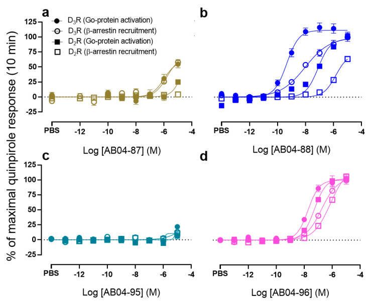
SPs and linkers are key to achieving subtype selectivity by binding to the SBPs unique to each receptor subtype [38,50,51,52]. To investigate whether these ligands exhibit D3R over D2R subtype selectivity, their ability to promote D3R and D2R mediated Go protein activation and β-arrestin recruitment were compared. In Go protein activation, all the compounds except AB04-88 and quinpirole showed < 10-fold D3R over D2R selectivity (Table 3). While the cis-stereochemistry of the PP, i.e., AB04-88 exhibited ~123-fold D3R over D2R selectivity in Go protein activation, selectivity with the trans-isomer could not be quantified (Figure 3a–b and Table 3) due to loss of Go protein activation at D2R (Table 2).
Table 3.
D3R over D2R selectivity and Go protein vs. β-arrestin bias factors of bitopic ligands.
| Selectivity (D2R/D3R) | Bias Factors | |||
|---|---|---|---|---|
| Compounds | Go Protein Activation | β-arrestin Recruitment | D3R | D2R |
| Quinpirole | 11.7 | 26.9 | ND | ND |
| rac-FOB02-04 | 3.5 | 11.5 | 0.9 | 1.6 |
| FOB02-04A | 8.1 | 5.8 | 1.4 | 1.4 |
| FOB02-04B | 6.5 | 2.6 | 1.1 | 1.1 |
| AB04-87 | ND | ND | 0.2 | ND |
| AB04-88 | 123.0 | 15.5 | 2.3 | 1.5 |
| rac-AB04-35 | 5.8 | 3.7 | 0.9 | 0.6 |
| AB04-95 | ND | ND | ND | ND |
| AB04-96 | 2.8 | 4.2 | 1.0 | 1.1 |
D3R over D2R selectivity and Go protein versus β-arrestin bias factors are reported. The value > 100 for D3R over D2R selectivity is highlighted in green. Bias factors were calculated as described in Section 2. Bias factor value > 2.0 is highlighted in orange. ND = not determined.
Figure 3.
Pharmacological comparison of cis and trans pairs in both D3R and D2R mediated G protein activation and β-arrestin recruitment at 10 min. CRCs of AB04-87 (a), AB04-88 (b), AB04-95 (c) and AB04-96 (d)-induced BRET at 10 min between Gαo-RLuc and Gγ-Venus and D3R or D2R-Rluc and β-arrestin2-Venus. CRCs are plotted as percentage of maximal response by quinpirole and presented as means ± SEM of n ≥ 3 independent experiments.
AB04-96 is the bitopic ligand with the same cis-stereochemistry of PP and butyl linker as AB04-88 but with a different SP. Interestingly, this cis-isomer only exhibited a 2.8-fold selectivity (Figure 3d and Table 3) with its corresponding trans-isomer showing near abolishment of activity at both D3R and D2R (Figure 3c and Table 1 and Table 2).
In β-arrestin recruitment, quinpirole, rac-FOB02-04, and AB04-88 exhibited > 10-fold D3R over D2R selectivity. Conversely, bitopic ligands FOB02-04A, FOB02-04B, rac-AB04-35, and AB04-96 exhibited < 10-fold selectivity. D3R over D2R selectivities in β-arrestin recruitment could not be determined for AB04-87 and AB04-96 because of their near abolishment of activity at either D2R only (AB04-87) or at both D3R and D2R (AB04-96) (Figure 3a,c and Table 1, Table 2 and Table 3).
3.3. The cis-AB04-88 But Not the trans-AB04-87 Shows D3R Selective Go Protein Bias
Previous studies have shown that bitopic ligands can not only improve subtype potency and selectivity but can also promote functional selectivity [42,52]. There is an increasing interest in understanding G protein versus β-arrestin biased agonism at GPCRs because of potential therapeutic utility upon activating one pathway over the other. To evaluate whether the compounds shown in Figure 1 can promote D3R specific G protein versus β-arrestin functional selectivity, bias factors were derived using the model that incorporates both efficacy and potency differences into a single index [1].
The biased activity equation used to evaluate G protein versus β-arrestin bias is described in the Section 2. An arbitrary but stringent cut-off of > ± 2.0 (in logarithmic scale) was chosen to identify biased ligands, where values > 2.0 represent bias towards G protein while values < −2.0 represent bias towards β-arrestin. The calculated biased factors are presented in Table 3.
Among all the ligands tested for bias at both D3R and D2R (Table 3), only the cis-isomer AB04-88 exhibited a biased factor equal to 2.3 (Figure 3b and Table 3) at D3R, suggesting that this ligand promotes D3R mediated Go protein bias. Upon inversion at the chiral center in the 2-position to generate trans-isomer AB04-87, the D3R mediated Go protein bias is completely lost (Figure 3a and Table 3). A similar trend can be observed at D2R, whereby the bias factor for AB04-88 was calculated to be 1.5 whereas the bias factor for AB04-87 could not be quantified (Figure 3a–b and Table 3), suggesting that AB04-88 exhibits a D3R selective Go protein bias.
In case of the second set of cis-trans pairs, the bias factor for the cis-AB04-96 was calculated to be 1.0 and 1.1 for D3R and D2R, respectively. However, the bias factors for its corresponding trans-isomer AB04-95 could not be quantified at both D3R and D2R because its activities were nearly abolished for both Go protein activation and β-arrestin recruitment. Although AB04-88 and AB04-96 were designed using the same cis form of the PP and the same butyl linker, they differ in their SPs. The observed D3R mediated Go protein bias using AB04-88 but not AB04-96 further stresses the role of SPs in conferring unique pharmacological profiles.
3.4. Time-Dependent Pharmacological Analysis Reveals Higher Go Protein Activation Emax for AB04-88 at Later Time Points
Time-dependent pharmacological profile, often called kinetic context, can be determined by the ligand-binding and receptor-signaling kinetics collectively and can impact biased agonism and its quantification [53,54]. Since our bitopic agonist AB04-88 exhibited D3R specific bias towards Go protein, we evaluated the kinetic profiles of AB04-88 in comparison to that of quinpirole. We assessed the kinetic profiles of both efficacies and potencies that constitute the two components used in our D3R biased agonism quantification [1], based on the data points collected every 2 min up to 46 min (see Section 2).
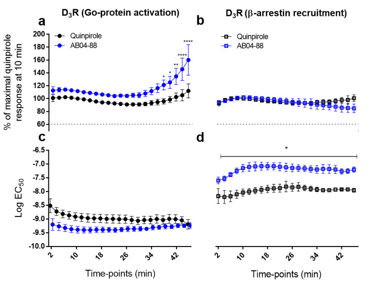
As shown in Figure 4, there were no significant differences between the AB04-88 and quinpirole efficacies at D3R until the 36 min time-point. Interestingly, after the 36 min time-point, AB04-88 exhibits significantly higher efficacies compared to quinpirole (in all cases p < 0.05; two-way ANOVA followed by Sidak’s multiple comparisons test) with maximal increases of > 40% observed at 44 and 46-min time-points (Figure 4a and Table 4). In contrast, there were no statistically significant differences observed between AB04-88 and quinpirole for EC50 of Go protein activation (Figure 4c and Table 4) or the efficacy component for β-arrestin recruitment (Figure 4b and Table 4). In contrast, in the β-arrestin recruitment assay, the Emax profiles for both AB04-88 and quinpirole nearly overlap up to 36 min, with AB04-88 showing slightly decreased Emax afterwards. The potency of AB04-88 remains significantly and consistently lower compared to quinpirole at all time-points ranging from 4–46 min (in all cases p < 0.05; two-way ANOVA followed by Sidak’s multiple comparisons test) (Figure 4d and Table 4).
Figure 4.
Time-dependent pharmacological profiles of AB04-88 and quinpirole on D3R mediated G protein activation and β-arrestin recruitment. a-c. Efficacy (a,b) and potency (c,d) profiles of D3R mediated G protein activation (a,c) and β-arrestin recruitment (b,d) measured with 2 min interval from 2 to 46 min. The Emax values are plotted as percentage of maximal response by quinpirole at 10 min. Plots are presented as means ± SEM of n ≥ 3 independent experiments. Statistical significances were calculated using GraphPad Prism’s ordinary two-way ANOVA (in each case, **** p < 0.0001) followed by Sidak’s multiple comparisons tests with ‘*’ representing significance of p < 0.05; ‘**’ of p < 0.01 and ‘****’ of p < 0.0001 compared to that of quinpirole.
Table 4.
Time dependent D3R pharmacological profile of quinpirole and AB04-88.
| Quinpirole | AB04-88 | |||||||||
|---|---|---|---|---|---|---|---|---|---|---|
| Go Protein Activation | β-arrestin Recruitment | Bias Factors | Go Protein Activation | β-arrestin Recruitment | Bias Factors | |||||
| Time-points (min) | pEC50 ± SEM | Emax (% of quinpirole) | pEC50 ± SEM | Emax (% of quinpirole) | pEC50 ± SEM | Emax (% of quinpirole) | pEC50 ± SEM | Emax (% of quinpirole) | ||
| 2 | 8.51 ± 0.25 | 101 ± 5.6 | 8.20 ± 0.26 | 92.8 ± 3.9 | 0.4 | 9.20 ± 0.21 | 112.7 ± 5.8 | 7.64 ± 0.12 | 94.4 ± 1.4 | 1.6 |
| 4 | 8.72 ± 0.14 | 101.4 ± 2.0 | 8.23 ± 0.23 | 97.9 ± 1.9 | 0.5 | 9.27 ± 0.14 | 113.8 ± 3.3 | 7.57 ± 0.08 * | 96.4 ± 0.2 | 1.8 |
| 6 | 8.81 ± 0.13 | 102.3 ± 1.0 | 8.21 ± 0.19 | 100.3 ± 1 | 0.6 | 9.32 ± 0.12 | 114.0 ± 1.6 | 7.43 ± 0.07 ** | 99.1 ± 0.2 | 1.9 |
| 8 | 8.86 ± 0.13 | 101.4 ± 0.4 | 8.13 ± 0.14 | 100.6 ± 0.3 | 0.7 | 9.37 ± 0.12 | 112.9 ± 0.6 | 7.30 ± 0.09 *** | 101.3 ± 0.1 | 2.1 |
| 10 | 8.91 ± 0.14 | 100.0 ± 0.0 | 8.07 ± 0.12 | 100.0 ± 0 | 0.8 | 9.39 ± 0.12 | 111.7 ± 0.0 | 7.20 ± 0.13 *** | 102.2 ± 0 | 2.3 |
| 12 | 8.94 ± 0.14 | 98.9 ± 0.3 | 8.04 ± 0.10 | 99.4 ± 0.3 | 0.9 | 9.40 ± 0.11 | 110.0 ± 0.5 | 7.16 ± 0.15 *** | 101.8 ± 0.9 | 2.3 |
| 14 | 8.96 ± 0.15 | 97.3 ± 0.6 | 8.01 ± 0.11 | 98.3 ± 0.6 | 0.9 | 9.40 ± 0.10 | 108.9 ± 0.8 | 7.14 ± 0.16 *** | 100.7 ± 1.8 | 2.3 |
| 16 | 8.97 ± 0.15 | 95.6 ± 0.9 | 7.97 ± 0.11 | 97.0 ± 0.9 | 1.0 | 9.39 ± 0.10 | 107.6 ± 1.2 | 7.13 ± 0.14 *** | 100.2 ± 2.8 | 2.3 |
| 18 | 8.98 ± 0.15 | 94.2 ± 1.1 | 7.94 ± 0.12 | 95.9 ± 1.3 | 1.0 | 9.39 ± 0.10 | 106.4 ± 1.5 | 7.13 ± 0.13 *** | 99.1 ± 3.3 | 2.3 |
| 20 | 8.99 ± 0.14 | 93.5 ± 1.2 | 7.92 ± 0.14 | 95.5 ± 1.5 | 1.0 | 9.39 ± 0.08 | 105.3 ± 1.7 | 7.14 ± 0.12 ** | 98.4 ± 3.5 | 2.3 |
| 22 | 8.99 ± 0.15 | 92.6 ± 1.4 | 7.92 ± 0.14 | 94.5 ± 1.7 | 1.0 | 9.40 ± 0.08 | 104.0 ± 1.9 | 7.15 ± 0.14** | 97.1 ± 3.8 | 2.3 |
| 24 | 9.00 ± 0.15 | 91.8 ± 1.6 | 7.86 ± 0.18 | 93.9 ± 1.9 | 1.1 | 9.38 ± 0.07 | 104.6 ± 2.1 | 7.17 ± 0.14 ** | 96.2 ± 4.5 | 2.2 |
| 26 | 9.00 ± 0.14 | 90.9 ± 1.6 | 7.89 ± 0.14 | 93.1 ± 1.9 | 1.1 | 9.36 ± 0.09 | 104.2 ± 2.3 | 7.20 ± 0.13 ** | 94.6 ± 5.1 | 2.2 |
| 28 | 9.03 ± 0.13 | 91.1 ± 1.9 | 7.87 ± 0.15 | 93.4 ± 1.9 | 1.1 | 9.35 ± 0.09 | 105.3 ± 3.1 | 7.20 ± 0.11 * | 94.0 ± 6.1 | 2.2 |
| 30 | 9.06 ± 0.12 | 91.0 ± 2.3 | 7.91 ± 0.10 | 93.1 ± 2.2 | 1.1 | 9.36 ± 0.08 | 105.3 ± 4.0 | 7.21 ± 0.11 ** | 93.0 ± 6.4 | 2.2 |
| 32 | 9.03 ± 0.13 | 91.6 ± 2.6 | 7.94 ± 0.08 | 93.1 ± 2.5 | 1.1 | 9.35 ± 0.06 | 108.0 ± 4.8 | 7.24 ± 0.11 ** | 92.1 ± 7.6 | 2.2 |
| 34 | 9.03 ± 0.13 | 93.3 ± 3.5 | 7.96 ± 0.07 | 94.3 ± 3.2 | 1.1 | 9.30 ± 0.08 | 111.6 ± 5.5 | 7.25 ± 0.13 ** | 90.6 ± 6.9 | 2.1 |
| 36 | 9.00 ± 0.15 | 94.7 ± 4.3 | 7.99 ± 0.06 | 94.7 ± 3.9 | 1.0 | 9.30 ± 0.07 | 116.2 ± 6.6 | 7.23 ± 0.15 ** | 89.7 ± 6.7 | 2.2 |
| 38 | 9.03 ± 0.15 | 95.8 ± 4.1 | 7.99 ± 0.05 | 95.1 ± 3.1 | 1.0 | 9.26 ± 0.09 | 121.3 ± 7.7 * | 7.23 ± 0.15 ** | 88.5 ± 6.0 | 2.2 |
| 40 | 8.99 ± 0.16 | 98.6 ± 4.8 | 7.97 ± 0.05 | 96.2 ± 3.5 | 1.0 | 9.27 ± 0.07 | 125.3 ± 9.4 * | 7.25 ± 0.14 ** | 87.6 ± 6.8 | 2.2 |
| 42 | 9.00 ± 0.15 | 102.5 ± 6.2 | 7.96 ± 0.03 | 97.6 ± 4.3 | 1.1 | 9.24 ± 0.07 | 134.6 ± 12.5 ** | 7.32 ± 0.09 * | 85.9 ± 6.9 | 2.1 |
| 44 | 9.05 ± 0.13 | 105.2 ± 8.7 | 7.96 ± 0.04 | 98.0 ± 6.5 | 1.1 | 9.24 ± 0.06 | 145.6 ± 17.3 **** | 7.31 ± 0.08 * | 85.4 ± 6.8 | 2.2 |
| 46 | 9.19 ± 0.17 | 112.0 ± 10.9 | 7.99 ± 0.07 | 100.6 ± 5.2 | 1.3 | 9.23 ± 0.09 | 160.0 ± 23.6 **** | 7.25 ± 0.09 ** | 84.9 ± 6.6 | 2.3 |
pEC50 ± SEM values and Emax values at both D3R mediated G protein activation and β-arrestin recruitment assays at time-points ranging from 2–46 min are reported. Bias factors, calculated as described in Section 2, are also reported. The bias factor values ≥ 2.0 are highlighted in orange. Using Sidak’s multiple comparisons tests, statistical significance are reported as ‘*’ representing significance of p < 0.05; ‘**’ of p < 0.01; ‘***’ of p < 0.001 and ‘****’ of p < 0.0001 compared to that of quinpirole.
We also calculated the bias factors at all time-points, AB04-88 exhibited a bias at timepoints ranging from 8–46 min (> ±2.0) but without major changes.
These data collectively suggest that, in the case of AB04-88, kinetics analysis revealed a significant Emax increase for Go protein activation at later time points, which, however, did not significantly affect the bias factor.
4. Discussion
The discovery of D3R selective agonists is of particular interest because such compounds would help ascertain D3R specific physiological and pathological functions from that of D2R in neurodegenerative disorders such as PD [55]. Furthermore, such agonists may exhibit improved clinical utility in the treatment of such disorders. The bitopic D3R ligands evaluated in this study exhibit varying pharmacological profiles compared to the reference D2R/D3R full agonist quinpirole. The PPs of the bitopic ligands in this study are based on the D3R-preferential agonist, PF592,379 and are either a mixture of diastereoisomers or one of the two cis- and trans- isomers based on the chirality of its morpholine ring. In addition, the bitopic ligands differ in their SPs, linkers, and linker stereochemistry.
Comparing the diastereomer pairs within the same ligand structures, the (2S, 5S) enantiomer of the PP morpholine ring shows more efficacious and potent D3R G protein signaling. This observation confirms the significance of enantiospecificity in the morpholine ring in achieving D3R activation efficacy.
In D3R Go protein activation BRET, cis-(2S, 5S)-AB04-88 presenting a 2-indole amide SP connected by a butyl linker, showed a 123-fold selectivity for D3R over D2R. Interestingly, another compound AB04-96 that has the same PP and linker but with a 3,4-dihydroquinolin-2(1H)-one SP only shows a 2.8-fold D3R over D2R selectivity. This observation highlights the importance of the SP in promoting subtype selectivity where the 2-indole amide, but not 3,4-dihydroquinolin-2(1H)-one promotes D3R subtype selectivity. This is consistent with previous findings where the presence of a 3,4-dihydroquinolin-2(1H)-one moiety improves D2R subtype binding [42,56,57]. Unlike the previous radioligand binding study in which FOB02-04A showed a 47.5-fold D3R over D2R selectivity, we only observed an eight-fold selectivity in Go protein activation BRET. The binding affinities and selectivities observed in radioligand binding studies may not always correlate with cell-based functional assays. The observed discrepancies in these two types of assays may be explained by inherent differences of probe dependency and specific assay conditions [58].
Given the increasing evidence supporting therapeutic utility of functional selectivity in GPCRs, we evaluated the D3R bitopic agonists for their signaling bias. AB04-88, which showed the highest potency in D3R Go protein activation among all bitopic ligands studied, also exhibited a Go protein activation over β-arrestin recruitment bias by two log units. While the (2S, 5S) cis stereoisomer of the PP appears favorable for Go protein activation over β-arrestin recruitment, AB04-96 with the same cis conformation of PP and linker, but a different SP, does not exhibit functional bias. Similar to the D3R over D2R selectivity, this difference highlights the significance of the SP in conferring functional bias as well. Future computational studies with the two diastereoisomeric (based on PP morpholine ring) sets of D3R bitopic agonists will provide clues as to the interplay between chirality and SP chemistry to provide unique pharmacology.
Increasing evidence suggests the influence of ligand binding kinetics and receptor signaling in biased agonism [53,54]. Since AB04-88 exhibited D3R specific Go protein bias at 10 min, we investigated whether this bias would change in a time-dependent manner. The only kinetic profile affected was that of the efficacy of AB04-88 in D3R Go protein activation, where it significantly increased compared to quinpirole after 36 min. However, the bias factor was not affected due to the largely stable potencies in both Go protein activation and β-arrestin assays, the difference of which would be the dominant component in the factor when AB04-88 remained a strong partial agonist at the later time-points of β-arrestin recruitment.
Together, our newly developed D3R bitopic ligands provide novel tools to further probe the unique pharmacology, specific cellular signaling, and therapeutic potential of D3R.
Author Contributions
P.A., H.Y. and L.S. designed the study. P.A. and H.Y. carried out the experiments; B.X. and L.S. developed the in-house analysis program; A.B., F.O.B. and A.H.N. designed and synthesized the tested compounds; all authors were involved in analyzing the results. P.A., H.Y. and L.S. wrote the initial draft, with contributions from all other authors in finalizing the manuscript. All authors have read and agreed to the published version of the manuscript.
Funding
Support for this research was provided by the National Institute on Drug Abuse—Intramural Research Program, Z1A DA000389 (A.H.N) and Z1A DA000606 (L.S.), and the Brain and Behavior Research Foundation Young Investigator Award (H.Y.).
Data Availability Statement
Not applicable.
Conflicts of Interest
The authors declare no conflict of interest.
Footnotes
Publisher’s Note: MDPI stays neutral with regard to jurisdictional claims in published maps and institutional affiliations.
References
- 1.Free R.B., Chun L.S., Moritz A.E., Miller B.N., Doyle T.B., Conroy J.L., Padron A., Meade J.A., Xiao J., Hu X., et al. Discovery and characterization of a G protein-biased agonist that inhibits beta-arrestin recruitment to the D2 dopamine receptor. Mol. Pharmacol. 2014;86:96–105. doi: 10.1124/mol.113.090563. [DOI] [PMC free article] [PubMed] [Google Scholar]
- 2.Beaulieu J.M., Gainetdinov R.R. The physiology, signaling, and pharmacology of dopamine receptors. Pharmacol. Rev. 2011;63:182–217. doi: 10.1124/pr.110.002642. [DOI] [PubMed] [Google Scholar]
- 3.Heidbreder C.A., Newman A.H. Current perspectives on selective dopamine D (3) receptor antagonists as pharmacotherapeutics for addictions and related disorders. Ann. N. Y. Acad. Sci. 2010;1187:4–34. doi: 10.1111/j.1749-6632.2009.05149.x. [DOI] [PMC free article] [PubMed] [Google Scholar]
- 4.Yang P., Perlmutter J.S., Benzinger T.L.S., Morris J.C., Xu J. Dopamine D3 receptor: A neglected participant in Parkinson Disease pathogenesis and treatment? Ageing Res. Rev. 2020;57:100994. doi: 10.1016/j.arr.2019.100994. [DOI] [PMC free article] [PubMed] [Google Scholar]
- 5.Murray A.M., Ryoo H.L., Gurevich E., Joyce J.N. Localization of dopamine D3 receptors to mesolimbic and D2 receptors to mesostriatal regions of human forebrain. Proc. Natl. Acad. Sci. USA. 1994;91:11271–11275. doi: 10.1073/pnas.91.23.11271. [DOI] [PMC free article] [PubMed] [Google Scholar]
- 6.Gonsai N.H., Amin V.H., Mendpara C.G., Speth R., Hale G.M. Effects of dopamine receptor antagonist antipsychotic therapy on blood pressure. J. Clin. Pharm. Ther. 2018;43:1–7. doi: 10.1111/jcpt.12649. [DOI] [PubMed] [Google Scholar]
- 7.Ballon J.S., Pajvani U., Freyberg Z., Leibel R.L., Lieberman J.A. Molecular pathophysiology of metabolic effects of antipsychotic medications. Trends Endocrinol. Metab. 2014;25:593–600. doi: 10.1016/j.tem.2014.07.004. [DOI] [PubMed] [Google Scholar]
- 8.Bezard E., Ferry S., Mach U., Stark H., Leriche L., Boraud T., Gross C., Sokoloff P. Attenuation of levodopa-induced dyskinesia by normalizing dopamine D3 receptor function. Nat. Med. 2003;9:762–767. doi: 10.1038/nm875. [DOI] [PubMed] [Google Scholar]
- 9.Solis O., Garcia-Montes J.R., Gonzalez-Granillo A., Xu M., Moratalla R. Dopamine D3 Receptor Modulates l-DOPA-Induced Dyskinesia by Targeting D1 Receptor-Mediated Striatal Signaling. Cereb. Cortex. 2017;27:435–446. doi: 10.1093/cercor/bhv231. [DOI] [PMC free article] [PubMed] [Google Scholar]
- 10.Ferre S., Lluis C., Lanciego J.L., Franco R. Prime time for G-protein-coupled receptor heteromers as therapeutic targets for CNS disorders: The dopamine D(1)-D(3) receptor heteromer. CNS Neurol. Disord. Drug Targets. 2010;9:596–600. doi: 10.2174/187152710793361603. [DOI] [PMC free article] [PubMed] [Google Scholar]
- 11.Leggio G.M., Bucolo C., Platania C.B., Salomone S., Drago F. Current drug treatments targeting dopamine D3 receptor. Pharmacol. Ther. 2016;165:164–177. doi: 10.1016/j.pharmthera.2016.06.007. [DOI] [PubMed] [Google Scholar]
- 12.Keck T.M., John W.S., Czoty P.W., Nader M.A., Newman A.H. Identifying Medication Targets for Psychostimulant Addiction: Unraveling the Dopamine D3 Receptor Hypothesis. J. Med. Chem. 2015;58:5361–5380. doi: 10.1021/jm501512b. [DOI] [PMC free article] [PubMed] [Google Scholar]
- 13.Das B., Modi G., Dutta A. Dopamine D3 agonists in the treatment of Parkinson's disease. Curr. Top. Med. Chem. 2015;15:908–926. doi: 10.2174/156802661510150328223428. [DOI] [PubMed] [Google Scholar]
- 14.Lao C.L., Kuo Y.H., Hsieh Y.T., Chen J.C. Intranasal and subcutaneous administration of dopamine D3 receptor agonists functionally restores nigrostriatal dopamine in MPTP-treated mice. Neurotox. Res. 2013;24:523–531. doi: 10.1007/s12640-013-9408-1. [DOI] [PubMed] [Google Scholar]
- 15.Levant B. The D3 dopamine receptor: Neurobiology and potential clinical relevance. Pharmacol. Rev. 1997;49:231–252. [PubMed] [Google Scholar]
- 16.Sokoloff P., Giros B., Martres M.P., Bouthenet M.L., Schwartz J.C. Molecular cloning and characterization of a novel dopamine receptor (D3) as a target for neuroleptics. Nature. 1990;347:146–151. doi: 10.1038/347146a0. [DOI] [PubMed] [Google Scholar]
- 17.Chien E.Y., Liu W., Zhao Q., Katritch V., Han G.W., Hanson M.A., Shi L., Newman A.H., Javitch J.A., Cherezov V., et al. Structure of the human dopamine D3 receptor in complex with a D2/D3 selective antagonist. Science. 2010;330:1091–1095. doi: 10.1126/science.1197410. [DOI] [PMC free article] [PubMed] [Google Scholar]
- 18.Pou C., Mannoury la Cour C., Stoddart L.A., Millan M.J., Milligan G. Functional homomers and heteromers of dopamine D2L and D3 receptors co-exist at the cell surface. J. Biol. Chem. 2012;287:8864–8878. doi: 10.1074/jbc.M111.326678. [DOI] [PMC free article] [PubMed] [Google Scholar]
- 19.Scarselli M., Novi F., Schallmach E., Lin R., Baragli A., Colzi A., Griffon N., Corsini G.U., Sokoloff P., Levenson R., et al. D2/D3 dopamine receptor heterodimers exhibit unique functional properties. J. Biol. Chem. 2001;276:30308–30314. doi: 10.1074/jbc.M102297200. [DOI] [PubMed] [Google Scholar]
- 20.Fiorentini C., Busi C., Gorruso E., Gotti C., Spano P., Missale C. Reciprocal regulation of dopamine D1 and D3 receptor function and trafficking by heterodimerization. Mol. Pharmacol. 2008;74:59–69. doi: 10.1124/mol.107.043885. [DOI] [PubMed] [Google Scholar]
- 21.Maggio R., Scarselli M., Capannolo M., Millan M.J. Novel dimensions of D3 receptor function: Focus on heterodimerisation, transactivation and allosteric modulation. Eur. Neuropsychopharmacol. 2015;25:1470–1479. doi: 10.1016/j.euroneuro.2014.09.016. [DOI] [PubMed] [Google Scholar]
- 22.Maggio R., Scarselli M., Novi F., Millan M.J., Corsini G.U. Potent activation of dopamine D3/D2 heterodimers by the antiparkinsonian agents, S32504, pramipexole and ropinirole. J. Neurochem. 2003;87:631–641. doi: 10.1046/j.1471-4159.2003.02038.x. [DOI] [PubMed] [Google Scholar]
- 23.Mailman R.B. GPCR functional selectivity has therapeutic impact. Trends Pharmacol. Sci. 2007;28:390–396. doi: 10.1016/j.tips.2007.06.002. [DOI] [PMC free article] [PubMed] [Google Scholar]
- 24.Urban J.D., Clarke W.P., von Zastrow M., Nichols D.E., Kobilka B., Weinstein H., Javitch J.A., Roth B.L., Christopoulos A., Sexton P.M., et al. Functional selectivity and classical concepts of quantitative pharmacology. J. Pharmacol. Exp. Ther. 2007;320:1–13. doi: 10.1124/jpet.106.104463. [DOI] [PubMed] [Google Scholar]
- 25.Kenakin T. Functional selectivity and biased receptor signaling. J. Pharmacol. Exp. Ther. 2011;336:296–302. doi: 10.1124/jpet.110.173948. [DOI] [PubMed] [Google Scholar]
- 26.Weiwer M., Xu Q., Gale J.P., Lewis M., Campbell A.J., Schroeder F.A., Van de Bittner G.C., Walk M., Amaya A., Su P., et al. Functionally Biased D2R Antagonists: Targeting the beta-Arrestin Pathway to Improve Antipsychotic Treatment. ACS Chem. Biol. 2018;13:1038–1047. doi: 10.1021/acschembio.8b00168. [DOI] [PubMed] [Google Scholar]
- 27.Allen J.A., Yost J.M., Setola V., Chen X., Sassano M.F., Chen M., Peterson S., Yadav P.N., Huang X.P., Feng B., et al. Discovery of beta-arrestin-biased dopamine D2 ligands for probing signal transduction pathways essential for antipsychotic efficacy. Proc. Natl. Acad. Sci. USA. 2011;108:18488–18493. doi: 10.1073/pnas.1104807108. [DOI] [PMC free article] [PubMed] [Google Scholar]
- 28.Park S.M., Chen M., Schmerberg C.M., Dulman R.S., Rodriguiz R.M., Caron M.G., Jin J., Wetsel W.C. Effects of beta-Arrestin-Biased Dopamine D2 Receptor Ligands on Schizophrenia-Like Behavior in Hypoglutamatergic Mice. Neuropsychopharmacology. 2016;41:704–715. doi: 10.1038/npp.2015.196. [DOI] [PMC free article] [PubMed] [Google Scholar]
- 29.Xu W., Wang X., Tocker A.M., Huang P., Reith M.E., Liu-Chen L.Y., Smith A.B., 3rd, Kortagere S. Functional Characterization of a Novel Series of Biased Signaling Dopamine D3 Receptor Agonists. ACS Chem. Neurosci. 2017;8:486–500. doi: 10.1021/acschemneuro.6b00221. [DOI] [PMC free article] [PubMed] [Google Scholar]
- 30.Simms S.L., Huettner D.P., Kortagere S. In vivo characterization of a novel dopamine D3 receptor agonist to treat motor symptoms of Parkinson’s disease. Neuropharmacology. 2016;100:106–115. doi: 10.1016/j.neuropharm.2015.04.004. [DOI] [PubMed] [Google Scholar]
- 31.Harris S.S., Urs N.M. Targeting beta-Arrestins in the Treatment of Psychiatric and Neurological Disorders. CNS Drugs. 2021;35:253–264. doi: 10.1007/s40263-021-00796-y. [DOI] [PubMed] [Google Scholar]
- 32.Xu W., Reith M.E.A., Liu-Chen L.Y., Kortagere S. Biased signaling agonist of dopamine D3 receptor induces receptor internalization independent of beta-arrestin recruitment. Pharmacol. Res. 2019;143:48–57. doi: 10.1016/j.phrs.2019.03.003. [DOI] [PubMed] [Google Scholar]
- 33.Zheng M., Zhang X., Sun N., Min X., Acharya S., Kim K.M. A novel molecular mechanism responsible for phosphorylation-independent desensitization of G protein-coupled receptors exemplified by the dopamine D3 receptor. Biochem. Biophys. Res. Commun. 2020;528:432–439. doi: 10.1016/j.bbrc.2020.05.197. [DOI] [PubMed] [Google Scholar]
- 34.Michino M., Boateng C.A., Donthamsetti P., Yano H., Bakare O.M., Bonifazi A., Ellenberger M.P., Keck T.M., Kumar V., Zhu C., et al. Toward Understanding the Structural Basis of Partial Agonism at the Dopamine D3 Receptor. J. Med. Chem. 2017;60:580–593. doi: 10.1021/acs.jmedchem.6b01148. [DOI] [PMC free article] [PubMed] [Google Scholar]
- 35.Michino M., Donthamsetti P., Beuming T., Banala A., Duan L., Roux T., Han Y., Trinquet E., Newman A.H., Javitch J.A., et al. A single glycine in extracellular loop 1 is the critical determinant for pharmacological specificity of dopamine D2 and D3 receptors. Mol. Pharmacol. 2013;84:854–864. doi: 10.1124/mol.113.087833. [DOI] [PMC free article] [PubMed] [Google Scholar]
- 36.Newman A.H., Beuming T., Banala A.K., Donthamsetti P., Pongetti K., LaBounty A., Levy B., Cao J., Michino M., Luedtke R.R., et al. Molecular determinants of selectivity and efficacy at the dopamine D3 receptor. J. Med. Chem. 2012;55:6689–6699. doi: 10.1021/jm300482h. [DOI] [PMC free article] [PubMed] [Google Scholar]
- 37.Fronik P., Gaiser B.I., Sejer Pedersen D. Bitopic Ligands and Metastable Binding Sites: Opportunities for G Protein-Coupled Receptor (GPCR) Medicinal Chemistry. J. Med. Chem. 2017;60:4126–4134. doi: 10.1021/acs.jmedchem.6b01601. [DOI] [PubMed] [Google Scholar]
- 38.Newman A.H., Battiti F.O., Bonifazi A. 2016 Philip, S. Portoghese Medicinal Chemistry Lectureship: Designing Bivalent or Bitopic Molecules for G-Protein Coupled Receptors. The Whole Is Greater Than the Sum of Its Parts. J. Med. Chem. 2020;63:1779–1797. doi: 10.1021/acs.jmedchem.9b01105. [DOI] [PMC free article] [PubMed] [Google Scholar]
- 39.Newman A.H., Ku T., Jordan C.J., Bonifazi A., Xi Z.X. New Drugs, Old Targets: Tweaking the Dopamine System to Treat Psychostimulant Use Disorders. Annu. Rev. Pharmacol. Toxicol. 2021;61:609–628. doi: 10.1146/annurev-pharmtox-030220-124205. [DOI] [PMC free article] [PubMed] [Google Scholar]
- 40.Draper-Joyce C.J., Michino M., Verma R.K., Klein Herenbrink C., Shonberg J., Kopinathan A., Scammells P.J., Capuano B., Thal D.M., Javitch J.A., et al. The structural determinants of the bitopic binding mode of a negative allosteric modulator of the dopamine D2 receptor. Biochem. Pharmacol. 2018;148:315–328. doi: 10.1016/j.bcp.2018.01.002. [DOI] [PMC free article] [PubMed] [Google Scholar]
- 41.Rossi M., Fasciani I., Marampon F., Maggio R., Scarselli M. The First Negative Allosteric Modulator for Dopamine D2 and D3 Receptors, SB269652 May Lead to a New Generation of Antipsychotic Drugs. Mol. Pharmacol. 2017;91:586–594. doi: 10.1124/mol.116.107607. [DOI] [PMC free article] [PubMed] [Google Scholar]
- 42.Bonifazi A., Yano H., Ellenberger M.P., Muller L., Kumar V., Zou M.F., Cai N.S., Guerrero A.M., Woods A.S., Shi L., et al. Novel Bivalent Ligands Based on the Sumanirole Pharmacophore Reveal Dopamine D2 Receptor (D2R) Biased Agonism. J. Med. Chem. 2017;60:2890–2907. doi: 10.1021/acs.jmedchem.6b01875. [DOI] [PMC free article] [PubMed] [Google Scholar]
- 43.Battiti F.O., Cemaj S.L., Guerrero A.M., Shaik A.B., Lam J., Rais R., Slusher B.S., Deschamps J.R., Imler G.H., Newman A.H., et al. The Significance of Chirality in Drug Design and Synthesis of Bitopic Ligands as D3 Receptor (D3R) Selective Agonists. J. Med. Chem. 2019;62:6287–6314. doi: 10.1021/acs.jmedchem.9b00702. [DOI] [PMC free article] [PubMed] [Google Scholar]
- 44.Allerton C.M.N., Cook A.S., Hepworth D., Miller D.C. WO 115985 Al. Aminopyridine Derivatives as Selective Dopamine D3 Agonists. 2005 Aug 12;
- 45.Ackley M.A. Morpholine Dopamine Agonists for The Treatment of Pain. WO 087512 Al. 2008 Jul 24;
- 46.Battiti F.O., Newman A.H., Bonifazi A. Exception That Proves the Rule: Investigation of Privileged Stereochemistry in Designing Dopamine D3R Bitopic Agonists. ACS Med. Chem. Lett. 2020;11:1956–1964. doi: 10.1021/acsmedchemlett.9b00660. [DOI] [PMC free article] [PubMed] [Google Scholar]
- 47.Moritz A.E., Bonifazi A., Guerrero A.M., Kumar V., Free R.B., Lane J.R., Verma R.K., Shi L., Newman A.H., Sibley D.R. Evidence for a stereoselective mechanism for bitopic activity by extended-length antagonists of the D3 dopamine receptor. ACS Chem. Neurosci. 2020;11:3309–3320. doi: 10.1021/acschemneuro.0c00425. [DOI] [PMC free article] [PubMed] [Google Scholar]
- 48.Virtanen P., Gommers R., Oliphant T.E., Haberland M., Reddy T., Cournapeau D., Burovski E., Peterson P., Weckesser W., Bright J., et al. SciPy 1.0: Fundamental algorithms for scientific computing in Python. Nat. Methods. 2020;17:261–272. doi: 10.1038/s41592-019-0686-2. [DOI] [PMC free article] [PubMed] [Google Scholar]
- 49.Brooks W.H., Guida W.C., Daniel K.G. The significance of chirality in drug design and development. Curr. Top Med. Chem. 2011;11:760–770. doi: 10.2174/156802611795165098. [DOI] [PMC free article] [PubMed] [Google Scholar]
- 50.Kumar V., Bonifazi A., Ellenberger M.P., Keck T.M., Pommier E., Rais R., Slusher B.S., Gardner E., You Z.B., Xi Z.X., et al. Highly Selective Dopamine D3 Receptor (D3R) Antagonists and Partial Agonists Based on Eticlopride and the D3R Crystal Structure: New Leads for Opioid Dependence Treatment. J. Med. Chem. 2016;59:7634–7650. doi: 10.1021/acs.jmedchem.6b00860. [DOI] [PMC free article] [PubMed] [Google Scholar]
- 51.Kumar V., Moritz A.E., Keck T.M., Bonifazi A., Ellenberger M.P., Sibley C.D., Free R.B., Shi L., Lane J.R., Sibley D.R., et al. Synthesis and Pharmacological Characterization of Novel trans-Cyclopropylmethyl-Linked Bivalent Ligands That Exhibit Selectivity and Allosteric Pharmacology at the Dopamine D3 Receptor (D3R) J. Med. Chem. 2017;60:1478–1494. doi: 10.1021/acs.jmedchem.6b01688. [DOI] [PMC free article] [PubMed] [Google Scholar]
- 52.Bonifazi A., Yano H., Guerrero A.M., Kumar V., Hoffman A.F., Lupica C.R., Shi L., Newman A.H. Novel and Potent Dopamine D2 Receptor Go-Protein Biased Agonists. ACS Pharmacol. Transl. Sci. 2019;2:52–65. doi: 10.1021/acsptsci.8b00060. [DOI] [PMC free article] [PubMed] [Google Scholar]
- 53.Lane J.R., May L.T., Parton R.G., Sexton P.M., Christopoulos A. A kinetic view of GPCR allostery and biased agonism. Nat. Chem. Biol. 2017;13:929–937. doi: 10.1038/nchembio.2431. [DOI] [PubMed] [Google Scholar]
- 54.Klein Herenbrink C., Sykes D.A., Donthamsetti P., Canals M., Coudrat T., Shonberg J., Scammells P.J., Capuano B., Sexton P.M., Charlton S.J., et al. The role of kinetic context in apparent biased agonism at GPCRs. Nat. Commun. 2016;7:10842. doi: 10.1038/ncomms10842. [DOI] [PMC free article] [PubMed] [Google Scholar]
- 55.Moritz A.E., Free R.B., Sibley D.R. Advances and challenges in the search for D2 and D3 dopamine receptor-selective compounds. Cell. Signal. 2018;41:75–81. doi: 10.1016/j.cellsig.2017.07.003. [DOI] [PMC free article] [PubMed] [Google Scholar]
- 56.Urs N.M., Gee S.M., Pack T.F., McCorvy J.D., Evron T., Snyder J.C., Yang X., Rodriguiz R.M., Borrelli E., Wetsel W.C., et al. Distinct cortical and striatal actions of a beta-arrestin-biased dopamine D2 receptor ligand reveal unique antipsychotic-like properties. Proc. Natl. Acad. Sci. USA. 2016;113:E8178–E8186. doi: 10.1073/pnas.1614347113. [DOI] [PMC free article] [PubMed] [Google Scholar]
- 57.Chen X., Sassano M.F., Zheng L., Setola V., Chen M., Bai X., Frye S.V., Wetsel W.C., Roth B.L., Jin J. Structure-functional selectivity relationship studies of beta-arrestin-biased dopamine D (2) receptor agonists. J. Med. Chem. 2012;55:7141–7153. doi: 10.1021/jm300603y. [DOI] [PMC free article] [PubMed] [Google Scholar]
- 58.Zou M.F., Keck T.M., Kumar V., Donthamsetti P., Michino M., Burzynski C., Schweppe C., Bonifazi A., Free R.B., Sibley D.R., et al. Novel Analogues of (R)-5-(Methylamino)-5,6-dihydro-4H-imidazo[4,5,1-ij]quinolin-2(1H)-one (Sumanirole) Provide Clues to Dopamine D2/D3 Receptor Agonist Selectivity. J. Med. Chem. 2016;59:2973–2988. doi: 10.1021/acs.jmedchem.5b01612. [DOI] [PMC free article] [PubMed] [Google Scholar]
Associated Data
This section collects any data citations, data availability statements, or supplementary materials included in this article.
Data Availability Statement
Not applicable.


