Abstract
Amygdalin (d-Mandelonitrile 6-O-β-d-glucosido-β-d-glucoside) is a natural cyanogenic glycoside occurring in the seeds of some edible plants, such as bitter almonds and peaches. It is a medically interesting but controversial compound as it has anticancer activity on one hand and can be toxic via enzymatic degradation and production of hydrogen cyanide on the other hand. Despite numerous contributions on cancer cell lines, the clinical evidence for the anticancer activity of amygdalin is not fully confirmed. Moreover, high dose exposures to amygdalin can produce cyanide toxicity. The aim of this review is to present the current state of knowledge on the sources, toxicity and anticancer properties of amygdalin, and analytical methods for its determination in plant seeds.
Keywords: amygdalin, hydrogen cyanide, cyanogenic glycosides, analytical procedures, almond, anticancer activity, toxicity
1. Introduction
Diseases related to industrial civilization are one of the biggest problems in developing and highly developed countries. Technological progress and the resulting environmental pollution are related to the increase in rates of diseases, such as cancer, diabetes, osteoporosis, overweight as well as cardiovascular, neurodegenerative and autoimmune diseases [1]. Cancer is a group of diseases involving unregulated cell growth with the potential to invade or spread to other tissues in the body [2]. In general, about 5–10% of cancers can be attributed to genetic defects whereas 90–95% are attributed to environment and lifestyle including smoking, diet (fried foods, red meat), obesity, low physical activity, excessive alcohol consumption, sun exposure, environmental pollution, infections and stress [3]. Although cancer is largely considered a preventable disease, the number of cancer-related deaths continues to increase worldwide [2]. In response, the World Health Organization (WHO) has increased campaigns focusing on research, early detection and prevention in order to identify life-style changes and medical interventions for the treatment of cancer [4]. At present, there are about 400,000 people in Poland diagnosed with various types of cancer [5] and more than 10 million people worldwide [2].
The most common medical approaches for treating cancer include surgical procedures, radiotherapy, chemotherapy, as well as several methods that are often used simultaneously to achieve a synergistic effect. The most common alternative approaches include modified diets, acupuncture, hypnotherapy and bioenergotherapy as well as the use of natural products including amygdalin [6,7].
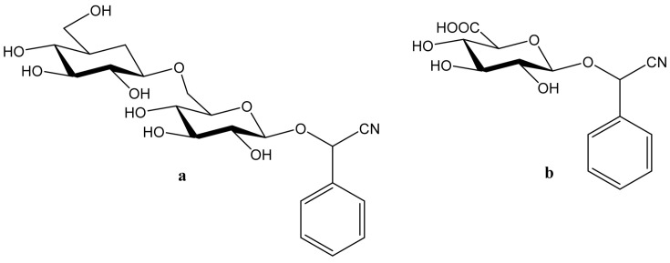
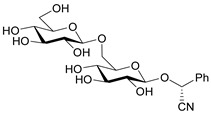
Amygdalin (d-Mandelonitrile 6-O-β-d-glucosido-β-d-glucoside) is a naturally occurring disaccharide, a source of HCN, highly concentrated in fruit kernels from Rosaceae species, for example, in bitter almonds, apricot and peach [1]. Bitter almonds have been used since ancient times to treat fevers, headache (via their purging activity) and as a diuretic [8]. Amygdalin is composed of two molecules of glucose, benzaldehyde and hydrogen cyanide and can exist in the form of two R and S epimers (Figure 1a) [9]. R-Amygdalin is natural amygdalin and S-amygdalin is called neoamygdalin.
Figure 1.
Chemical structures of amygdalin (a) and Laetrile (b).
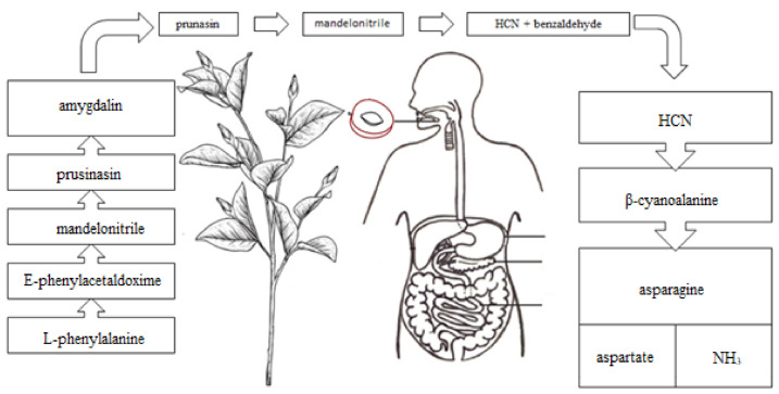
Beta-glucosidase stored in compartments of plant cells is also present in the human small intestine [10] and degrades amygdalin into prunasin, mandelonitrile, glucose, benzaldehyde and hydrogen cyanide (Figure 2). Hydrogen cyanide (HCN), benzaldehyde, prunasin and mandelonitrile, can be absorbed into the lymph and portal circulations [11]. The anticancer activity of amygdalin is thought to be related to the cytotoxic effects of enzymatically released HCN and non-hydrolyzed cyanogenic glycosides [12].
Figure 2.
Hydrogen cyanide formation as a result of hydrolysis of amygdalin.
Laetrile (d-Mandelonitrile-β-glucuronide), which is derived from amygdalin, has been used as a complementary and alternative natural medicine (CAM) in the treatment of cancer for over 30 years [13] (Figure 1b). Studies of amygdalin on various cancer cell lines demonstrated their anticancer activity [14], but the statements related to a patient study, made by the U.S. Food and Drug Administration (FDA) in the late 1970s [15] did not confirm this. Since then, however, many publications have been presented confirming both the toxicity occurring with excessive consumption of amygdalin contained in bitter almonds and the therapeutic, especially anticancer, properties of amygdalin [16]. Many papers have also been published describing methods for the determination of amygdalin in food products, which is of crucial importance in the context of the ambivalent effects of these compound. Therefore, the aim of this review is to present the current state of knowledge on the sources and toxicity of cyanogenic glycosides and analytical methods for determination of amygdalin in plant seeds [2].
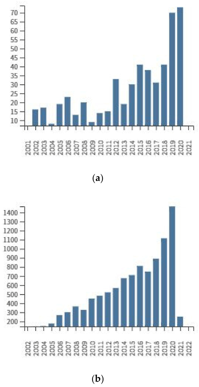
Growing interest in the biological activity of amygdaline and related research problems (Figure 3) are difficult to estimate accurately due to the many similar keywords that are simultaneously or alternatively used in literature of the subject. According to the Web of Science® database (Accessed on: 24 March 2021), the number of hits for individual entries was: vitamin B17 (26), Laetrile (315), amygdalin (725), cyanogenic glycoside (957). The following number of quotations have been found for the descriptor of amygdalin and: analytical procedures (14), anticancer (26), toxicity (83), almond (93), cancer (123), seeds (156).
Figure 3.
Total publications by year (a) and sum of times cited by year (b) for amygdalin as a topic (Web of Science®, accessed on 3rd March 2021).
2. Amygdalin as a Member of Cyanogenic Glycosides—Their Sources, Toxicity and Anticancer Activity
Amygdalin belongs to the cyanogenic glycosides (CGs), which are group of organic chemical compounds composed of sugar(s) and an aglycon containing 1-cyanobenzyl moiety. The 1-cyanobenzyl moiety is linked to the hemiacetal OH group located at the anomeric carbon atom of the sugar moiety (Table 1). CGs can be classified not only as cyanohydrin derivatives where OH group is functionalized with sugar moiety, but also as a group of organic cyanides (nitriles) of the RCN type. Sometimes, nitriles are also included in the group of pseudohalogenes [17]. To the group of primary CGs belong also: prunasin, linamarin, dhurrin, vicianin, prulaursin, sambunigrin, neolinustatin, taxifylline, lotaustralin and linustatin [18].
Table 1.
Information on selected cyanogenic glycosides.
2.1. Sources
Cyanogenic glycosides-containing plants occur in about 2000 species belonging to 110 families (e.g., Rosaceae, Poaceae, Papilionaceae, Euphorbiaceae, Scrophulariaceae), including many plants and seeds of edible fruits, such as peach and edible kernels of almond (Table 2). The natural function of cyanogenic glycosides is to protect the plant against insects and larger herbivores [19]. The content of amygdalin usually increases during the fruit enlargement stage and remains constant or minimally decreases during ripening. In the peach seed, the amygdalin content is greater in the endocarp than in the mesocarp. Bitterness in the almond kernel is determined by the content of the cyanogenic amygdalin diglucoside [20].
Table 2.
Concentration of hydrogen cyanide released in the process of enzymatic hydrolysis of amygdalin in various parts of plant tissues [21].
| Plant | Cyanogenic Potential [mg HCN/kg Plant Material] |
|
|---|---|---|
| Peach | Kernel | 710 |
| Plum | Kernel | 696 |
| Nectarine | Kernel | 196 |
| Apricot | Kernel | 785 |
| Apple | Seed | 690 |
Biosynthesis of amygdalin involves the initial conversion of L-phenylalanine into mandelonitrile catalyzed by cytochrome P450 and CYP71AN24. By the action of UDP-glucosyltransferase, mandelonitrile is converted to prunasin. The glucosyltransferase catalyzes conversion of prunasin into amygdalin [22]. Plants containing CGs usually contain degradation enzymes, such as β-glycosidases (E.C. 3.2.1.21) which hydrolyze α-glucosidic bonds and lead to formation of α-hydroxy nitriles (cyanohydrins) and sugar moieties. Hydroxynitrile lyases (E.C. 4.1.2.47) catalyze further dissociation of cyanohydrins to carbonyl compounds (benzaldehyde) and hydrogen cyanide (Figure 4). The HCN release occurs when tissues of a cyanogenic plant are macerated, e.g., when eaten by herbivores, resulting in contact of CGs with enzymes that hydrolyze them. These enzymes can be deactivated by thermal denaturation (e.g., hot water, high temperature). In plants that do not have β-glycosidase enzymes, but contain CGs, hydrolysis can be achieved in the digestive tract of animals and humans, provided that gastrointestinal endosymbiotes produce β-glycosidase [18]. In humans, the decisive formation of HCN is probably caused by the bacterial flora of the intestine that is able to produce β-glycosidase in the brush border of the small intestine [10,12,23].
Figure 4.
Hydrogen cyanide formation as a result of hydrolysis of amygdalin.
2.2. Toxicity
Compounds with the CN group, both of the organic (RCN) and inorganic (HCN, CN-anions) origin are absorbed into the body through the gastrointestinal tract, as well as through the respiratory system and skin. They lead to inactivation of enzymes containing ferric ions (Fe3+). For example, a key enzyme of the respiratory chain-cytochrome oxidase, binding to the active site of cytochrome c oxidase, inhibits oxygen metabolism, especially in myocardium and brain cells [24]. In animals, hydrogen cyanide reacts with methemoglobin in the blood, but most cyanide metabolism occurs in tissues [25]. A significant (80%) part of cyanides is detoxified in liver. This is due to a thiosulfate sulfur-transferase enzyme (i.e., rhodanase [E.C. 2.8.1.1]), which is present in the liver mitochondria. Sulfur, which is necessary for this reaction, is taken from biological compounds, e.g., thiosulfates. Rhodanase transforms cyanides into thiocyanates, which are quickly excreted in urine. The process of the cyanide metabolism in the living organism may take place in various ways (Figure 3). One example is combining cyanide with hydroxocobalamin (vitamin B12a), to obtain cyanocobalamin (i.e., vitamin B12). The remaining cyanide ions are oxidized to formates and carbon dioxide. Formates are excreted in urine and carbon dioxide, where together with hydrocyanic acid are excreted through the lungs. A small amount of cyanides also combine with cysteine to form 2-iminothiazolidine-4-carboxylic acid [26].
The toxic dose of hydrogen cyanide released by enzymatic hydrolysis of CGs in plant tissues is defined as the dose exceeding 20 mg of hydrogen cyanide per 100 g of fresh weight [27]. Excessive consumption of seeds may have a negative effect on the body, causing a number of adverse reactions of the following types: diarrhea, vomiting, abdominal pain and in extreme cases may lead to death (Table 3). Human lethal dose of intravenous injection of amygdalin is 5 g [28]. No data are available for other fruits of Poland’s climate zone. It is believed that the consumption of 50 bitter almonds in a short period of time can be a lethal dose for an adult and that a dose of 5–10 bitter almonds can be poisonous for a child. The adult lethal dose of amygdalin is estimated to be 0.5–3.5 mg/kg body weight [1,29].
Table 3.
Information on amygdalin poisoning.
| Patient | Dose | Effects | Ref |
|---|---|---|---|
| Child (2 years) | 500 mg | vomiting, apathy, diarrhea, accelerated breathing | [30] |
| Child (4 year) | 500 mg | diarrhea, accelerated breath, blood cyanide concentration 163 µg/L | [31] |
| Adult woman (80 years) | 300 mL | dyspnea, vertigo and vomiting. | [32] |
| Adult woman | 9 g | vomiting, dizziness, blood cyanide concentration 143 µg/L | [33] |
2.3. The Anticancer and Other Biological Activities
CGs medical applications are mainly related to amygdalin, discovered in 1830 by French chemists Pierre-Jean Robiquet and Antoine François Boutron-Charlard. A theory by Dr. Ernst T. Krebs, Sr., that amygdalin could be an effective drug against cancer, but is too toxic for humans, was announced in 1920. Despite this statement, his son Ernst Theodore Krebs, Jr., synthesized in 1952 a less harmful amygdalin derivative with one subunit of glucose, which he called Laetrile [34]. The mixture of amygdalin and its modified form was described by Krebs as “vitamin B17” [35,36] although in the literal sense neither amygdalin nor Laetrile are vitamins. In 1977, the FDA (USA) issued a statement indicating that there was no evidence of the Laetrile safety and efficacy [2].
While it is forbidden to sell amygdalin and Laetrile in the U.S. and Europe, there are laboratories and clinics in Mexico offering amygdalin preparations and therapies for many years (e.g., Cyto Pharma De Mexico, 40 years on the market) [37]. However, there is no solid clinical data to support the efficacy of these therapies on patients [38]. In contrast, in vitro cell culture studies show, a number of amygdalin activities that would be beneficial in cancer treatment (Table 4). For example, amygdalin has the capacity to control apoptotic proteins and signaling molecules, which may be a justification for a decrease in tumor proliferation. Amygdalin treatment increased expression of Bax, decreased expression of Bcl-2 and induced caspase-3 activation in human DU145 and LNCaP prostate cancer cells [9], induced apoptosis of HeLa cervical cancer cells mediated by endogenous mitochondrial pathway [39] and reduced adhesion and migration of UMUC-3 and RT112 bladder cancer cells through activation of focal adhesion kinase (FAK) and modulation of β1 integrin [40]. Amygdalin has also the ability to inhibit anti-apoptotic expression of genes including Survivin, and XIAP genes [13]. Other biological activities of amygdalin have also been demonstrated and they include antibacterial [41,42,43], antioxidant [44,45], anti-atherosclerotic [46], anti-asthmatic [47], preventing lung [48] and liver fibrosis [49]. Amygdalin also improves microcirculatory disturbance, attenuates pancreatic fibrosis [50], possesses anti-inflammatory and analgesic activity [51], stimulates muscle cell growth [39] and finally may serve as a beneficial agent in treating a dry eye disease [52].
Table 4.
Examples of in vitro cytotoxicity studies on cancer cells.
| Cell Lines Used for Testing | Amygdalin Concentration [mg mL−1] |
Results Observed | Ref. | |
|---|---|---|---|---|
| Bladder cancer | RT 112 UMUC-3 TCCSUP | 1.25–10 | limited proliferative capacity and apoptosis. decrease in cdk4 expression level in RT112 and TCCSUP lines. |
[40] |
| Cervical cancer | HeLa | 1.25–20 | initiation of the cell apoptosis, reduction of Bcl-2 expression level, increase of Bax expression level. | [39] |
| Colon cancer | SNU-C4 | 0.25–5 | reduction of the expression level of many genes associated with following cell functions: growth, apoptosis, transmission. | [53] |
| Breast cancer | MDA-MB-231, MCF-7 | 2.5–80 | reduction of proliferative activity of the cells | [54] |
| MDA-MB-231 | 10 | growth rate of cancer cells was inhibited | [55] | |
| Kidney cancer | Caki-1 A498 KTC-26 xds |
10 | -reduced ability collagen and fibronectin. -reduced cell mobility. |
[47] |
3. Determination of Amygdalin in Plant Seeds
3.1. Collection, Transport and Storage of Plant Seeds
The first steps of any analytical procedure are sampling, transport and storage of the material for further analysis. If these steps are not performed properly, the time and cost and value of the analysis may be increased or limited. Additionally, samples may degrade or change, and incorrect chemical identification and quantification errors may occur. Typically, all fruits, vegetables and food products should be obtained using a logically thought out random sampling plan, collected and stabilized (e.g., freezing, refrigeration, drying, etc.) as soon as possible.
The maturation state of the fruit should be defined at the time of harvest and aligned with the analytical goals (e.g., determining a specific state of maturation or commercial maturity). Transportation to the laboratory should be under controlled or defined conditions (e.g., refrigeration). For the CG analysis, a fruit should be separated into peel, flesh and kernel. The fruit can be dried at room temperature [56] or lyophilized [57]. The plant material is next fragmented and homogenized using a mortar [58] or by blender [59] and sieved to a specific and defined particle size. In case of almonds, the skins is removed by blanching (i.e., immersion in hot water) [60]. Controlling the storage conditions is critical for maintaining the integrity of the samples prior to extraction and analysis. Samples are usually stored at −80 °C until they are analyzed to inhibit enzymatic degradation [56,61].
3.2. Sample Preparation
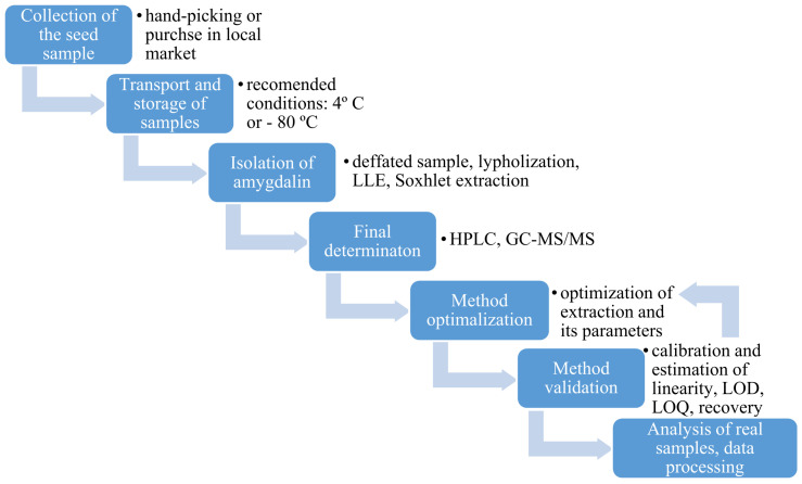
Seed samples have a complex matrix, therefore they may require additional preparation for analysis (Figure 5). Many problems are associated with the fatty matrix and the low concentrations of compounds present in the samples. Most samples will require multiple extraction steps with both aqueous and organic solvents. The solubility of amygdalin in water and ethanol is 83 g·L−1 and 1 g·L−1, respectively. In water, amygdalin hydrolyzes into benzaldehyde, hydrocyanic acid, glucose [59] and can be converted into S-amygdalin (neoamygdalin), during extraction, refluxing and/or storage making it ineffective against cancer [6] Amygdalin is easily hydrolyzed by acids and bases, so control of pH is critical. Amygdalin epimerization occurs in boiling water and especially under alkaline conditions due to the weakly acidic character of the benzyl proton. It is also important to note that due to the tendency of amygdalin to epimerize at higher temperatures, extractions should be performed at temperatures lower than 100 °C [62].
Figure 5.
General workflow during the analysis of seed samples.
When fruit samples are collected, their amount is expressed in kg, but after preparation of a suitable representative sample the amount needed for analysis can be 0.1 to 5 g. After fruit sample collection and inactivation of enzymes, samples are homogenized and a suitable extraction solvent is selected. Amygdalin is extracted from seeds with polar solvents, such as ethanol, methanol, ethyl acetate and water. It has been observed that polar solvents give low amygdalin recovery due to conversion of the natural isomer into the S-amygdalin. In the presence of water and weak bases, the epimerization of stereogenic carbon occurs and S-amygdalin is formed. Neoamygdalin may also be transformed into amygdalin during processing [63,64,65].
If the seed samples contain a lot of fat, one may consider use of diethyl ether [19], diethyl ether [66] or n-hexane [67] to remove the fat first without losses in amygdalin and other CGs. Organic solvents are removed through drying before aqueous extractions are performed. In order to minimize the amount of the solvents used and improve extraction efficiency, dynamic extraction can be performed with refluxing using a Soxhlet extractor [19,56]. The efficiency of a static extraction can be improved with an ultrasonic bath [68]. The most widely used extraction method for amygdalin is the solid-phase extraction (SPE) using a C18 extraction column [69,70,71]. To avoid the epimerization of amygdalin, extractions are performed at temperatures lower than 100 °C, usually at 35–40 °C [59,66,68]. While amygdalin is easily soluble in methanol and ethanol, it can also be extracted into water containing 0.1% citric acid under reflux, which may be a more environmentally green option [58]. Finally, the obtained supernatant is filtered through cartridge and/or syringe filter, and the diluted sample is analyzed.
3.3. Analytical Techniques for Plant Seeds Analysis
After sample preparation, the next step in a general analytical procedure is to select the relevant analytical technique for the final determination. The choice depends on several factors, such as concentration of analytes in the sample, composition of the matrix and the concentration of interfering agents. Raman spectroscopy or FT-IR can be used to check the cyanogenic glycoside distribution in the fruit stone sample because organic compounds and functional groups can be identified by their unique vibrational pattern [72]. The presence of broad peaks at 3150–3600 cm−1 represents the stretching vibration of OH groups in the amygdalin structure. Aliphatic C-H stretching vibrations and the vibrations of aromatic ring appear at 2885–2927 cm−1. Amygdalin can be probed by the Raman spectroscopy due to a characteristic band of the nitrile group at 2245 cm−1 in a part of the spectrum that is free from interference of frequencies due to other chemicals. The peaks at 1620 cm−1 and 864 cm−1 are due to aromatic C=C and aromatic C-H bending, respectively [72,73]. Raman linear mapping studies on bitter almond seeds showed that the amygdalin content increased from the seed center to the margin [74]. In apricot seeds, amygdalin is unevenly distributed and its location does not follow the same pattern for all seeds [75].
A review of literature shows that amygdalin is measured in plant seeds samples primarily using high-performance liquid chromatography (HPLC) [56,66,67] and gas chromatography (GC) methods [76,77]. However, HPLC is more convenient than gas chromatography because of its ability to separate overlapping signals and eliminate background. The primary type of chromatographic column used to resolve amygdalin in seed samples is C18 which is filled with ultra-pure silica modified with low polarity octadecyl groups. The predominant detectors used in the HPLC technique include UV–Vis [60], diode array (DAD) [68], mass spectrometry (MS) [58] and MS/MS ones [78]. The HPLC-MS identification of cyanogenic glucosides is usually performed in the positive ionization mode. The SIM MS spectrum of amygdalin contains a peak at m/z 458 corresponding to the [M+H]+ ion of amygdalin. It is possible to eliminate any potential false positives, by monitoring the 475–325 transitions [22]. However, in MS/MS cases, negative ionization resulted in a better sensitivity as compared to the positive ionization. Quantification of amygdalin was achieved using the transition of 456–323 [79]. The purity and structural identification of the extracted amygdalin was verified spectroscopically using the UV–Vis technique [80,81]. The presence of signal peaks at wavelengths of 1370–1400 nm indicates the O-H stretching modes of water absorption, while regions of 1100–1600 nm and 1700–2300 nm corresponded to sugar display bands [60]. The maximum absorption of amygdalin was detected at 214 nm using a photodiode array detector [66]. However, the use of methanol in separation of amygdalin from the extract may have an unsatisfactory resolution [81]. Moreover, NMR spectroscopy was used for additional structural characteristics of amygdalin. The 1H-NMR and 13C-NMR nuclear resonances of amygdalin diluted in DMSO-d6 was performed [73]. A summary of the recent literature on analytical procedures for determination of amygdalin in seed samples is summarized in Table 5.
Table 5.
Total amygdalin content in different samples.
| Analytical Technique | Sample | Recovery [%] | Intra/Inter-Day Variation [%] | LOD | LOQ | Detected Compounds in Real Samples | Ref |
|---|---|---|---|---|---|---|---|
| LC-DAD | apricot seeds | 91 ± 10 | 0.8/3.8 | 1.2 mg·L−1 | 4.0 mg·L−1 | bitter seeds 26 ± 14 mg·g−1 sweet seeds 0.16 ± 0.09 mg·g−1 |
[82] |
| apricot liqueur | - | - | - | - | 38.79 µg·mL−1 | [83] | |
| cherry liqueur | 16.08 µg·mL−1 | ||||||
| HPLC–MS/MS | almonds | - | - | 200 µg·g−1 | - | <LOD | [22] |
| HPLC-UV | plum seeds | - | - | - | - | 25.30 g 100 g−1 | [59] |
| almonds | - | 0.13/0.75 | 2 µg·mL−1 | - | 4.51 ± 0.04% | ||
| loquat fruit kernel | - | - | - | - | 7.58 ± 0.76 mg·g−1 | ||
| almonds | 98.4–102.9 | 0.25/0.31 | 0.02 mg·L−1 | 0.07 mg·L-1 | sweet: <350 mg·kg−1 bitter: 14,700–50,400 mg·kg−1 |
[60] | |
| peach seeds | 99.05 | 0.19 | 0.03 mg 100 g−1 | 0.09 mg 100 g−1 | 6.3 ± 0.2 g 100 g−1 | [84] | |
| plum seeds | 0.439 ± 0.001 g 100 g−1 | ||||||
| apricot seeds | 7.9 ± 0.2 g 100 g−1 | ||||||
| peach seeds | - | - | - | - | seed: 12.14 ± 4.80 mg 100g−1 | [19] | |
| citrullus colocynth kernels | 97.34 ± 0.58 | - | 0.88 mg·L−1 | 2.93 mg·L−1 | 0.27 ± 0.03 100 g−1 | [72] | |
| apples | - | 0.095 | 0.0505 mg·g−1 | 0.0548 mg·g−1 | 0.28–1.40 mg·g−1 | [73] | |
| Armeniacae semen | 98.0–102.6 | - | - | - | 45.42 ± 1.21 mg·g−1 | [85] | |
| bitter almond oil | 96.0–102.0 | 4.8/7.2 | 0.07 µg·mL−1 | - | 0.092 ± 0.003 mg·g−1 | [86] | |
| wild almond oil | - | - | - | - | 12.8–12.9 mg/100 mL oil | [87] | |
| sweet apricot kernels | - | - | - | - | 5.0 ± 0.23 mg·g−1 | [88] | |
| HPLC-DAD | apricot seeds | - | - | - | - | 0.861 g·100 g−1 | [56] |
| almonds | - | - | - | - | 0.37–1.46 g·kg−1 | [89] | |
| apple seeds | 98 | - | 0.1 µg·mL−1 | - | 1–3.9 mg·g−1 | [66] | |
| apricots | - | - | 0.1 µg·mL−1 | 0.3 µg·mL- | 14.37 ± 0.28 mg·g−1 | [67] | |
| cherries | 2.68 ± 0.02 mg·g−1 | ||||||
| peaches | 6.81 ± 0.02 mg·g−1 | ||||||
| pears | 1.29 ± 0.04 mg·g−1 | ||||||
| cucumbers | 0.07 ± 0.02 mg·g−1 | ||||||
| courgettes | 0.21 ± 0.13 mg·g−1 | ||||||
| melons | 0.12 ± 0.07 mg·g−1 | ||||||
| apricot kernels | - | - | 0.2 µg·mL−1 | - | - | [69] | |
| apricot kernels | 99.08 | 2.4/3.5 | - | - | 0.217–0.284 mg·mL−1 | [58] | |
| plum seeds | - | - | 1.06 μg·mL−1 | 3.49 μg·mL−1 | 25.30 g 100 g−1 | [65] | |
| bayberry kernels | 77.9 | - | - | - | 129.13–358.68 mg·L−1 | [57] | |
| food suplements | 94.81 | 0.57/1.52 | 0.13 mg·L−1 | 0.40 mg·L−1 | 20.68 ± 1.58 mg·g−1 | [86] | |
| apricot kernels | 91 | 0.8/3.8 | 1.2 mg·L−1 | 4.0 mg·L−1 | 26 ± 14 mg·g−1 | [90] | |
| UHPLC-(ESI)QqQ MS/MS | nonbitter almonds | - | - | 0.1 ng·mL−1 | 0.33 ng·mL−1 | 63.13 ± 57.54 mg·kg−1 | [64] |
| semibitter almonds | 992.24 ± 513.04 mg·kg−1 | ||||||
| bitter almonds | 40,060.34 ± 7855.26 mg·kg−1 | ||||||
| alomnds | - | - | - | - | 1.62–76.50 mg·kg−1 | [67] | |
| Spectrophotometric method | cassava root | - | - | - | - | 3.40 mg·L−1 | [80] |
| cassava roots | - | - | - | - | 8.84–48.33 mg·g−1 | [91] | |
| sorghum seeds | 122.31 mg·g−1 | ||||||
| mango seeds | 4.41 mg·g−1 | ||||||
| watermelon seeds | 3.97 mg·g−1 | ||||||
| almond seeds | 3.91 mg·g−1 | ||||||
| ELISA | black cherries | 99 ± 1.2 | - | 200 ± 0.05 pg·mL−1 | - | 2.14 ± 0.15 mg·g−1 | [92] |
| yellow plums | 2.30 ± 0.90 mg·g−1 | ||||||
| peaches | 5.79 ± 0.83 mg·g−1 | ||||||
| black plums | 9.75 ± 1.32 mg·g−1 |
- no data.
4. Future Trends and Conclusions
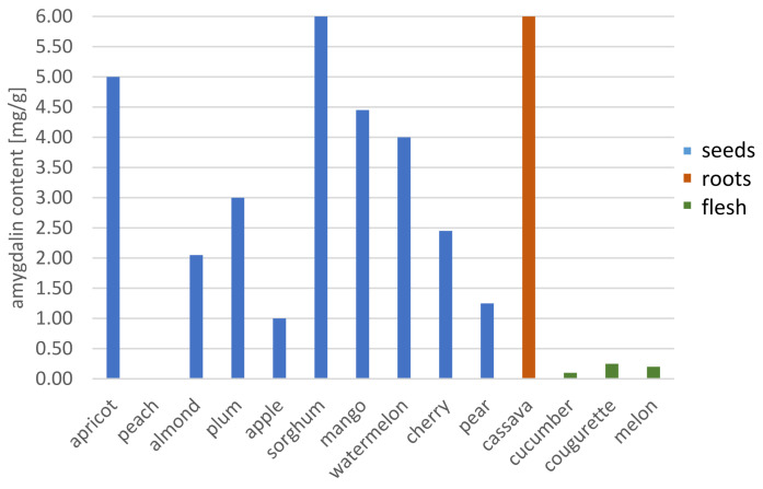
Research on cyanogenic glycosides has increased dramatically over the past 10 years. Much of this interest centers on the cytotoxic effect of amygdalin on cancer cells in vitro and understanding the distribution of amygdalin in plants that are commonly consumed in the human diet. The amygdalin content of various edible plants differs depending on the kind and the region where they are cultivated (Figure 6). As amygdalin is synthesized in response to environmental stress, agronomic and environmental factors such as latitude, climate and variety of the plant can influence its levels in plant tissues. Understanding the distribution and levels of cyanogenic glycosides in plants is important as poisoning with cyanides have usually occurred by accident and due to a lack of awareness of levels in a food or natural products. To date, there have been numerous cases of cyanide poisoning resulting from the ingestion of too many seeds containing amygdalin and as the result of the Laetrile treatment, but there are no reports of people cured of cancer by consuming the seeds containing amygdalin.
Figure 6.
Changes in the amygdalin content in edible plants.
Research into the use of amygdalin in the treatment of cancer continues. There is evidence confirming the cytotoxic effect of amygdalin on cancer cells in vitro, however, these results are not yet demonstrated in clinical studies. Nevertheless, there is still a need for quantifying the levels of amygdalin and other CGs in plant materials in order to support the clinical trials, and to better understand their intake in the human diet. New studies on amygdalin isolation and characterization should employ principles of green chemistry, and eliminate the use of toxic solvents, such as methanol. Moreover, novel spectrophotometric techniques can be explored for determining CG content in real time (e.g., FTIR spectroscopy).
Author Contributions
Conceptualization, Ż.P. and P.B.; investigation, M.K., K.O. and E.J.-W.; writing—original draft preparation, E.J.-W., M.K. and K.O.; writing—review and editing P.B., Ż.P. and A.E.M.; visualization, M.K. and E.J.-W.; supervision, Ż.P. and P.B. All authors have read and agreed to the published version of the manuscript.
Funding
This research received no external funding.
Institutional Review Board Statement
Not applicable.
Informed Consent Statement
Not applicable.
Data Availability Statement
No new data were created or analyzed in this study. Data sharing is not applicable to this article.
Conflicts of Interest
The authors declare no conflict of interest.
Sample Availability
Samples of the compounds are not available from the authors.
Footnotes
Publisher’s Note: MDPI stays neutral with regard to jurisdictional claims in published maps and institutional affiliations.
References
- 1.Flies E.J., Mavoa S., Zosky G.R., Mantzioris E., Williams C., Eri R., Brook B.W., Buettel J.C. Urban-associated diseases: Candidate diseases, environmental risk factors, and a path forward. Environ. Int. 2019;133:105187. doi: 10.1016/j.envint.2019.105187. [DOI] [PubMed] [Google Scholar]
- 2.Bray F., Ferla J., Soerjomatarm I., Siegel R., Torre L., Jemal A. Global Cancer Statistics 2018: GLOBOCAN Estimates of Incidence and Mortality Worldwide for 36 Cancers in 185 Countries. Cancer J. Clin. 2018;68:394–424. doi: 10.3322/caac.21492. [DOI] [PubMed] [Google Scholar]
- 3.World Cancer Research Fund/American Institute for Cancer Research Diet . Nutrition, Physical Activity and Cancer: A Global Perspective. International Peace Research Association; Solona, Sweden: 2018. [Google Scholar]
- 4.Sirota H., Rubovits D.R., Cousins J.H., Weinberg A.D., Laufman L., Lane M. Cancer control: Prevention. Health Values. 2007;12:33–36. [PubMed] [Google Scholar]
- 5.Wojciechowska U., Didkowska J., Zatoński W. Nowotwory złośliwe w Polsce w 2010 roku. Kraj. Rejestr Nowotworów. 2010;2012:44–45. doi: 10.3389/fnins.2016.00562. [DOI] [Google Scholar]
- 6.Takayama Y., Kwai S. Study on the prevention of racemization of amygdalin. Chem. Pharm. Bull. 1984;32:778–781. doi: 10.1248/cpb.32.778. [DOI] [Google Scholar]
- 7.Santos S.B., Sousa Lobo J.M., Silva A.C. Biosimilar medicines used for cancer therapy in Europe: A review. Drug Discov. Today. 2018;24:293–299. doi: 10.1016/j.drudis.2018.09.011. [DOI] [PubMed] [Google Scholar]
- 8.Albala K. Adaptation of Ideas from West to East. Petits Propos Culin. 2009;88:19–34. [Google Scholar]
- 9.Chang H.-K., Shin M.-S., Yang H.-Y., Lee J.-W., Kim Y.-S., Lee M.-H., Kim J., Kim K.-H., Kim C.-J. Amygdalin induces apoptosis through regulation of Bax and Bcl-2 expressions in human DU145 and LNCaP prostate cancer cells. Biol. Pharm. Bull. 2006;29:1597–1602. doi: 10.1248/bpb.29.1597. [DOI] [PubMed] [Google Scholar]
- 10.Shim S.M., Kwon H. Metabolites of amygdalin under simulated human digestive fluids. Int. J. Food Sci. Nutr. 2010;61:770–779. doi: 10.3109/09637481003796314. [DOI] [PubMed] [Google Scholar]
- 11.Chang J., Zhang Y. Catalytic degradation of amygdalin by extracellular enzymes from Aspergillus niger. Process. Biochem. 2012;47:195–200. doi: 10.1016/j.procbio.2011.10.030. [DOI] [Google Scholar]
- 12.Nowak A., Zielińska A. Aktywność przeciwnowotworowa amigdaliny Anticancer activity of amygdalin. Postępy Fitoter. 2016;17:282–292. [Google Scholar]
- 13.Arshi A., Hosseini S.M., Hosseini F.S.K., Amiri Z.Y., Hosseini F.S., Sheikholia Lavasani M., Kerdarian H., Dehkordi M.S. The anti-cancer effect of amygdalin on human cancer cell lines. Mol. Biol. Rep. 2019;46:2059–2066. doi: 10.1007/s11033-019-04656-3. [DOI] [PubMed] [Google Scholar]
- 14.Sireesha D., Reddy B.S., Reginald B.A., Samatha M., Kamal F. Effect of amygdalin on oral cancer cell line: An in vitro study. J. Oral Maxillofac. Pathol. 2019;23:104–107. doi: 10.4103/jomfp.JOMFP. [DOI] [PMC free article] [PubMed] [Google Scholar]
- 15.Rosen G., Shorr R. Laetrile: End play around the FDA. A review of legal developments. Ann. Intern. Med. 1979;90:418–423. doi: 10.7326/0003-4819-90-3-418. [DOI] [PubMed] [Google Scholar]
- 16.Jukes T.H. Laetrile for cancer. JAMA. 1976;236:1284–1289. doi: 10.1001/jama.1976.03270120056033. [DOI] [PubMed] [Google Scholar]
- 17.Achmatowicz O., PTChem . Przewodnik do Nomenklatury Związków Organicznych. PTChem; Warszawa, Poland: 1994. [Google Scholar]
- 18.Siegień Irena Cyjanogeneza u roslin i jej efektywność w ochronie roślin przed atakiem roślinożerców i patogenów. Kosmos. Probl. Nauk Biol. 2007;2:155–166. [Google Scholar]
- 19.Zagrobelny M., Bak S., Rasmussen A.V., Jørgensen B., Naumann C.M., Møller B.L. Cyanogenic glucosides and plant-insect interactions. Phytochemistry. 2004;65:293–306. doi: 10.1016/j.phytochem.2003.10.016. [DOI] [PubMed] [Google Scholar]
- 20.Lee S.H., Oh A., Shin S.H., Kim H.N., Kang W.W., Chung S.K. Amygdalin contents in peaches at different fruit development stages. Prev. Nutr. Food Sci. 2017;22:237–240. doi: 10.3746/pnf.2017.22.3.237. [DOI] [PMC free article] [PubMed] [Google Scholar]
- 21.Rezaul Haque M., Howard Bradbury J. Total cyanide determination of plants and foods using the picrate and acid hydrolysis methods. Food Chem. 2002;77:107–114. doi: 10.1016/S0308-8146(01)00313-2. [DOI] [Google Scholar]
- 22.Del Cueto J., Ionescu I.A., Pičmanová M., Gericke O., Motawia M.S., Olsen C.E., Campoy J.A., Dicenta F., Møller B.L., Sánchez-Pérez R. Cyanogenic Glucosides and Derivatives in Almond and Sweet Cherry Flower Buds from Dormancy to Flowering. Front. Plant. Sci. 2017;8:1–16. doi: 10.3389/fpls.2017.00800. [DOI] [PMC free article] [PubMed] [Google Scholar]
- 23.Rietjens I.M.C.M., Martena M.J., Boersma M.G., Spiegelenberg W., Alink G.M. Molecular mechanisms of toxicity of important food-borne phytotoxins. Mol. Nutr. Food Res. 2005;49:131–158. doi: 10.1002/mnfr.200400078. [DOI] [PubMed] [Google Scholar]
- 24.Jaszczak E., Polkowska Ż., Narkowicz S., Namieśnik J. Cyanides in the environment—analysis—problems and challenges. Environ. Sci. Pollut. Res. 2017;24:15929–15948. doi: 10.1007/s11356-017-9081-7. [DOI] [PMC free article] [PubMed] [Google Scholar]
- 25.Abraham K., Buhrke T., Lampen A. Bioavailability of cyanide after consumption of a single meal of foods containing high levels of cyanogenic glycosides: A crossover study in humans. Arch. Toxicol. 2016;90:559–574. doi: 10.1007/s00204-015-1479-8. [DOI] [PMC free article] [PubMed] [Google Scholar]
- 26.Simeonova F., Fishbein L. Hydrogen Cyanide and Cyanides: Human Health Aspects. [(accessed on 20 February 2021)];World Health Organ. Geneva. 2004 Available online: https://apps.who.int/iris/handle/10665/42942. [Google Scholar]
- 27.Kołaciński Z., Burda P., Łukasik-Głębocka M., Sein Anand J. Postępowanie w ostrych zatruciach cyjankami-stanowisko Sekcji Toksykologii Klinicznej Polskiego Towarzystwa Lekarskiego. Przegl. Lek. 2011;68:8–11. [PubMed] [Google Scholar]
- 28.Qadir M., Fatima K. Review on Pharmacological Activity of Amygdalin. Arch. Cancer Res. 2017;5:10–12. doi: 10.21767/2254-6081.100160. [DOI] [Google Scholar]
- 29.Blaheta R.A., Nelson K., Haferkamp A., Juengel E. Amygdalin, quackery or cure? Phytomedicine. 2016;23:367–376. doi: 10.1016/j.phymed.2016.02.004. [DOI] [PubMed] [Google Scholar]
- 30.Sahin S., Kirel B., Carman K. Fatal cyanide poisoning in a child, caused by eating apricot seeds. Am. J. Case Rep. 2011;12:70–72. doi: 10.12659/AJCR.881827. [DOI] [Google Scholar]
- 31.Sauer H., Wollny C., Oster I., Tutdibi E., Gortner L., Gottschling S., Meyer S. Severe cyanide poisoning from an alternative medicine treatment with amygdalin and apricot kernels in a 4-year-old child. Wien. Med. Wochenschr. 2015;165:185–188. doi: 10.1007/s10354-014-0340-7. [DOI] [PubMed] [Google Scholar]
- 32.Drankowska J., Kos M., Kościuk A., Tchórz M. Cyanide poisoning from an alternative medicine treatment with apricot kernels in a 80- year-old female. J. Educ. Heal. Sport. 2018;8:19–26. [Google Scholar]
- 33.Tatli M., Eyüpoğlu G., Hocagil H. Acute cyanide poisoning due to apricot kernel ingestion. J. Acute Dis. 2017;6:87–88. doi: 10.12980/jad.6.2017JADWEB-2016-0075. [DOI] [Google Scholar]
- 34.Unproven methods of cancer management: Laetrile. CA. Cancer J. Clin. 1991;41:187–192. doi: 10.3322/canjclin.41.3.187. [DOI] [PubMed] [Google Scholar]
- 35.Edward J.C. Possible adverse side effects from treatment with laetrile. Med. Hypotheses. 1979;5:1045–1049. doi: 10.1016/0306-9877(79)90053-7. [DOI] [PubMed] [Google Scholar]
- 36.Lewis J.P. Laetrile. West. J. Med. 1977;127:55–62. [PMC free article] [PubMed] [Google Scholar]
- 37.Moss R.W. Patient perspectives: Tijuana cancer clinics in post-NATFA era. Integr. Cancer Ther. 2005;4:65–86. doi: 10.1177/1534735404273918. [DOI] [PubMed] [Google Scholar]
- 38.Kamil A., Chen C.Y.O. Health benefits of almonds beyond cholesterol reduction. J. Agric. Food Chem. 2012;60:6694–6702. doi: 10.1021/jf2044795. [DOI] [PubMed] [Google Scholar]
- 39.Milazzo S., Lejeune S., Ernst E. Laetrile for cancer: A systematic review of the clinical evidence. Support. Care Cancer. 2007;15:583–595. doi: 10.1007/s00520-006-0168-9. [DOI] [PubMed] [Google Scholar]
- 40.Chen Y., Ma J., Wang F., Hu J., Cui A., Wei C., Yang Q., Li F. Amygdalin induces apoptosis in human cervical cancer cell line HeLa cells. Immunopharmacol. Immunotoxicol. 2013;35:43–51. doi: 10.3109/08923973.2012.738688. [DOI] [PubMed] [Google Scholar]
- 41.Makarević J., Rutz J., Juengel E., Kaulfuss S., Reiter M., Tsaur I., Bartsch G., Haferkamp A., Blaheta R.A. Amygdalin blocks bladder cancer cell growth in vitro by diminishing cyclin A and cdk2. PLoS ONE. 2014;9:e0105590. doi: 10.1371/journal.pone.0105590. [DOI] [PMC free article] [PubMed] [Google Scholar]
- 42.Al-Bakri S.A., Nima Z.A., Jabri R.R., Ajeel E.A. Antibacterial activity of apricot kernel extract containing Amygdalin. Iraqi J. Sci. 2010;51:571–576. [Google Scholar]
- 43.Savić I.M., Nikolić V.D., Savić-Gajić I.M., Kundaković T.D., Stanjković T.P., Najman S.J. Chemical composition and biological activity of agroindustrial residues. Adv. Technol. 2016;5:38–45. doi: 10.5937/savteh1602038S. [DOI] [Google Scholar]
- 44.Yashunsky D.V., Kulakovskaya E.V., Kulakovskaya T.V., Zhukova O.S., Kiselevskiy M.V., Nifantiev N.E. Synthesis and Biological Evaluation of Cyanogenic Glycosides. J. Carbohydr. Chem. 2015;34:460–474. doi: 10.1080/07328303.2015.1105249. [DOI] [Google Scholar]
- 45.Walia M., Rawat K., Bhushan S., Padwad Y.S., Singh B. Fatty acid composition, physicochemical properties, antioxidant and cytotoxic activity of apple seed oil obtained from apple pomace. J. Sci. Food Agric. 2014;94:929–934. doi: 10.1002/jsfa.6337. [DOI] [PubMed] [Google Scholar]
- 46.Waszkowiak K., Gliszczyńska-Świgło A., Barthet V., Skręty J. Effect of Extraction Method on the Phenolic and Cyanogenic Glucoside Profile of Flaxseed Extracts and their Antioxidant Capacity. JAOCS J. Am. Oil Chem. Soc. 2015;92:1609–1619. doi: 10.1007/s11746-015-2729-x. [DOI] [PMC free article] [PubMed] [Google Scholar]
- 47.Luo H., Li L., Tang J., Zhang F., Zhao F., Sun D., Zheng F., Wang X. Amygdalin inhibits HSC-T6 cell proliferation and fibrosis through the regulation of TGF-β/CTGF. Mol. Cell. Toxicol. 2016;12:265–271. doi: 10.1007/s13273-016-0031-0. [DOI] [Google Scholar]
- 48.Guo J., Wu W., Sheng M., Yang S., Tan J. Amygdalin inhibits renal fibrosis in chronic kidney disease. Mol. Med. Rep. 2013;7:1453–1457. doi: 10.3892/mmr.2013.1391. [DOI] [PubMed] [Google Scholar]
- 49.Hwang H.J., Lee H.J., Kim C.J., Shim I., Hahm D.H. Inhibitory effect of amygdalin on lipopolysaccharide-inducible TNF-α and IL-1β mRNA expression and carrageenan-induced rat arthritis. J. Microbiol. Biotechnol. 2008;18:1641–1647. [PubMed] [Google Scholar]
- 50.Baroni A., Paoletti I., Greco R., Satriano R.A., Ruocco E., Tufano M.A., Perez J.J. Immunomodulatory effects of a set of amygdalin analogues on human keratinocyte cells. Exp. Dermatol. 2005;14:854–859. doi: 10.1111/j.1600-0625.2005.00368.x. [DOI] [PubMed] [Google Scholar]
- 51.Yang C., Li X., Rong J. Amygdalin isolated from Semen Persicae (Tao Ren) extracts induces the expression of follistatin in HepG2 and C2C12 cell lines. Chin. Med. 2014;9:23. doi: 10.1186/1749-8546-9-23. [DOI] [PMC free article] [PubMed] [Google Scholar]
- 52.Heikkila R.E., Cabbat F.S. The prevention of alloxan-induced diabetes by amygdalin. Life Sci. 1980;27:659–662. doi: 10.1016/0024-3205(80)90006-5. [DOI] [PubMed] [Google Scholar]
- 53.Kim C.S., Jo K., Lee I.S., Kim J. Topical application of apricot kernel extract improves dry eye symptoms in a unilateral exorbital lacrimal gland excision mouse. Nutrients. 2016;8:750. doi: 10.3390/nu8110750. [DOI] [PMC free article] [PubMed] [Google Scholar]
- 54.Park H.J., Yoon S.H., Han L.S., Zheng L.T., Jung K.H., Uhm Y.K., Lee J.H., Jeong J.S., Joo W.S., Yim S.V., et al. Amygdalin inhibits genes related to cell cycle in SNU-C4 human colon cancer cells. World J. Gastroenterol. 2005;11:5156–5161. doi: 10.3748/wjg.v11.i33.5156. [DOI] [PMC free article] [PubMed] [Google Scholar]
- 55.Lee H.M., Moon A. Amygdalin regulates apoptosis and adhesion in Hs578T triple-negative breast cancer cells. Biomol. Ther. 2016;24:62–66. doi: 10.4062/biomolther.2015.172. [DOI] [PMC free article] [PubMed] [Google Scholar]
- 56.Moon J.Y., Kim S.W., Yun G.M., Lee H.S., Kim Y.D., Jeong G.J., Ullah I., Rho G.J., Jeon B.G. Inhibition of cell growth and down-regulation of telomerase activity by amygdalin in human cancer cell lines. Animal Cells Syst. (Seoul) 2015 doi: 10.1080/19768354.2015.1060261. [DOI] [Google Scholar]
- 57.Yildrim F., Askin M.A. Variability of amygdalin content in seeds of sweet and bitter apricot cultivars in Turkey. African J. Biotechnol. 2010;9:6522–6524. doi: 10.5897/AJB10.884. [DOI] [Google Scholar]
- 58.Wang T., Lu S., Xia Q., Fang Z., Johnson S. Separation and purification of amygdalin from thinned bayberry kernels by macroporous adsorption resins. J. Chromatogr. B Anal. Technol. Biomed. Life Sci. 2015;975:52–58. doi: 10.1016/j.jchromb.2014.10.038. [DOI] [PubMed] [Google Scholar]
- 59.Bolarinwa I.F., Olaniyan S.A., Olatunde S.J., Ayandokun F.T., Olaifa I.A. Effect of processing on amygdalin and cyanide contents of some Nigerian foods. J. Chem. Pharm. Res. 2016;8:106–113. doi: 10.2306/scienceasia1513-1874.2013.39.444. [DOI] [Google Scholar]
- 60.Savic I.M., Nikolic V.D., Savic-Gajic I.M., Nikolic L.B., Ibric S.R., Gajic D.G. Optimization of technological procedure for amygdalin isolation from plum seeds (Pruni domesticae semen) Front. Plant. Sci. 2015;6:1–11. doi: 10.3389/fpls.2015.00276. [DOI] [PMC free article] [PubMed] [Google Scholar]
- 61.Cortés V., Talens P., Barat J.M., Lerma-García M.J. Potential of NIR spectroscopy to predict amygdalin content established by HPLC in intact almonds and classification based on almond bitterness. Food Control. 2018;91:68–75. doi: 10.1016/j.foodcont.2018.03.040. [DOI] [Google Scholar]
- 62.Del Cueto J., Møller B.L., Dicenta F., Sánchez-pérez R. Plant Physiology and Biochemistry β -Glucosidase activity in almond seeds. Plant. Physiol. Biochem. 2018;126:163–172. doi: 10.1016/j.plaphy.2017.12.028. [DOI] [PubMed] [Google Scholar]
- 63.Wahab M.F., Breitbach Z.S., Armstrong D.W., Strattan R., Berthod A. Problems and Pitfalls in the Analysis of Amygdalin and Its Epimer. J. Agric. Food Chem. 2015;63:8966–8973. doi: 10.1021/acs.jafc.5b03120. [DOI] [PubMed] [Google Scholar]
- 64.Burns A.E., Bradbury J.H., Cavagnaro T.R., Gleadow R.M. Total cyanide content of cassava food products in Australia. J. Food Compos. Anal. 2012;25:79–82. doi: 10.1016/j.jfca.2011.06.005. [DOI] [Google Scholar]
- 65.Lee J., Zhang G., Wood E., Rogel Castillo C., Mitchell A.E., Wu H., Cao C., Zhou C. Quantification of amygdalin in nonbitter, semibitter, and bitter almonds (prunus dulcis) by UHPLC-(ESI)QqQ MS/MS. J. Agric. Food Chem. 2013;61:7754–7759. doi: 10.1021/jf402295u. [DOI] [PubMed] [Google Scholar]
- 66.Xu S., Xu X., Yuan S., Liu H., Liu M., Zhang Y., Zhang H., Gao Y., Lin R., Li X. Identification and analysis of amygdalin, neoamygdalin and amygdalin amide in different processed bitter almonds by HPLC-ESI-MS/MS and HPLC-DAD. Molecules. 2017;22:1425. doi: 10.3390/molecules22091425. [DOI] [PMC free article] [PubMed] [Google Scholar]
- 67.Bolarinwa I.F., Orfila C., Morgan M.R.A. Determination of amygdalin in apple seeds, fresh apples and processed apple juices. Food Chem. 2015;170:437–442. doi: 10.1016/j.foodchem.2014.08.083. [DOI] [PubMed] [Google Scholar]
- 68.Bolarinwa I.F., Orfila C., Morgan M.R.A. Amygdalin content of seeds, kernels and food products commercially- available in the UK. Food Chem. 2014;152:133–139. doi: 10.1016/j.foodchem.2013.11.002. [DOI] [PubMed] [Google Scholar]
- 69.Karsavuran N., Charehsaz M., Celik H., Asma B.M., Yakıncı C., Aydın A. Amygdalin in bitter and sweet seeds of apricots. Toxicol. Environ. Chem. 2014;96:1564–1570. doi: 10.1080/02772248.2015.1030667. [DOI] [Google Scholar]
- 70.Luo K.K., Kim D.A., Mitchell-Silbaugh K.C., Huang G., Mitchell A.E. Comparison of amygdalin and benzaldehyde levels in California almond (Prunus dulcis) varietals. Acta Hortic. 2018;1219:1–7. doi: 10.17660/ActaHortic.2018.1219.1. [DOI] [Google Scholar]
- 71.Rogel-Castillo C., Luo K., Huang G., Mitchell A.E. Effect of Drying Moisture Exposed Almonds on the Development of the Quality Defect Concealed Damage. J. Agric. Food Chem. 2017;65:8948–8956. doi: 10.1021/acs.jafc.7b03680. [DOI] [PubMed] [Google Scholar]
- 72.Thygesen L.G., Løkke M.M., Micklander E., Engelsen S.B. Vibrational microspectroscopy of food. Raman vs. FT-IR. Trends Food Sci. Technol. 2003;14:50–57. doi: 10.1016/S0924-2244(02)00243-1. [DOI] [Google Scholar]
- 73.Muhammad S.S., Abbas S.M., Khammas Z.A.-A. Extraction and Determination of Amygdaline in Iraqi Plant Seeds Using the Combined Simple Extraction Procedure and High-Performance Liquid Chromatography. Baghdad Sci. J. 2013;10:350–361. doi: 10.21123/bsj.10.2.350-361. [DOI] [Google Scholar]
- 74.Amaya-Salcedo J.C., Cárdenas-González O.E., Gómez-Castaño J.A. Solid-to-liquid extraction and HPLC/UV determination of amygdalin of seeds of apple (Malus pumila Mill): Comparison between traditional-solvent and microwave methodologies. Acta Agron. 2018;67:381–388. doi: 10.15446/acag.v67n3.67186. [DOI] [Google Scholar]
- 75.Micklander E., Brimer L., Engelsen S.B. Noninvasive Assay for Cyanogenic Constituents in Plants by Raman Spectroscopy: Content and Distribution of Amygdalin in Bitter Almond (Prunus amygdalus) Appl. Spectrosc. 2002;56:1139–1146. doi: 10.1366/000370202760295368. [DOI] [Google Scholar]
- 76.Krafft C., Cervellati C., Paetz C., Schneider B., Popp J. Distribution of amygdalin in apricot (Prunus armeniaca) seeds studied by Raman microscopic imaging. Appl. Spectrosc. 2012;66:644–649. doi: 10.1366/11-06521. [DOI] [PubMed] [Google Scholar]
- 77.Isozaki T., Matano Y., Yamamoto K., Kosaka N., Tani T. Quantitative determination of amygdalin epimers by cyclodextrin-modified micellar electrokinetic chromatography. J. Chromatogr. A. 2001;923:249–254. doi: 10.1016/S0021-9673(01)00969-4. [DOI] [PubMed] [Google Scholar]
- 78.Zhang N., Zhang Q.-A., Yao J.-L., Zhang X.-Y. Changes of amygdalin and volatile components of apricot kernels during the ultrasonically-accelerated debitterizing. Ultrason. Sonochem. 2019;58:104614. doi: 10.1016/j.ultsonch.2019.104614. [DOI] [PubMed] [Google Scholar]
- 79.Toomey V.M., Nickum E.A., Flurer C.L. Cyanide and Amygdalin as Indicators of the Presence of Bitter Almonds in Imported Raw Almonds. J. Forensic Sci. 2012;57:1313–1317. doi: 10.1111/j.1556-4029.2012.02138.x. [DOI] [PubMed] [Google Scholar]
- 80.Wu H., Cao C., Zhou C. Determination of amygdalin in the fruit of Eriobotrya japonica Lindl by high performance liquid chromatography. Biomed. Res. 2017;28:8827–8831. [Google Scholar]
- 81.Tivana L.D., Da Cruz Francisco J., Zelder F., Bergenståhl B., Dejmek P. Straightforward rapid spectrophotometric quantification of total cyanogenic glycosides in fresh and processed cassava products. Food Chem. 2014;158:20–27. doi: 10.1016/j.foodchem.2014.02.066. [DOI] [PubMed] [Google Scholar]
- 82.Savic I.M., Nikolic V.D., Savic I.M., Nikolic L.B., Stankovic M.Z. Development and validation of HPLC method for the determination of amygdalin in the plant extract of plum kernel. Res. J. Chem. Environ. 2012;16:80–86. [Google Scholar]
- 83.Senica M., Stampar F., Veberic R., Mikulic-Petkovsek M. Fruit Seeds of the Rosaceae Family: A Waste, New Life, or a Danger to Human Health? J. Agric. Food Chem. 2017;65:10621–10629. doi: 10.1021/acs.jafc.7b03408. [DOI] [PubMed] [Google Scholar]
- 84.Senica M., Stampar F., Veberic R., Mikulic-Petkovsek M. Transition of phenolics and cyanogenic glycosides from apricot and cherry fruit kernels into liqueur. Food Chem. 2016;203:483–490. doi: 10.1016/j.foodchem.2016.02.110. [DOI] [PubMed] [Google Scholar]
- 85.García M.C., González-García E., Vásquez-Villanueva R., Marina M.L. Apricot and other seed stones: Amygdalin content and the potential to obtain antioxidant, angiotensin i converting enzyme inhibitor and hypocholesterolemic peptides. Food Funct. 2016;7:4693–4701. doi: 10.1039/C6FO01132B. [DOI] [PubMed] [Google Scholar]
- 86.Koo J.Y., Hwang E.Y., Cho S., Lee J.H., Lee Y.M., Hong S.P. Quantitative determination of amygdalin epimers from armeniacae semen by liquid chromatography. J. Chromatogr. B Anal. Technol. Biomed. Life Sci. 2005;814:69–73. doi: 10.1016/j.jchromb.2004.10.019. [DOI] [PubMed] [Google Scholar]
- 87.Hosseini M., Heydari R., Alimoradi M. Reversed-phase vortex-assisted liquid-liquid microextraction: A new sample preparation method for the determination of amygdalin in oil and kernel samples. J. Sep. Sci. 2015;38:663–669. doi: 10.1002/jssc.201401172. [DOI] [PubMed] [Google Scholar]
- 88.Mirzaei H., Rezaei K. Amygdalin Contents of Oil and Meal from Wild Almond: Effect of Different Heat Pretreatment and Extraction Methods. JAOCS J. Am. Oil Chem. Soc. 2019;96:1163–1171. doi: 10.1002/aocs.12257. [DOI] [Google Scholar]
- 89.Pavlović N., Vidović S., Vladić J., Popović L., Moslavac T., Jakobović S., Jokić S. Recovery of Tocopherols, Amygdalin, and Fatty Acids From Apricot Kernel Oil: Cold Pressing Versus Supercritical Carbon Dioxide. Eur. J. Lipid Sci. Technol. 2018;120 doi: 10.1002/ejlt.201800043. [DOI] [Google Scholar]
- 90.Yildirim A.N., Akinci-Yildirim F., Polat M., Şan B., Sesli Y. Amygdalin content in kernels of several almond cultivars grown in Turkey. HortScience. 2014;49:1268–1270. doi: 10.21273/HORTSCI.49.10.1268. [DOI] [Google Scholar]
- 91.Lv W.-F., Ding M.-Y., Zheng R. Isolation and Quantitation of Amygdalin in Apricot-kernel and Prunus Tomentosa Thunb. by HPLC with Solid -Phase Extraction. J. Chromatogr. Sci. 2005;43:383–387. doi: 10.1093/chromsci/43.7.383. [DOI] [PubMed] [Google Scholar]
- 92.Folashade B.I., Olaniyan S.A., Temilade A.F., Ifasegun O., Salawu S. Effect of Processing on Amygdalin and Cyanide Contents of Some Nigerian Foods; Proceedings of the Fourth International Conference on Engineering and Technology Research; Ogbomoso, Nigeria. 23–25 February 2016. [Google Scholar]
Associated Data
This section collects any data citations, data availability statements, or supplementary materials included in this article.
Data Availability Statement
No new data were created or analyzed in this study. Data sharing is not applicable to this article.


