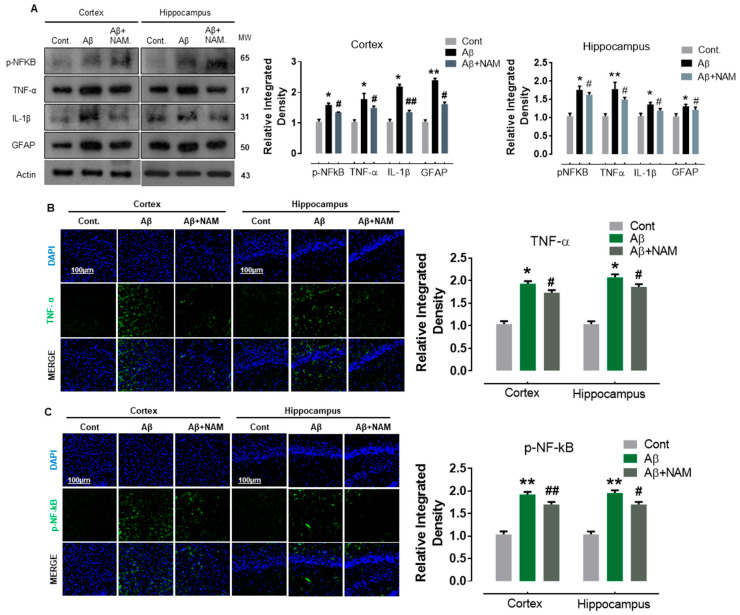
Figure 3.
NAM decreases the expression level of proinflammatory cytokines in Aβ1–42-induced Mouse Brains (A) Shows the Western blot results of inflammatory cytokines, i.e., p-NF-kB, TNF-α, GFAP and IL-1β both in cortex and hippocampus. 6 animals were kept per group. (B,C) indicating the confocal results of TNF-α, and p-NF-kB. Experiments were repeated 3 times and the scale bar was kept 100 µm. “Asterisk sign (*) indicated significant difference between control and Aβ injection group; hash sign (#) indicated significant difference between Aβ injection group and Aβ + NAM treated group. Significance: (*) # = p ≤ 0.05, (**) ## = p ≤ 0.01.”.

