Abstract
Aims
LMNA-cardiomyopathy is often associated with pathology in the cardiac conduction system necessitating device implantations. The aim was to study the timing and types of device implantations and need for re-implantations in LMNA mutation carriers.
Methods
We studied the hospital records of 60 LMNA mutation carriers concerning device implantations and re-implantations and their indications. Data were collected until April 2019.
Results
The median follow-up time from the first ECG recording to the last clinical follow-up, transplantation, or death was 7.7 (IQR=9.1) years. Altogether 61.7% (n=37) of the LMNA mutation carriers received a pacemaker or an implantable cardioverter defibrillator (ICD), and of them 27.0% (n=10) needed a device upgrade. Notably, in some patients the upgrade took place very soon after the first implantation. The first device was implanted at an average age of 47.9 years (SD=9.5), whereas the upgrade took place at an average age of 50.3 years (SD=8.1). Most upgrades were ICD implantations. Male patients underwent device upgrade more often and at a younger age than women. By the end of follow-up, 35.0% (n=21) of the patients fulfilled echocardiographic criteria for dilated cardiomyopathy, and 90.5% of them (n=19) needed pacemaker implantation.
Conclusion
Most LMNA mutation carriers underwent pacemaker implantation in this study. Due to the progressive nature of LMNA-cardiomyopathy, device upgrades are quite common. An ICD should be considered when the initial device implantation is planned in an LMNA mutation carrier.
Keywords: arrhythmias, cardiac, cardiomyopathies, pacemaker, artificial, defibrillators, implantable
Key questions.
What is already known about this subject?
LMNA-cardiomyopathy patients are at high risk for atrioventricular block, atrial and ventricular arrhythmias, and often need electrical pacing.
What does this study add?
Due to the progressive nature of LMNA-cardiomyopathy, device upgrades are often indicated, sometimes soon after the initial implantation. Nearly all LMNA mutation carriers with dilated cardiomyopathy eventually need a pacemaker.
How might this impact on clinical practice?
Choosing an implantable cardioverter defibrillator at the initial device implantation needs to be considered in LMNA mutation carriers.
Introduction
LMNA mutations cause a variety of phenotypes such as lipodystrophy, muscular disease, neuropathy, progeria and cardiomyopathy.1 Cardiomyopathy caused by LMNA mutations, or LMNA-cardiomyopathy, is typically inherited in an autosomal dominant manner.2 The cardiac phenotype typically first manifests as disturbances in the electrical system in early adulthood.3 An even earlier clinical abnormality seen in cardiolaminopathy is an elevated level of high sensitivity troponin T.4 Characteristic findings include progressive atrioventricular block (AVB), and both atrial and ventricular arrhythmias.5 The most typical macroscopic cardiomyopathy phenotype is dilated cardiomyopathy (DCM) with mainly mild dilatation of the left ventricle, although the ensuing heart failure can be severe.3 6 A more recently described rare phenotype is right predominant cardiomyopathy resembling arrhythmogenic right ventricular cardiomyopathy.7 8 Cardiac magnetic resonance studies of LMNA mutation carriers have shown a localisation of late gadolinium enhancement in the interventricular septum, while a similar scarring pattern was described in autopsy studies.9 10 Furthermore, a multicentre study of LMNA mutation carriers with drug-refractory ventricular arrhythmias found that these arrhythmias typically originate from the basal septal area.11 We have previously introduced an ECG entity, septal remodelling, as a simple and sensitive tool to detect pathology in the septal region in LMNA mutation carriers.12
Considering the range of electrical disturbances seen in LMNA-cardiomyopathy, and the progressive nature of the disease, it is not always apparent what type of cardiac pacing is appropriate, if an implantable cardioverter defibrillator (ICD) is indicated, and when the device should be upgraded. Given the increased risk of ventricular arrhythmias in cardiomyopathy-causing LMNA mutation carriers, the European Society of Cardiology guidelines recommend considering more liberal indications than usual for ICD implantation in LMNA mutation carriers with additional risk factors: reduced left ventricular ejection fraction (LVEF) of ≤45%, AVB, male sex or non-missense mutations.13 14 It has also been suggested that when an LMNA mutation carrier needs a pacemaker, an ICD should be chosen. Similarly, when cardiac resynchronisation therapy (CRT) is appropriate, a CRT-D device has been proposed.5
The aim of this study was to review the timing and type of device implantations and need for re-implantations in a cohort of LMNA mutation carriers.
Methods
This is a retrospective study based on available hospital charts. We included 60 Finnish LMNA mutation carriers (31 men and 29 women) identified in clinical practice or in previous studies.15 16 Data were collected until April 2019. The variants are listed in table 1. The most common variant was the Finnish founder mutation c.427T>C, p.(Ser143Pro).15
Table 1.
The LMNA variants, their prevalence, and the prevalence of pacemakers among the variant carriers
| LMNA variant | Carriers (n=60) | Patients with pacemakers (n=37) |
| c.1086delT, p.(Leu363Trpfs*117) | 6 | 6 |
| c.1380G>C, p.(Glu460Asp) | 12 | 5 |
| c.1442dupA, p.(Tyr481*) | 1 | 1 |
| c.1493delG, p.(Ala499Leufs) | 1 | 1 |
| c.1517A>C, p.(His506Pro) | 2 | 1 |
| c.394G>C, p.(Ala132Pro) | 4 | 2 |
| c.427T>C, p.(Ser143Pro) | 19 | 13 |
| c.481G>A, p.(Glu161Lys) | 1 | 1 |
| c.497G>C, p.(Arg166Pro) | 1 | 1 |
| c.568C>T, p.(Arg190Trp) | 6 | 3 |
| c.710T>C, p.(Phe237Ser) | 7 | 3 |
The diagnostic criteria used for DCM were left ventricular end-diastolic diameter >27 mm/m2 and LVEF <45%.17 Favourable response to CRT was defined as LVEF improvement of 10 units or more. More moderate improvement in LVEF, reduction in the levels of B-type natriuretic peptide (BNP) or N-terminal pro-BNP (ProBNP), and/or QRS complex shortening in ECG were considered signs of possibly favourable response to CRT treatment.
The Shapiro-Wilk test was used to assess whether the data were normally distributed. Normally distributed continuous variables were given as mean and SD and non-parametric as median and IQR. Independent samples t-test was used to compare the means of normally distributed parameters. Frequencies were compared with the χ2 test when appropriate, and otherwise with the Fisher’s exact test. SPSS V.25 and V.27 were used for data analysis.
Results
The median follow-up time from the first ECG recording to the last clinical follow-up, transplantation or death was 7.7 years (IQR=9.1 years). Of all the patients, 35.0% (n=21) fulfilled the diagnostic criteria set for DCM at some point during follow-up. Coronary artery disease was excluded using angiography or coronary CT in 41.7% of the patients; the indication for the procedure was cardiomyopathy, except in one patient with known LMNA mutation, where the indication was ventricular tachycardia (VT). Heart transplantation was required in 16.7% (n=10) of the patients, and seven individuals (11.7%) died during follow-up, all of them due to cardiomyopathy.
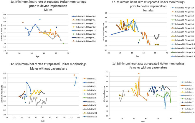
The mean patient age at the time of the first available ECG recording was 39.4 years, while the mean age at the time of the last ECG recording was 45.7 years. Table 2 shows the respective PR intervals and QRS complex durations in ECG. Table 3 shows the highest level of AVB and the mean age of the study population at the time of the detection of the conduction disorder. Figure 1A, B shows the minimum heart rate during one or more Holter monitorings in men and women prior to pacemaker implantation. Figure 1C, D shows the corresponding values in individuals, who did not have a pacemaker implanted during the follow-up. In 75.0% of the men (6/8) and 84.6% (11/13) of the women, the pacemaker implantation took place within 1 year of the preceding Holter monitoring. One female individual received a pacemaker nearly a decade after the preceding Holter recording, but the device was a CRT-D, which was implanted due to reduced LVEF. One female and two male individuals received the devices within 2 years of the preceding Holter monitoring.
Table 2.
The mean age at the time of the first (ECG 1) and last (ECG 2) available ECG recordings and the respective median PR intervals and QRS complex durations in ECG
| Mean age | SD | Median PR (ms) | Median QRS (ms) | |
| ECG 1 | 39.4 (n=57) | 12.3 | 200 (IQR=107) (n=43) |
96 (IQR=17) (n=37) |
| ECG 2 | 45.7 (n=37) | 12.3 | 226 (IQR=120) (n=31) |
100 (IQR=25) (n=32) |
PR, PR interval in ECG; QRS, QRS complex duration in ECG.
Table 3.
The presence and detection ages of AVBs
| Highest AVB detected | Frequency | Per cent | Mean age, years (n) | Min age | Max age | SD |
| 1st degree | 17 | 28.3 | 44.4 (n=26)* | 21.4 | 65.5 | 11.0 |
| 2nd degree | 14 | 23.3 | 45.7 (n=15)* | 30.5 | 65.6 | 10.7 |
| Mobitz 1 | 10 | |||||
| Mobitz 2 | 3 | |||||
| Both | 1 | |||||
| 3rd degree | 5 | 8.3 | 49.1 (n=5) | 41.2 | 61.6 | 8.3 |
| Any AVB | 36 | 60.0 | 44.0 (n=36) | 21.4 | 65.5 | 10.6 |
| No AVB | 24 | 40.0 | 39.7† (n=24) | 20.7 | 70.5 | 15.0 |
*Includes individuals with an initial lower level AVB followed by a higher-level block.
†At last follow-up.
AVB, atrioventricular block.
Figure 1.
(A, B) The lowest heart rate at single or repeated Holter monitorings in men and women prior to pacemaker implantation. Age at pacemaker implantation is given. (C, D) The lowest heart rate at single or repeated Holter monitorings in men and women who did not have a pacemaker implanted by the end of the data collection. PM, pacemaker.
Timing and type of devices
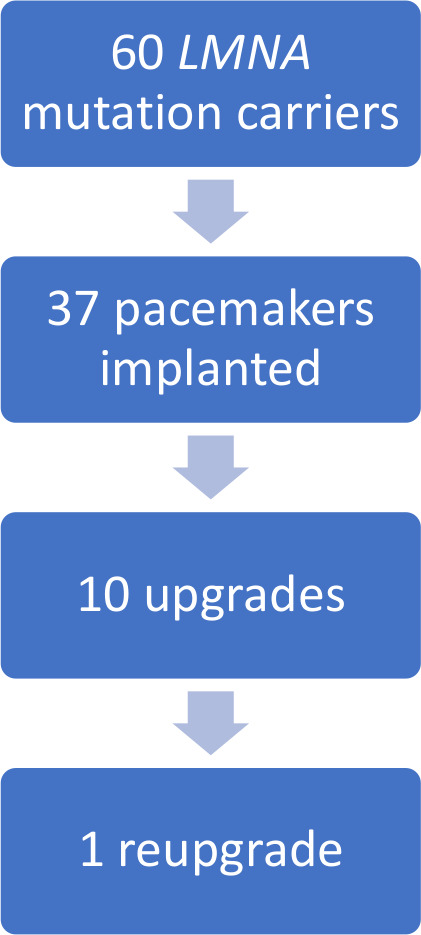
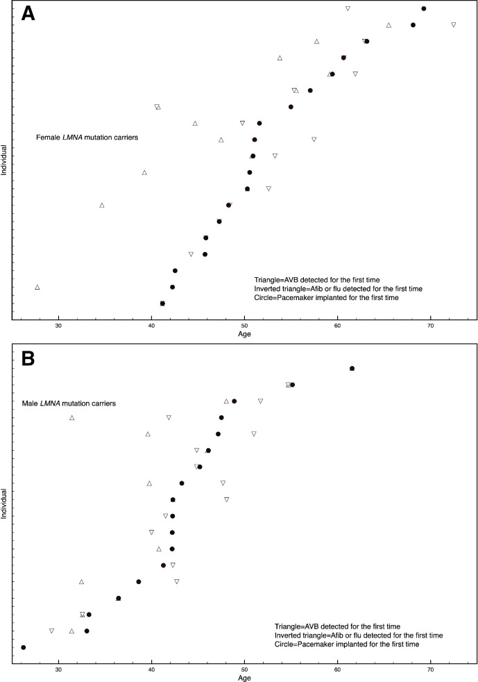
The majority of the patients, 61.7% (n=37), received a pacemaker or an ICD at some point of the follow-up. At the time of the device implantation, 11 individuals (29.7%) fulfilled the echocardiographic DCM criteria, whereas 26 individuals (70.3%) did not. On the other hand, of the 21 patients who fulfilled the DCM criteria by the end of the follow-up, 19 (90.5%) underwent device implantation at some point. The initial pacemaker types are listed in table 4. Of the patients with pacemakers, 27.0% (n=10) needed a device upgrade. The upgrades tended to be more common in men (38.9%, 7/18) than in women (15.8%, 3/19), but the difference was not statistically significant. One upgrade was performed at the time of elective pacemaker generator replacement. Of the 13 individuals with the Ser143Pro LMNA variant who received a device, no one required a device upgrade. A flow chart of pacemaker implantations and upgrades is given in figure 2. The device upgrade types are listed in table 5, and the clinical characteristics of patients who did or did not undergo a device update are shown in table 6. The mean interval from the first pacemaker implantation to the upgrade was 5.1 years (SD=5.1 years), but ranged from less than a month to 14.8 years. Of note, in five cases the upgrade took place less than 2 years after the initial pacemaker implantation. The first device was implanted at an average age of 47.9 years (SD=9.5), whereas the upgrade took place at an average age of 50.3 years (SD=8.1). One individual received a second upgrade at age 59.2 years. By the end of data collection, 58.1% (18/31) of the men, and 65.5% (19/29) of the women (the difference was not statistically significant) had undergone device implantation. Men received the first device almost 10 years earlier (mean age 42.9 years, SD=8.1) than women (mean age 52.6 years, SD=8.4, p=0.001). Regarding device upgrade, the mean age for men was 48.3 (SD 9.1), and for women, 54.8 years (SD=2.5, statistically non-significant difference). Altogether 18 individuals received an ICD, 12 as the first device and 6 as an upgrade. Three of the ICDs were implanted after cardiac resuscitation, and one due to sustained VT as secondary prophylaxis, seven as primary prophylaxis, but with documented NSVT (non-sustained ventricular tachycardia), and the remaining seven as primary prophylaxis without known previous VT. At the time of the ICD implantation, 12 (66.7%) individuals fulfilled the echocardiographic DCM criteria whereas 6 (33.3%) individuals did not. Figure 3 shows the incidences of first recordings of AVB, atrial fibrillation or flutter, and the timing of device implantation.
Table 4.
The initial pacemaker types
| AAI | 1 |
| VVI | 8 |
| DDD | 14 |
| VVI+ICD | 6 |
| DDD+ICD | 3 |
| CRT-P | 2 |
| CRT-D | 3 |
Figure 2.

A flow chart of pacemaker implantations and upgrades.
Table 5.
Pacemaker upgrades
| First PM type | Second PM type | N=10 | Upgrade indication |
| AAI | DDD | 1 | DAV 3, syncope |
| VVI | VVI-ICD | 2 | Sustained VT, resuscitation* |
| VVI | DDD-ICD | 1 | Heart failure |
| DDD | DDD-ICD | 1 | Presyncope and documented NSVT |
| DDD | CRT-P | 1 | Generator replacement, dyssynchrony and heart failure |
| DDD | CRT-D | 2 | Heart failure (n=2), symptomatic NSVT (n=1) |
| VVI-ICD | DDD-ICD | 1 | Symptomatic bradycardia |
| VVI-ICD | CRT-D | 1 | Heart failure |
*Additionally, one individual received a second upgrade later from VVI-ICD to CRT-D.
CRT, cardiac resynchronisation therapy; DAV, distal atrioventricular block; ICD, implantable cardioverter defibrillator; NSVT, non-sustained ventricular tachycardia; PM, pacemaker.
Table 6.
Comparison of clinical characteristics of patients having or having not having undergone a pacemaker upgrade
| PM upgrade (n=10) |
No PM upgrade (n=27) | Statistical significance | |
| Males | 7 (70%) | 11 (40.7%) | ns (males vs females) |
| Ser143Pro | 0 (0%) | 13 (76.5%) | p=0.007 |
| Transplantation | 1 (10%) | 6 (22.2%) | ns |
| Afib or flutter | 9 (90%) | 21 (77.8%) | ns |
| DCM by the end of follow-up | 7 (70%) | 12 (44.4%) | ns |
| Ventricular arrhythmia requiring treatment | 4 (40%) | 6 (22.2%) | ns |
Afib, atrial fibrillation; DCM, dilated cardiomyopathy; PM, pacemaker.
Figure 3.
(A) Women (n=19) and (B) men (n=18). The incidence of a first recording of atrioventricular block (AVB), atrial fibrillation (Afib)/flutter (flu) and device implantation. Each line represents an LMNA mutation carrier. Each mutation carrier, who received a device is shown.
CRT response
Ten patients received a CRT-P (n=3) or a CRT-D (n=7) pacemaker, five as their initial device and five as an upgrade (see tables 4 and 5). The mean CRT implantation age was 51.6 years (SD=10.0). Seven of these patients (77.8%) had a favourable response to the device, and two were non-responders. The CRT responses are listed in table 7. Two individuals were followed up elsewhere, and data concerning CRT response was available from only one of them.
Table 7.
CRT responses
| Individual (M/F) | QRS shortening | LVEF improvement | Biomarker reduction (BNP or ProBNP) | Overall response |
| F | no | yes | yes | Favourable |
| M | no | Slight improvement | NA | Possibly favourable |
| M | NA | NA | NA | Non-responder* |
| F | yes | NA | NA | Possibly favourable† |
| F | NA | NA | NA | NA |
| M | yes | no | no | Possibly favourable |
| M | NA | yes | yes | Favourable |
| M | yes | yes | no | Favourable |
| M | no | yes | no | Favourable |
| F | no | no | no | Non-responder |
*Patient died 3 years after the CRT implantation.
†Subjectively favourable, later follow-up elsewhere.
BNP, B-type natriuretic peptide; CRT, cardiac resynchronisation therapy; LVEF, left ventricular ejection fraction; QRS, QRS complex duration.
Pacemaker complications
Device-related complications occurred in 7 (18.9%) of the 37 individuals, 26.3% (5/19) of the women and 11.1% (2/18) of the men (statistically non-significant difference). As the overall number of device implantations, including the upgrades, was 48, the overall complication rate in all of the implantations was 14.6%. Five of the complications took place after the first or only pacemaker implantation and two after an upgrade. The complication rates were 13.5% for first implantations and 18.2% for upgrades. The device-related complications included one infection leading to pacemaker removal and re-implantation, two cases of thrombosis requiring anticoagulation, one myocardial perforation, one pacemaker pocket haematoma and two cases of broken pacemaker leads leading to lead replacement.
Ventricular arrhythmias
During follow-up 16.7% (n=10) of the LMNA mutation carriers had ventricular arrhythmias requiring treatment; of those 33.3% (n=3) had ventricular fibrillation and 66.7% (n=7) had VT. Amiodarone treatment was reported in 15.0% (n=9) of the mutation carriers, in four patients to treat ventricular arrhythmias and in six for atrial fibrillation; one patient was initially treated with amiodarone for atrial fibrillation and later on for VT.
Discussion
This is a descriptive, retrospective study dealing with the need for, timing and type of pacemaker implantations in LMNA mutation carriers. We found that the majority (61.7%) of the 60 studied patients needed pacemaker implantation. In addition, a quarter of the patients with devices needed a device upgrade, which sometimes occurred quite soon after the initial implantation. Most upgrades were devices with a defibrillator, thus supporting the view that when a device is needed in an LMNA mutation carrier, the need for an ICD should always be considered.5 This strategy has previously been studied in a prospective manner with encouraging results.18 A significant proportion of the pacemaker implantations in this study took place before the current recommendations concerning ICD implantation in LMNA mutation carriers were available. This probably explains to some extent the extensive need of devise upgrades that was seen.
Device upgrade tended to be more common in men, and patient age at the first device implantation was almost 10 years lower in men than in women. This is in line with the previously identified higher risk for malignant ventricular arrhythmias in men.14 At the time of device implantation, a third of the patients fulfilled the diagnostic criteria for DCM. On the other hand, 90.5% of the patients who fulfilled the DCM criteria at any point during follow-up underwent device implantation. Most of the ICD implantations were primary prophylactic, and at the time of ICD implantation two-thirds of the patients fulfilled the DCM criteria. All deaths during follow-up were related to cardiomyopathy, but none due to sudden cardiac death.
The overall complication rate related to pacemakers—18.9% of the patients with devices and 14.6% of all implantations—was rather high, compared with the rates reported in other studies. A nationwide Danish study reported complications in 9.5% of their patients.19 The same study reported a larger complication risk in females, and a larger complication rate concerning device upgrades. Similar tendencies were seen in the present study.
Our observations regarding CRT responses are not fully comprehensive, because this is a retrospective study based on hospital records, not designed to assess CRT responses. Of patients with available follow-up data after CRT implantation, 77.8% showed signs of a favourable response. This is fairly well in line with the response rates reported in CRT studies. However, it should be acknowledged that the reported response rates vary with the criteria used to assess the response.20 The relatively small number of patients as well as the retrospective setting are limitations to this study particularly concerning our observations regarding CRT responses.
Typically, the first indication for device implantation in this population of LMNA mutation carriers was progressive bradycardia, but as shown in repeated Holter recordings both in individuals requiring a pacemaker implantation and those who had not yet needed one, the progression is sometimes very slow. The appropriate timing of pacemaker implantation is therefore still a challenge and repeated monitoring of individuals carrying disease-causing LMNA mutations is needed. As the majority of device upgrades involved ICDs, it is important to assess the need for an ICD when device implantation is planned for an LMNA mutation carrier.
Footnotes
Contributors: LHO collected the data and participated in data analysis and interpretation, and manuscript writing. KN participated in data interpretation and manuscript writing. HP participated in planning the study, data interpretation and manuscript writing. SW participated in patient recruitment and data collection. TH participated in planning the study, data interpretation and manuscript writing.
Funding: This work was supported by The Finnish Foundation for Cardiovascular Research, Aarne Koskelo Foundation, Special Government Subsidy (Y2019SK005 and TYH2014208) and Government Research Funding. Open access funded by Helsinki University Library.
Competing interests: None declared.
Provenance and peer review: Not commissioned; externally peer reviewed.
Data availability statement
No data are available. The data underlying this article cannot be shared publicly due to privacy of the individuals that participated in the study.
Ethics statements
Patient consent for publication
Not required.
Ethics approval
The study patients gave written informed consent, and the study was approved by the Ethics Committee of the Helsinki University Central Hospital (HUS/24/2017 and HUS/60/1019). The data underlying this article cannot be shared publicly due to privacy of the individuals that participated in the study.
References
- 1.Brayson D, Shanahan CM. Current insights into LMNA cardiomyopathies: existing models and missing LINCs. Nucleus 2017;8:17–33. 10.1080/19491034.2016.1260798 [DOI] [PMC free article] [PubMed] [Google Scholar]
- 2.Burkett EL, Hershberger RE. Clinical and genetic issues in familial dilated cardiomyopathy. J Am Coll Cardiol 2005;45:969–81. 10.1016/j.jacc.2004.11.066 [DOI] [PubMed] [Google Scholar]
- 3.van Berlo JH, de Voogt WG, van der Kooi AJ, et al. Meta-analysis of clinical characteristics of 299 carriers of LMNA gene mutations: do lamin A/C mutations portend a high risk of sudden death? J Mol Med 2005;83:79–83. 10.1007/s00109-004-0589-1 [DOI] [PubMed] [Google Scholar]
- 4.Chmielewski P, Michalak E, Kowalik I, et al. Can Circulating Cardiac Biomarkers Be Helpful in the Assessment of LMNA Mutation Carriers? J Clin Med 2020;9. 10.3390/jcm9051443. [Epub ahead of print: 12 05 2020]. [DOI] [PMC free article] [PubMed] [Google Scholar]
- 5.Peretto G, Sala S, Benedetti S, et al. Updated clinical overview on cardiac laminopathies: an electrical and mechanical disease. Nucleus 2018;9:380–91. 10.1080/19491034.2018.1489195 [DOI] [PMC free article] [PubMed] [Google Scholar]
- 6.Hershberger RE, Morales A, Siegfried JD. Clinical and genetic issues in dilated cardiomyopathy: a review for genetics professionals. Genet Med 2010;12:655–67. 10.1097/GIM.0b013e3181f2481f [DOI] [PMC free article] [PubMed] [Google Scholar]
- 7.Quarta G, Syrris P, Ashworth M, et al. Mutations in the lamin A/C gene mimic arrhythmogenic right ventricular cardiomyopathy. Eur Heart J 2012;33:1128–36. 10.1093/eurheartj/ehr451 [DOI] [PubMed] [Google Scholar]
- 8.Ollila L, Kuusisto J, Peuhkurinen K, et al. Lamin A/C mutation affecting primarily the right side of the heart. Cardiogenetics 2013;3. 10.4081/cardiogenetics.2013.e1 [DOI] [Google Scholar]
- 9.Holmström M, Kivistö S, Heliö T, et al. Late gadolinium enhanced cardiovascular magnetic resonance of lamin A/C gene mutation related dilated cardiomyopathy. J Cardiovasc Magn Reson 2011;13:30. 10.1186/1532-429X-13-30 [DOI] [PMC free article] [PubMed] [Google Scholar]
- 10.Raman SV, Sparks EA, Baker PM, et al. Mid-myocardial fibrosis by cardiac magnetic resonance in patients with lamin A/C cardiomyopathy: possible substrate for diastolic dysfunction. J Cardiovasc Magn Reson 2007;9:907–13. 10.1080/10976640701693733 [DOI] [PubMed] [Google Scholar]
- 11.Kumar S, Androulakis AFA, Sellal J-M, et al. Multicenter experience with catheter ablation for ventricular tachycardia in lamin A/C cardiomyopathy. Circ Arrhythm Electrophysiol 2016;9. 10.1161/CIRCEP.116.004357 [DOI] [PubMed] [Google Scholar]
- 12.Ollila L, Nikus K, Holmström M, et al. Clinical disease presentation and ECG characteristics of LMNA mutation carriers. Open Heart 2017;4:e000474. 10.1136/openhrt-2016-000474 [DOI] [PMC free article] [PubMed] [Google Scholar]
- 13.Priori SG, Blomström-Lundqvist C, Mazzanti A, et al. 2015 ESC guidelines for the management of patients with ventricular arrhythmias and the prevention of sudden cardiac death: the task force for the management of patients with ventricular arrhythmias and the prevention of sudden cardiac death of the European Society of cardiology (ESC). endorsed by: association for European paediatric and congenital cardiology (AEPC). Eur Heart J 2015;36:2793–867. 10.1093/eurheartj/ehv316 [DOI] [PubMed] [Google Scholar]
- 14.van Rijsingen IAW, Arbustini E, Elliott PM, et al. Risk factors for malignant ventricular arrhythmias in lamin a/c mutation carriers a European cohort study. J Am Coll Cardiol 2012;59:493–500. 10.1016/j.jacc.2011.08.078 [DOI] [PubMed] [Google Scholar]
- 15.Kärkkäinen S, Heliö T, Miettinen R, et al. A novel mutation, Ser143Pro, in the lamin A/C gene is common in finnish patients with familial dilated cardiomyopathy. Eur Heart J 2004;25:885–93. 10.1016/j.ehj.2004.01.020 [DOI] [PubMed] [Google Scholar]
- 16.Kärkkäinen S, Reissell E, Heliö T, et al. Novel mutations in the lamin A/C gene in heart transplant recipients with end stage dilated cardiomyopathy. Heart 2006;92:524–6. 10.1136/hrt.2004.056721 [DOI] [PMC free article] [PubMed] [Google Scholar]
- 17.Manolio TA, Baughman KL, Rodeheffer R, et al. Prevalence and etiology of idiopathic dilated cardiomyopathy (summary of a National Heart, Lung, and Blood Institute workshop. Am J Cardiol 1992;69:1458–66. 10.1016/0002-9149(92)90901-A [DOI] [PubMed] [Google Scholar]
- 18.Anselme F, Moubarak G, Savouré A, et al. Implantable cardioverter-defibrillators in lamin A/C mutation carriers with cardiac conduction disorders. Heart Rhythm 2013;10:1492–8. 10.1016/j.hrthm.2013.06.020 [DOI] [PubMed] [Google Scholar]
- 19.Kirkfeldt RE, Johansen JB, Nohr EA, et al. Complications after cardiac implantable electronic device implantations: an analysis of a complete, nationwide cohort in Denmark. Eur Heart J 2014;35:1186–94. 10.1093/eurheartj/eht511 [DOI] [PMC free article] [PubMed] [Google Scholar]
- 20.Tomassoni G. How to define cardiac resynchronization therapy response. J Innov Card Rhythm Manag 2016;7:S1–7. 10.19102/icrm.2016.070003 [DOI] [Google Scholar]
Associated Data
This section collects any data citations, data availability statements, or supplementary materials included in this article.
Data Availability Statement
No data are available. The data underlying this article cannot be shared publicly due to privacy of the individuals that participated in the study.


