

Abstract
Involvement of the external ear by PG is a rare encounter, and steroids remain to be the main stay in its treatment.
Keywords: external ear, limited setting, Pyoderma gangrenosum, Tanzania
Involvement of the external ear by PG is a rare encounter, and steroids remain to be the main stay in its treatment.

1. INTRODUCTION
Pyoderma gangrenosum (PG) being a primary sterile neutrophilic dermatosis, the etiology is still uncertain and it rarely affects children. A high index of suspicion is essential to execute its prompt treatment. To the best of our knowledge, this is the first reported case in our country.
Pyoderma gangrenosum refers to a primary sterile neutrophilic dermatosis with an idiopathic etiology that rarely affects children. 1 , 2 Involvement of the external ear is a rare encounter though its rarity does not seem to determine its inherent behavior with regard to characteristics of the ulcer and response to treatment. 3 , 4 , 5 The diagnosis of PG remains to be clinical especially in resource‐limited settings, and there is no consensus to date on its gold‐standard treatment. A high index of suspicion is essential to execute its prompt treatment. The objective of this case report was to report an unusual case of involvement of external ear by PG in a 3‐year‐old child which went unestablished for a couple of months and to the best of our knowledge this is so far the first reported case in our country and the aim is to lay emphasis on the importance of prompt diagnosis of PG and its treatment to halt its destructive behavior.
2. CASE REPORT
A 3‐year‐old male child presented to one of the remote dispensaries with a history of 6‐month ulcerative lesions involving the external ear. Lesions were painful and were accompanied by a history of recurrent nonfoul smelling ipsilateral otorrhoea. The kid had no history of fever or night sweats, history of trauma involving the external ear, or gastrointestinal complaints.
On examination, he was malnourished (had moderate acute malnutrition) and the ear had necrotic skin overlying the pinna and multiple papules with some being pus filled.
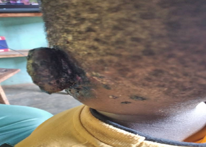
The patient was subjected to laboratory workups; hemoglobin was 13 g/dL, HIV negative. Histopathological analysis of the lesions and immunological workups such as purified protein derivative, venereal disease research laboratory was not done due to remoteness of health facility. Serum albumin and globulin were not ordered because of the same reason despite the child being malnourished. Clinical diagnosis of PG of the external ear was made. (Figures 1 and 2).
FIGURE 1.

Showing necrotic external ear with some pustules before treatment initiation
FIGURE 2.
Showing necrotic external ear with some mutilated sites prior treatment
The patient received: intramuscular triamcinolone 40 mg stat, prednisolone tablets where 10 mg once daily for 7 days was provided and then tapered to 5 mg once daily for 7 days, betamethasone cream which was applied topically over the ulcerated external ear, syrup ampicillin + cloxacillin 250 mg administered 8 hourly for 7 days and boric acid ear drops were applied 4 hourly for 7 days. The patient was followed up for 6 months with no recurrence. (Figure 3).
FIGURE 3.

Showing complete healing of the external ear after 6‐mo follow up
3. DISCUSSION
Pyoderma gangrenosum being a rare disease was first elaborated in 1930 and described as a chronic destructive inflammatory, noninfectious skin disease with recurrence tendency. 1 , 2 PG lesions typically present as painful, erythematous pustules, bullae, or nodules. 1 , 3 , 4 , 5 , 6 They may evolve into necrotic plaques with raised edges or into violaceous deep ulcers with undermined bluish borders, healing with atrophic or cribriform scars. Such description matches with what was seen in our case report. 1 , 2 , 4 , 5 , 6 , 7 , 8
PG affects mainly adults aged between 20 and 50 years with female predilection. It account for only 4% of cases in children and adolescents with a higher incidence of head and perineal involvement. PG has preponderance of pustular lesions and a frequent history of trauma. 1 , 2 , 3 , 5 , 6 , 8 , 9 , 10 The commonest presentation of PG in children is disseminated ulcerative lesions. 1 , 6 , 8
The actual pathogenesis of PG has remained to be idiopathic though it has been postulated to be a neutrophilic disorder characterized by dysregulation of the immune system, such as overexpression of growth factors and interleukins, especially interleukin (IL)‐1, (IL)‐6, and (IL)‐8 and tumor necrosis factor (TNF)‐a. 10
Though the etiology has been idiopathic in majority of cases, in about fifty percent of cases, it is associated with neoplasms and/or systemic inflammatory illnesses, such as Behcet' disease, ulcerative colitis and Crohn's disease. 4 , 7 More recently, PG has been associated with novel drug therapies like pegfilgrastim. 2
The diagnosis of pediatric PG is often delayed, with an average of 2 months due to its lower prevalence among children and because it can be misdiagnosed with other ulcerative diseases. 1 , 2 , 6 This correlate with what was seen in this case where the diagnosis was delayed for 6 months.
Though there are no histopathological findings specific to PG, but skin biopsy should be performed to rule out other etiologies. 1 , 3 , 4 , 6 In resource‐limited settings, its diagnosis remains to be clinical.
Once the diagnosis of pediatric PG is confirmed, clinicians are recommended to identify for an underlying etiology as the disease can be the initial presentation of a systemic disorder. 1 This was a challenge to our case since the patient was residing at a resource‐limited setting.
The head and neck region may be affected by PG, although to date only few cases of ear involvement have been reported. 1 , 6 , 8 This is the first novel case reported in our country involving the external ear.
To date, there is no gold‐standard treatment or published algorithm for choice of therapy for pediatric PG though treatment should be tailored according to the underlying etiology. It includes systemic steroids, corticosteroid sparing agents such as dapsone and cyclosporine, and TNF‐alpha inhibitors such as adalimumab and infliximab. Response to treatment is high with cure rates reaching 90%. 6 , 10
Our patient achieved a rapid response when treated with steroids with no recurrence after 6 months of follow up, thus laying emphasis on the role of steroids in PG. Auricular cases previously reported showed good response to the classical therapeutic strategy based on the use of corticosteroids or neutrophil chemotaxis inhibitors and thus use of these drugs as first choice. No cases of auricular PG treated with anti‐TNF‐α biologic agents have been reported from the available literatures. Thus, its value in managing refractory cases of auricular PG is unknown, as already shown in PG of other sites. 10 Despite the good response to steroids manifested in auricular PG cases, yet clinicians must expect episodes of flare‐up in some patients, as usually observed in most cases of typical pyoderma. Generally, the basis for diagnosis of PG in this case report met the minor criteria set by Maverakis et al 11 No flare has been reported in this case report.
4. CONCLUSION
A high index of suspicion is of paramount importance toward prompt diagnosis and treatment of PG. Clinical diagnosis remains to be the main stay in resource limited settings similar to what can be depicted from this case report. Steroids have been found be the cornerstone in management of PG in our case report similar to what has been reported in other parts of the world.
CONFLICT OF INTEREST
None declared.
AUTHOR CONTRIBUTIONS
ZSA: collected information for the case and drafted the initial version of the manuscript. EPT: drafted the initial version of the manuscript. AAK: contribute to critical feedback and editing of the manuscript. All authors: read and approved the final version to be published in Clinical Case Reports.
ETHICAL APPROVAL
This report is in accordance with the Declaration of Helsinki. The parent of the child gave a written informed consent prior inclusion of the child in this report.
ACKNOWLEDGMENT
The parent of the child is highly acknowledged for providing a written informed consent to publish the case report.
Abraham ZS, Thomas EP, Kahinga AA. Unilateral Pyoderma gangrenosum of the external ear in a resource‐limited setting: Case report and literature review. Clin Case Rep. 2021;9:2016–2018. 10.1002/ccr3.3930
DATA AVAILABILITY STATEMENT
The data that support the findings of this report are available from the corresponding author upon reasonable request.
REFERENCES
- 1. Sousa MC, Lemos ÉF, De Morais OO, Coutinho AS, Gomes CM. Pyoderma gangrenosum leading to bilateral involvement of ears. J Clin Aesthet Dermatol. 2014;7(1):41. [PMC free article] [PubMed] [Google Scholar]
- 2. Gameiro A, Pereira N, Cardoso JC, Gonçalo M. Pyoderma gangrenosum: challenges and solutions. Clin Cosmet Investig Dermatol. 2015;8:285. [DOI] [PMC free article] [PubMed] [Google Scholar]
- 3. Al‐Rimawi HS, Abuekteish FM, Daoud AS, Oboosi MM. Familial pyoderma gangrenosum presenting in infancy. Eur J Pediatr. 1996;155(9):759‐762. [DOI] [PubMed] [Google Scholar]
- 4. Wcisło‐Dziadecka D, Bergler‐Czop B, Brzezińska‐Wcisło L, Arasiewicz H. Extensive phlegmon and pyoderma gangrenosum: diagnostic difficulties. Adv Dermatol Allergol/Postȩpy Dermatol Alergol. 2015;32(1):46. [DOI] [PMC free article] [PubMed] [Google Scholar]
- 5. Kędzierska K, Sindrewicz K, Smektała T, et al. Wegener's granulomatosis and pyoderma gangrenosum‐rare causes of facial ulcerations. Adv Hyg Exp Med/Postepy Hig Med Dosw. 2016;70:210‐218. [DOI] [PubMed] [Google Scholar]
- 6. Paramkusam G, Meduri V, Gangeshetty N. Pyoderma gangrenosum with oral involvement–case report and review of the literature. Int J Oral Sci. 2010;2(2):111‐116. [DOI] [PMC free article] [PubMed] [Google Scholar]
- 7. Yamamoto T. Pyoderma gangrenosum: an important dermatologic condition occasionally associated with rheumatic diseases. World J Rheumatol. 2015;5(2):101‐107. [Google Scholar]
- 8. Snyder RA. Pyoderma gangrenosum involving the head and neck. Arch Dermatol. 1986;122(3):295‐302. [PubMed] [Google Scholar]
- 9. Khandpur S, Mehta S, Nagi Reddy BS. Pyoderma gangrenosum in two siblings: a familial predisposition. Pediatr Dermatol. 2001;18(4):308‐312. [DOI] [PubMed] [Google Scholar]
- 10. Wollina U. Pyoderma gangrenosum–a review. Orphanet J Rare Dis. 2007;2(1):19. [DOI] [PMC free article] [PubMed] [Google Scholar]
- 11. Maverakis E, Ma C, Shinkai K, et al. Diagnostic criteria of ulcerative pyoderma gangrenosum: a Delphi consensus of international experts. JAMA Dermatol. 2018;154(4):461‐466. [DOI] [PubMed] [Google Scholar]
Associated Data
This section collects any data citations, data availability statements, or supplementary materials included in this article.
Data Availability Statement
The data that support the findings of this report are available from the corresponding author upon reasonable request.