Abstract
Patient: Female, 29-year-old
Final Diagnosis: Graves’ disease
Symptoms: General malaise
Medication:—
Clinical Procedure: Laboratory checkup
Specialty: Endocrinology and Metabolism • Hematology
Objective:
Unusual clinical course
Background:
Atypical manifestations of Graves’ disease (GD) such as anemia have been noticed in the last decades. Anemia is present in up to 34% of patients with GD, yielding various anemia types such as GD anemia, pernicious anemia, iron deficiency anemia, and autoimmune hemolytic anemia (AIHA). So far, AIHA is the rarest manifestation of anemia in GD.
Case Report:
We report a case of 29-year-old woman with initial presentation of typical anemia. Further findings revealed GD signs and symptoms such as orbitopathy, increased appetite along with loss of weight, and hand tremors. Laboratory findings showed very low hemoglobin (3.9 g/dL), reticulocytosis, elevated indirect bilirubin, and positive direct Coomb’s test. Later, thyroid function testing showed decreased TSH, elevated fT4, and positive TrAb. The diagnosis of GD was made, with AIHA as initial presenting manifestation. The patient was treated using corticosteroids followed by anti-thyroid without any blood transfusion and responded well.
Conclusions:
In this case, typical AIHA was the initial presenting manifestation of GD and should not be overlooked since delayed diagnosis increases morbidity and mortality. Thyroid function assessment may be needed to search for etiologies of AIHA. Regardless of the exact underlying pathophysiology, AIHA under GD generally responds well to anti-thyroid and steroid treatment.
Keywords: Anemia, Hemolytic, Autoimmune; Graves Disease; Thyroid Diseases
Background
Hyperthyroidism is logically associated with elevated number of red blood cells since there is elevated tissue oxygen demand, hence erythropoietin secretion increases [1]. However, in up to 33% of cases, anemia was found as an atypical hematologic manifestation in GD patients. The anemia is of various types: GD anemia, pernicious anemia, iron deficiency anemia, and autoimmune hemolytic anemia (AIHA). Of these type, AIHA is the rarest type, with only 4 reported cases [2]. It appears that an association exists between hyperthyroidism and autoimmunity; the hypothesis had been reported, but not proven.
Here, we report the case of a GD patient with the initial manifestation of AIHA. Anti-thyroid therapy and steroid treatment effectively relieved symptoms and increased hemoglobin levels without blood transfusions. The scarcity of pure AIHA manifestations in GD potentially delays diagnosis or leads to unnecessary performance of examinations that negatively impact patient outcomes.
Case Report
A 29-year-old woman came to the ER with complaints of weakness since 2 months and worsened in the last 1 week before being admitted to the hospital. Symptoms of anemia (palpitations and pallor) were identified without any mucocutaneous bleeding. The patient denied previous weakness and menstrual disorders. The patient reported loss of weight (10 kilograms) along with increase in appetite in the past year. Symptoms of chronic liver and kidney diseases were not identified. The typical lupus complaint was also denied.
The patient was hospitalized 1 month ago with diagnosis of anemia (hemoglobin 6 g/dL), but did not receive any blood transfusions. Instead, she received ferrous sulfate tablets and PPIs. She denied a history of smoking. The patient had 3 children. She did not have a familial history of endocrine or liver disorders.
During the physical examination, an increased heart rate of 120 bpm with regular rhythm was recorded, while other vital signs were unremarkable. Important findings were BMI of 16.5, anemic conjunctival, exophthalmos, Dalrymple sign (+), a palpable bilateral diffuse goiter 3×3×1 cm, symmetrical, soft consistency, no thrill palpable, and no bruits. Her hands were warm, wet, and pale. Fine tremors were identified when a paper was placed on both hands. There was no pretibial edema. The color of urine collected in a bottle was clear yellow. Wayne’s and Newcastle thyrotoxicosis index were 31 (>20) and 44 (>39), respectively.
Laboratory findings at admission showed reduced hemoglobin, reticulocytosis, elevated indirect bilirubin, and elevated urobilin levels from urinalysis, but test results for lactate dehydrogenase and haptoglobin were not available (Table 1). Peripheral blood smears revealed normochromic normocytic anemia and anisopoikilocytosis. Polyspecific Direct Coomb’s test was positive, but the monospecific test could not be done due to resource limitations. The initial diagnosis was AIHA with suspected GD. The patient initially received corticosteroid (methylpredniso-lone), which was then tapered down according to the protocol.
Table 1.
Laboratory findings during hospitalization.
| Lab test | Admission | Normal range* | Lab test | Day 2 | Normal range* |
|---|---|---|---|---|---|
| Hemoglobin | 3.9 g/dL | 11.0–14.7g/dL | Hemoglobin | 6.1 g/dL | 11.0–14.7g/dL |
| Reticulocyte (%) | 32.77% | 0.80–2.21% | TSH | 0.005 mU/L | 0.55–4.78 mU/L |
| Reticulocyte# (106/uL) | 0.16 | 0.034–0.100.106/uL | fT4 | 2.03 ng/dL | 0.89–1.76 ng/dL |
| Direct bilirubin | 0.92 mg/dL | <0.20 mg/dL | TrAb | 4.82 IU/L | <1.75 IU/L |
| Total bilirubin | 1.86 mg/dL | 0.2–1.0 mg/dL | ANA | 28.57 AU/mL | <40 AU/mL |
| Urobilin urine | 2.0 | <1.0 | C3 | 49.2 mg/dL | 50–120 mg/dL |
| C4 | 11.1 mg/dL | 20–50 mg/dL |
Based on local laboratory references.
After 2 days of hospitalization, patient complaints were relieved along with increased hemoglobin level (6.1 g/dL). Thyroid function tests showed reduced TSH, elevated fT4 levels, along with positive TrAb and negative ANA test (Table 1). Thyroid ultrasound revealed bilateral diffuse goiter with normal parenchymal echo intensity, right lobe dimensions was 1.7×1.8×3.4 cm, while the left lobe was 1.8×1.3×3.4 cm. The patient was then diagnosed with GD and received additional therapy of methimazole 10 mg/day.
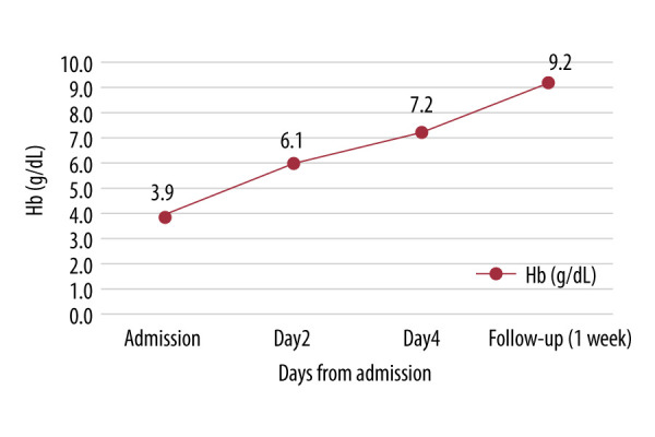
Improvements in hemoglobin level were recorded during discharge (7.2 g/dL) and 1-week follow-up (9.2 g/dL), presented in Figure 1. Additionally, oral propranolol was added to the previous therapy (oral methylprednisolone and methimazole) since the heart rate was 110 bpm, despite improved hemoglobin (9.2 g/dL). Unfortunately, the patient did not attend out-patient care because of the pandemic.
Figure 1.

Hemoglobin levels during disease course (without blood transfusion).
Discussion
GD is hyperthyroidism caused by thyroid stimulating immunoglobulin (TSI) or thyroid stimulating antibody (TSAb) which is synthesized by B lymphocytes in the thyroid gland and partly in the bone marrow and lymph nodes [3]. Signs and symptoms of GD usually are similar to any cause of thyrotoxicosis as well as those specific to Graves’ disease (orbitopathy and dermopathy). These clinical presentations depend on age, disease duration, severity, and individual susceptibility to thyroid hormone excess [4]. It may be cost-effective to use diagnostic indexes such as Wayne’s and Newcastle index to limit the number of investigations required [5,6].
Hyperthyroidism is associated with increased tissue oxygen demand, followed by increased erythropoietin secretion. Therefore, an increased number of circulating erythrocytes is logically plausible [4]. Interestingly, anemia was found in up to 34% of hyperthyroid cases and is considered an atypical hematologic manifestation [2]. This phenomenon is hypothesized to be due to altered iron metabolism, hemolysis, and oxidative stress leading to enhanced osmotic fragility of erythrocytes and lipid peroxidation, and hence shortened erythrocyte survival [1].
In GD, anemia has been reported in 33% of cases [7]. It is postulated that decreased erythrocyte circulation time and auto-immune mechanisms are responsible for anemia in GD [8]. It is quite challenging to detect GD if it is clinically presented as anemia since both conditions have overlapping symptoms. However, some specific types of anemia are directly or indirectly associated with GD, such as pernicious anemia, iron deficiency anemia due to celiac disease, and AIHA [9]. In addition, an unclassified type of anemia that occurs in GD which improves after anti-thyroid treatment is termed GD anemia [7].
AIHA is by far the rarest form of GD-related anemia. AIHA is a heterogenous disorder characterized by destruction of RBC through antibodies. AIHA is further classified based on auto-antibody type or the underlying disease. Warm AIHA accounts for 48–70% of patients with AIHA. Warm autoantibodies are dominated by IgG that strongly react toward erythrocytes at a temperature of 37°C, causing extravascular hemolysis through FcγRIII or C3b receptors on macrophages [10]. These autoanti-bodies are invariably polyclonal and flawed during T-cell regulation of humoral immune system; therefore, distinction function between self and non-self is defective. Gene polymorphism for signal substance CTLA-4, which activates regulatory T cells (Treg cells), increases risk of autoimmunity [11]. Therefore, it is not surprising that half of warm AIHA cases were secondary to lymphoproliferative disease (chronic lymphocytic leukemia) and other autoimmune-based conditions, particularly ulcerative colitis, rheumatoid arthritis, and systemic lupus erythematosus and GD [10].
Some patients develop signs and symptoms of anemia, while others with compensated hemolysis or mild anemia do not. Diagnosis includes evidence of hemolysis which includes reticulocytosis, elevated indirect bilirubin, elevated conditions lactate dehydrogenase (LDH) levels, decreased haptoglobin, and increased urinary urobilinogen. DAT testing is vital, preferably monospecific DAT, to further determine the autoantibody iso-types or complement [10].
According to Hegazi [2], only 4 cases of GD with AIHA have been reported. The exact pathophysiology of AIHA in GD is not fully understood, yet autoimmunity and hyperthyroidism might play major roles. One hypothesis is that the TSH receptor autoanti-bodies cross-react with the red blood cell surface, resulting in AIHA, mainly because the warm-type reactive antibody is the IgG antibody and possibly this occurs by polymorphism of the CTLA-4 gene [12]. In some cases, AIHA in Graves’ disease has been successfully treated with anti-thyroid without glucocorticoids, which emphasizes the role of hyperthyroidism [13,14]. Yet, in other cases, AIHA occurs in a euthyroid condition, leading to autoimmune processes as a possible mechanism [15].
Previous research concluded that anti-thyroid alone can reduce microsomal and TSH receptor antibodies. Methimazole regulates autoantibody levels, possibly through a direct effect on autoantibody synthesis and independent of serum thyroxine levels [16]. Meanwhile, another study emphasized the use of glucocorticoids as first-line agents for the treatment of idiopathic warm-type AIHA [17] and appears to be effective in GD as well. Naji et al observed a dramatic increase of hemoglobin in GD patients with AIHA during 3 weeks on glucocorticoid and methimazole treatment without blood transfusions, similar to this case [5].
Conclusions
This report describes a rare case of GD with AIHA manifestations. It is important to always identified etiologies of AIHA through thyroid function assessment. Regardless of the exact underlying pathophysiology, GD with AIHA generally responds well to anti-thyroid treatment and steroids.
Abbreviations
- AIHA
autoimmune hemolytic anemia;
- ANA
antinuclear antibodies;
- BMI
body mass index;
- CLL
chronic lymphocytic leukemia;
- CTLA-4
cytotoxic T-lymphocyte-associated protein 4;
- ER
Emergency Room;
- FcγR
Fc-gamma receptors;
- FT4
free thyroxine;
- GD
Graves’ disease;
- Hb
hemoglobin;
- RBC
red blood cell;
- RES
reticuloendothelial system;
- RF
rheumatoid factor;
- SLE
systemic lupus erythematosus;
- TrAb
TSH receptor autoantibodies;
- TsAb
thyroid stimulating antibody;
- TSH
thyroid stimulating hormone;
- TSI
thyroid stimulating immunoglobulin
Footnotes
Conflict of Interest
None.
References:
- 1.Szczepanek-Parulska E, Hernik A, Ruchała M. Anemia in thyroid diseases. Pol Arch Intern Med. 2017;127(5):352–60. doi: 10.20452/pamw.3985. [DOI] [PubMed] [Google Scholar]
- 2.Hegazi MO, Ahmed S. Atypical clinical manifestations of Graves; disease: An analysis in depth. J Thyroid Res. 2012;2012:768019. doi: 10.1155/2012/768019. [DOI] [PMC free article] [PubMed] [Google Scholar]
- 3.Diana T, Olivo PD, Kahaly GJ. Thyrotropin receptor blocking antibodies. Horm Metab Res. 2018;50(12):853–62. doi: 10.1055/a-0723-9023. [DOI] [PMC free article] [PubMed] [Google Scholar]
- 4.Jameson JL, Kasper DL, Longo DL, et al. 20th Edition Harrison’s Principles of Internal Medicine. 20th ed. USA: McGraw-Hill Education; 2018. p. 3790. [Google Scholar]
- 5.Naji P, Kumar G, Dewani S, et al. Graves’ disease causing pancytopenia and autoimmune hemolytic anemia at different time intervals: A case report and a review of the literature. Case Rep Med. 2013;2013:194542. doi: 10.1155/2013/194542. [DOI] [PMC free article] [PubMed] [Google Scholar]
- 6.The Indonesian Society of Endocrinology Task Force on Thyroid Diseases Indonesian Clinical Practice Guidelines for Hyperthyroidism. J ASEAN Fed Endocr Soc. 2014;27(1):34. [Google Scholar]
- 7.Gianoukakis AG, Leigh MJ, Richards P, et al. Characterization of the anaemia associated with Graves’ disease. Clin Endocrinol. 2009;70(5):781–87. doi: 10.1111/j.1365-2265.2008.03382.x. [DOI] [PMC free article] [PubMed] [Google Scholar]
- 8.Akoum R, Michel S, Wafic T, et al. Myelodysplastic syndrome and pancytopenia responding to treatment of hyperthyroidism: Peripheral blood and bone marrow analysis before and after antihormonal treatment. J Cancer Res Ther. 2007;3(1):43–46. doi: 10.4103/0973-1482.31972. [DOI] [PubMed] [Google Scholar]
- 9.Boelaert K, Newby PR, Simmonds MJ, et al. Prevalence and relative risk of other autoimmune diseases in subjects with autoimmune thyroid disease. Am J Med. 2010;123(2):183.e1–9. doi: 10.1016/j.amjmed.2009.06.030. [DOI] [PubMed] [Google Scholar]
- 10.Jäger U, Barcellini W, Broome CM, et al. Blood Reviews. Vol. 41. Churchill Livingstone; 2020. Diagnosis and treatment of autoimmune hemolytic anemia in adults: Recommendations from the First International Consensus Meeting; p. 100648. [DOI] [PubMed] [Google Scholar]
- 11.Berentsen S. Role of complement in autoimmune hemolytic anemia. Transfus Med Hemotherapy. 2015;42(5):303–10. doi: 10.1159/000438964. [DOI] [PMC free article] [PubMed] [Google Scholar]
- 12.DeGroot LJ. Graves’ disease and the manifestations of thyrotoxicosis. Endotext. MDText.com, Inc., 2000 [cited 2021 Feb 9] http://www.ncbi.nlm.nih.gov/pubmed/25905422.
- 13.Ushiki T, Masuko M, Nikkuni K, et al. Successful remission of Evans syndrome associated with Graves’ disease by using propylthiouracil mono-therapy. Intern Med. 2011;50(6):621–25. doi: 10.2169/internalmedicine.50.4319. [DOI] [PubMed] [Google Scholar]
- 14.Ogihara T, Katoh H, Yoshitakk H, et al. Hyperthyroidism associated with autoimmune hemolytic anemia and periodic paralysis: A report of a case in which antihyperthroid therapy alone was effective against hemolysis. Jpn J Med. 1987;26(3):401–3. doi: 10.2169/internalmedicine1962.26.401. [DOI] [PubMed] [Google Scholar]
- 15.Ikeda K, Maruyama Y, Yokoyama M, et al. Association of Graves’ disease with Evan’s syndrome in a patient with IgA nephropathy. Intern Med. 2001;40(10):1004–10. doi: 10.2169/internalmedicine.40.1004. [DOI] [PubMed] [Google Scholar]
- 16.McGregor AM, Petersen MM, McLachlan SM, et al. Carbimazole and the autoimmune response in Graves’ disease. N Engl J Med. 1980;303(6):302–7. doi: 10.1056/NEJM198008073030603. [DOI] [PubMed] [Google Scholar]
- 17.Crowther M, Chan YLT, Garbett IK, et al. Evidence-based focused review of the treatment of idiopathic warm immune hemolytic anemia in adults. Blood. 2011;118(15):4036–40. doi: 10.1182/blood-2011-05-347708. [DOI] [PubMed] [Google Scholar]


