In recent years, with increasingly advanced technology and greatly improved resolution, ultrasound has become an effective tool for fetal diagnosis and treatment, dubbed “the third eye” of obstetricians. The concept of treating “Fetus as Patient” was first proposed in 2008[1] and has flourished in this decade, while fetal treatments had already been widely used in European and American countries as early as the 1990s.
When an abnormality is noted in a fetus, the most frequently accepted options are terminating the pregnancy, or treating the neonate after birth, or performing preimplantation diagnosis for the next baby. As an alternative, in utero treatment is a relatively new concept that was introduced and practiced in recent years. Many parents initially express emotions of shock and sadness when first informed of an abnormality in the fetus, and on receipt of the same diagnostic results from other doctors, their first thought tends to be terminating the pregnancy. However, thanks to the spread and accessibility of advanced knowledge, more and more parents are asking whether it is possible to perform in utero treatment and even surgery. In fact, we, as obstetricians, are also persistently pursuing various fetal therapies in the hope of relieving fetal illness and accomplishing a smooth delivery.
No fetal therapy can be applied without the support of ultrasound. Currently, the Fetal Medical Center of Taipei Chang Gung Memorial Hospital is equipped with complete treatment devices, including the equipment for ultrasound-guided stent placement in fetuses and endoscopic bipolar electrocautery devices/radiofrequency ablation for the treatment of fetal selective growth restriction or acardiac fetuses. In addition, the Fetal Medical Center has been approved by the Ministry of Health and Welfare for a new technology I learned about in Brazil. From 2020, we will be engaged in the treatment of open spinal bifida with fetal endoscopic surgery. Experience in European and American countries has confirmed that open spinal bifida has a better prognosis through fetal surgery than surgery after birth. We are to cooperate with experts at the neurosurgery department and meet with patients face–to-face. Coupled with the active assistance of Tung-Yao Chang, the Dean of Taiji Clinic, in helping us diagnose patients, we hope that more benefits can be delivered to patients in the near future.
Just like amniocentesis, fetal therapy is an invasive technique with certain risks, including the escape of amniotic fluid, infection, and miscarriage. The incidence may be as high as 10% or even higher in open fetal surgery.[2] Moreover, the needle or endoscope used in fetal treatment is much thicker than that used for the amniocentesis, so it is predicted that the risk of complications is higher. Below, we describe several fetal therapies that are already in common use.
ULTRASOUND-GUIDED PLACEMENT OF STENT AND DRAINAGE CATHETER
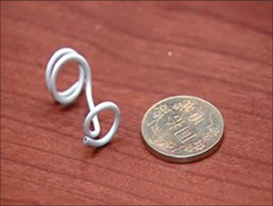
For treating conditions such as enlarged fetal bladder and pleural effusion [Figure 1], a stent or drainage catheter can be placed to drain fluids into the amniotic cavity.[3]
Figure 1.

Stent for the enlarged fetal bladder and pleural effusion in the fetus
ULTRASOUND-GUIDED FETAL ENDOSCOPY
Ultrasound-guided fetal endoscopy can be used to treat twin-twin transfusion syndrome, as well as open spina bifida and congenital diaphragmatic hernia. As mentioned above, this therapy is ready to treat fetal open spina bifida from 2020. Regarding congenital diaphragmatic hernia, due to the small number of patients, no one has applied for the introduction of this therapy in Taiwan. According to the standard practice in Western countries, which is similar to balloon angioplasty, this therapy places a balloon into the fetal trachea using a fetoscope to enlarge its trachea and keep the lungs dilated, and the balloon is broken and removed before delivery.[4] These procedures are all performed using a fetal endoscope.
ULTRASOUND-GUIDED PUNCTURE
Puncture, similar to amniocentesis, uses a long needle to treat fetuses under the guidance of ultrasound imaging. For example, for a fetus with anemia, blood can be transfused into the fetal umbilical cord using a long needle; for an oligohydramnios fetus with functioning kidneys or a pregnant woman experiencing escape of amniotic fluid for unknown reasons, a long needle can be inserted into the amniotic fluid cavity to either infuse amniotic fluid or to remove it.
ULTRASOUND-GUIDED FETAL CARDIAC CATHETERIZATION
Fetal cardiac catheterization has the highest fetal mortality rate, approximately 10%, although many patients from Catholic countries in Europe and South America have been treated with this surgical procedure.[5] I was surprised by this surgery when I first heard about it. The patients for whom this therapy is applicable are fetuses who have been diagnosed with aortic stenosis or pulmonary stenosis. By drawing on the principles of cardiac catheterization in adult patients, a puncture needle is directly inserted into the fetal heart under the guidance of ultrasound, in an attempt to widen the narrowed artery with angioplasty. It is a technology that has not yet been developed or applied in Taiwan. With extensive application of level II ultrasound examination, more fetal heart diseases would be diagnosed prenatally. The technology of cardiac catheterization is expected to be extensively applied in Taiwan in the near future.
Financial support and sponsorship
Nil.
Conflicts of interest
Dr. Steven W. Shaw, an editor at Journal of Medical Ultrasound, had no role in the peer review process of or decision to publish this article.
REFERENCES
- 1.Lyerly AD, Little MO, Faden RR. A critique of the 'fetus as patient'. Am J Bioeth. 2008;8:42–4. doi: 10.1080/15265160802331678. [DOI] [PMC free article] [PubMed] [Google Scholar]
- 2.Kabagambe SK, Jensen GW, Chen YJ, Vanover MA, Farmer DL. Fetal surgery for myelomeningocele: A systematic review and meta-analysis of outcomes in fetoscopic versus open repair. Fetal Diagn Ther. 2018;43:161–74. doi: 10.1159/000479505. [DOI] [PubMed] [Google Scholar]
- 3.Morris RK, Malin GL, Quinlan-Jones E, Middleton LJ, Hemming K, Burke D, et al. Percutaneous vesicoamniotic shunting versus conservative management for fetal lower urinary tract obstruction (PLUTO): A randomised trial. Lancet. 2013;382:1496–506. doi: 10.1016/S0140-6736(13)60992-7. [DOI] [PMC free article] [PubMed] [Google Scholar]
- 4.Deprest J, de Coppi P. Antenatal management of isolated congenital diaphragmatic hernia today and tomorrow: Ongoing collaborative research and development. Journal of Pediatric Surgery Lecture. J Pediatr Surg. 2012;47:282–90. doi: 10.1016/j.jpedsurg.2011.11.020. [DOI] [PubMed] [Google Scholar]
- 5.Arzt W, Tulzer G. Fetal surgery for cardiac lesions. Prenat Diagn. 2011;31:695–8. doi: 10.1002/pd.2810. [DOI] [PubMed] [Google Scholar]


