PLOS ONE logoLink to PLOS ONE
. 2021 Apr 29;16(4):e0251012. doi: 10.1371/journal.pone.0251012

Vinpocetine alleviates lung inflammation via macrophage inflammatory protein-1β inhibition in an ovalbumin-induced allergic asthma model

Won Seok Choi 1, Hyun Sik Kang 1, Hong Jo Kim 1, Wang Tae Lee 1, Uy Dong Sohn 1,*, Ji-Yun Lee 1,*
Editor: Saba Al Heialy2
PMCID: PMC8084130  PMID: 33914833

Abstract

Asthma is a well-known bronchial disease that causes bronchial inflammation, narrowing of the bronchial tubes, and bronchial mucus secretion, leading to bronchial blockade. In this study, we investigated the association between phosphodiesterase (PDE), specifically PDE1, and asthma using 3-isobutyl-1-methylxanthine (IBMX; a non-specific PDE inhibitor) and vinpocetine (Vinp; a PDE1 inhibitor). Balb/c mice were randomized to five treatment groups: control, ovalbumin (OVA), OVA + IBMX, OVA + Vinp, and OVA + dexamethasone (Dex). All mice were sensitized and challenged with OVA, except for the control group. IBMX, Vinp, or Dex was intraperitoneally administered 1 h before the challenge. Vinp treatment significantly inhibited the increase in airway hyper-responsiveness (P<0.001) and reduced the number of inflammatory cells, particularly eosinophils, in the lungs (P<0.01). It also ameliorated the damage to the bronchi and alveoli and decreased the OVA-specific IgE levels in serum, an indicator of allergic inflammation increased by OVA (P<0.05). Furthermore, the increase in interleukin-13, a known Th2 cytokine, was significantly decreased by Vinp (P<0.05), and Vinp regulated the release and mRNA expression of macrophage inflammatory protein-1β (MIP-1β) increased by OVA (P<0.05). Taken together, these results suggest that PDE1 is associated with allergic lung inflammation induced by OVA. Thus, PDE1 inhibitors can be a promising therapeutic target for the treatment of asthma.

Introduction

Asthma is a chronic inflammatory airway disease that leads to coughing, wheezing, and chest tightness [1]. These symptoms are caused by increased mucus secretion, airway hyper-reactivity, and functional and structural changes in lung tissue [2]. Treatment of asthma is usually dependent on corticosteroids and beta2-agonist as a bronchodilator [3]. Some biologic agents, including anti-IgE and anti-interleukin-5 (IL-5), have been developed recently and used in the treatment of severe asthma [4]. However, the specific mechanisms of asthma remain unclear. Although inhaled corticosteroids are the gold-standard therapy used to treat patients with asthma, long-term use of high-dose inhaled corticosteroids can cause adverse effects, such as hypothalamic–pituitary−adrenal axis suppression, reduced bone growth, and increased risk of opportunistic infections [5]. Therefore, there is still a need for the development of new asthma treatment.

Asthma has been associated with T helper cell 2 (Th2)-mediated immunity due to aberrant production of IL-4, IL5, and IL13. In one study, 50% of asthmatic patients showed Th2-related inflammation [6]. Atopic asthma and the genetic predisposition to produce immunoglobulin E (IgE) to common allergens is driven by IL-4-dependent Ig class switching in B cells [7]. Airway eosinophilia depends on both IL-5 and Stat6 signaling [8]. Each cytokine has distinct functional effects in the induction of disease, but IL-13 predominates in its contribution to the pathophysiology in asthma [9]. IL-13 is now thought to be especially critical, as it promotes goblet cell differentiation, mucus production, bronchial hyper-responsiveness, IgE synthesis, and eosinophil recruitment [10].

Eosinophils play a key role in numerous inflammatory diseases, including allergic disorders [11]. In most asthma phenotypes, there are increases in eosinophils in the tissues, blood, and bone marrow and in general, the numbers increase with disease severity [12]. The eosinophil is the central effector cell responsible for ongoing airway inflammation [12, 13]. When activated, eosinophils can release many mediators, such as eosinophil peroxidase, lysozyme, lipid mediators, cytokines, and chemokines, which contribute to airway hyper-reactivity [1416]. Excessive eosinophil airway infiltration causes serious symptoms, such as severe cough, dyspnea, and hypoxemia [17]. For eosinophil recruitment and activation, chemo-attractants play a key role in determining the increased tissue localization and activation during disease [18]. Macrophage inflammatory protein-1β (MIP-1β) is a well-known chemokine produced by various cells, such as neutrophils, epithelial cells, B cells, T cells, and eosinophils [17]. In a previous study, MIP-1β displayed chemo-attractant activity for murine eosinophils via C-C chemokine receptor type 5 (CCR5) and was shown to be involved in eosinophil recruitment during airway inflammation [15].

Phosphodiesterase (PDE) inhibitors prevent the inactivation of intracellular cyclic adenosine monophosphate (cAMP) and cyclic guanosine monophosphate (cGMP) [19]. Studies have demonstrated that cAMP and cGMP have a role in airway smooth muscle relaxation and down-regulate the airway inflammation and airway remodeling [20, 21]. Consequently, most PDEs are expressed in lung and immune cells [21]. The cAMP-specific PDE family negatively regulates the function of almost all pro-inflammatory and immune cells and exerts widespread anti-inflammatory activity in animal models of asthma. Some PDE inhibitors have been implicated in the treatment of chronic obstructive pulmonary disease and asthma [22]. For example, Roflumilast, a PDE4 inhibitor, is currently being used to treat chronic obstructive pulmonary disease and effectively improves asthma symptoms [10, 23]. Based on this evidence, we hypothesized that targeting PDEs has potential application in asthma treatment.

PDE1, as one of the subtypes in the PDE superfamily, is expressed in pulmonary arterial smooth muscle cells, epithelial cells, fibroblasts, macrophages, and lymphocytes [21, 24, 25]. It is well-known that PDE1 degrades both cAMP and cGMP [26]. Inhibition of cAMP and cGMP degradation can regulate airway inflammation and airway smooth muscle contraction [20]. Although the direct association between PDE1 and asthma remains unclear, recent studies have provided clues to the relationship between allergic lung inflammation and PDE1. PDE1A and PDE1C protein expression was detected in the isolated lung cells of mice, and it was increased in the inflammation state [27, 28]. A previous study also reported that PDE1A inhibition prevented lung fibrosis [29]. Other work demonstrated that PDE1 inhibition may dampen inflammatory responses of microglia in the disease state [26]. Moreover, PDE1B has been associated with the activation or differentiation of immune cells [30, 31]. It was found to be expressed in T lymphocytes and modulate the allergic response by regulating IL-13 production, which was closely related to allergic lung inflammation [3032]. In this context, we hypothesized that PDE1 inhibition could down-regulate allergic inflammation.

Vinpocetine (Vinp), a derivative of the alkaloid vincamine, is a PDE1 inhibitor [33] that exerts a neuroprotective effect by relaxing the cerebrovascular system [34]. It is also known to have anti-inflammatory activity by preventing the increase in tumor necrosis factor-alpha (TNF-α) [35]. However, the role of Vinp as a PDE1 inhibitor in lung diseases, such as asthma, remains unclear.

It is known that 3-isobutyl-1-methylxanthine (IBMX), a methylated xanthine derivative, acts as a non-competitive selective PDE inhibitor [36]. In order to understand the action of Vinp more completely, we investigated the relationship between asthma and PDE1 by examining the effect of Vinp on asthma in a murine model. We also compared the effects of IBMX and Vinp on ovalbumin (OVA)-induced allergic lung inflammation.

Materials and methods

Materials

OVA, grade V from hen egg white (lyophilized powder, ≥98%) and dexamethasone (Dex; catalog number D1756) were purchased from Sigma–Aldrich (St Louis, MO, USA). Aluminum hydroxide (Imject® Alum) was purchased from Thermo Scientific, Rockford, IL, USA. IBMX and Vinp were obtained from Tocris Bioscience, Bristol, UK. Stock solutions of IBMX, Vinp, and Dex were prepared at 3.5 mg/ml in DMSO. Before the intraperitoneal (i.p.) injection to the mice, IBMX, Vinp, and Dex were diluted to 1 mg/ml with normal saline. All chemicals used in this study were of analytical grade.

Animals

Balb/c male mice (5 weeks old, weighing 20–30 g) were obtained from Samtako Bio Korea, Gyeonggi-do, Republic of Korea, and housed in the mouse facility in the R&D Center of Chung-Ang University (Seoul, Republic of Korea) with sterilized bedding. The air condition was maintained at 24±2°C with 50±5% humidity, and the light/dark cycle was synchronized to 12:12 h. Pathogen-free food and water were provided. The mice were acclimated for 1 week, and 10 mice per group were randomized to five treatment groups (control, OVA, OVA + IBMX, OVA + Vinp, and OVA + Dex). Overall health was monitored twice a week. After the experiment, mice were euthanized by intramuscular injection with 40 mg/kg Zoletil 50 (125 mg tiletamine and 125 mg zolazepam; Virbac, Carros, France)–10 mg/kg xylazine (Sigma−Aldrich). All experimental procedures complied with the guidelines established by the Institutional Animal Care and Use Committee of Chung-Ang University, and the study design was approved by the appropriate ethics review board (IACUC 2018–00124).

OVA-induced asthma model

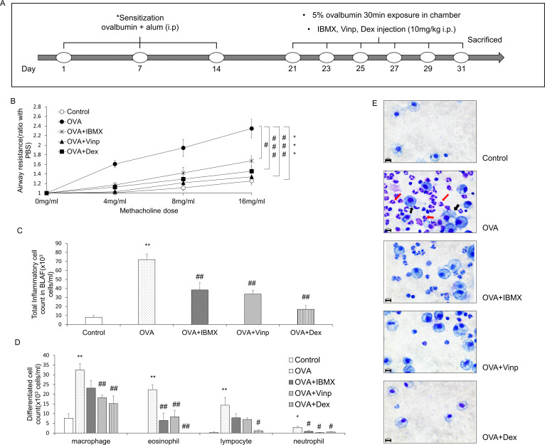
On days 1, 7, and 14, 100 μg OVA and 1 mg of aluminum hydroxide in 200 μl of normal saline were injected (i.p.) into the mice for sensitization. On days 21, 23, 25, 27, 29, and 31, mice were exposed to 5% OVA in normal saline for 30 min using a nebulizer (Aerogen®, Galway, Ireland). The control group was exposed to normal saline. In the drug-treated groups (OVA + IBMX, OVA + Vinp, OVA + Dex), IBMX, Vinp, or Dex was injected (i.p.). 1 h before 5% OVA exposure. The administration dosage was 10 mg/kg (the injection volume was 0.2 ml per mouse). In the OVA group, 0.2 ml of the vehicle was injected (i.p.) 1 h before 5% OVA exposure (Fig 1A).

Fig 1. Effects of IBMX and Vinp in an OVA-induced asthma model.

Fig 1

(A) Schedule for inducing asthma in mice model. (B) The methacholine test was performed to measure airway resistance. Mice in the OVA, OVA + IBMX, OVA + Vinp, and OVA + Dex groups were exposed to methacholine (4, 8, 16 mg/ml). After methacholine exposure, the airway resistance of the lungs was measured by plethysmography. Inflammatory cells in BALF were stained with Kwik-Diff. (C) Stained inflammatory cells were counted by a hemocytometer. (D) Differentiated cells (macrophages, eosinophils, lymphocytes, and neutrophils) were analyzed based on standard morphological criteria. (E) Stained cells were observed using a microscope (red arrows, eosinophils; black arrows, macrophages; magnification, 63×; scale bar, 10 μm). Data are expressed as mean ± SEM. Statistical analysis was performed using the Student’s t-test, one-way ANOVA, and two-way ANOVA (Data were considered significant at *P<0.05, **P<0.01, and ***P<0.001 compared with the control group and #P<0.05, ##P<0.01, and ###P<0.001 compared with the OVA group). IBMX, 3-isobutyl-1-methylxanthine; BALF, broncho-alveolar lavage fluid; OVA, ovalbumin; Vinp, vinpocetine; Dex, dexamethasone.

Measurement of airway resistance and tidal volume of lungs

Airway resistance is the resistance of the respiratory tract to airflow during inhalation and exhalation. Tidal volume is the lung volume representing the normal volume of air displaced between normal inhalation and exhalation when extra effort is not applied. Airway resistance and tidal volume can be measured by plethysmography. We also confirmed that the methacholine test did not affect the inflammatory parameter in this study. One day after the final exposure to OVA, mice in the OVA, OVA + IBMX, OVA + Vinp, and OVA + Dex groups were exposed to methacholine (4, 8, and 16 mg/ml) for 1 min. After methacholine exposure, the airway resistance of the lungs was measured for 3 min using a Buxco® non-invasive double-chamber plethysmograph (Data Sciences International, St. Paul, MN, USA). Measured airway resistance and tidal volume were automatically calculated by FinePointe software (Data Sciences International) and expressed as a ratio from the value at 0 mg/ml (n = 9).

Inflammatory cell-counting in broncho-alveolar lavage fluid (BALF)

After the methacholine test, mice were anesthetized and euthanatized as described above. BALF was obtained by lavage of the right lung, then centrifuged at 1,500g for 10 min. The supernatant was collected and stored at –78°C for further biochemical analysis (Measurement of cytokine release in BALF). The pellet was suspended in 250 μl PBS, and the suspended cells, representing total inflammatory cells, were counted using a hemocytometer. The differential inflammatory cells, such as eosinophils, macrophages, and neutrophils, were counted based on standard morphological criteria after staining on a glass slide using the ShandonTM Kwik-DiffTM Stain Kit (Thermo Fisher Scientific, Waltham, MA, USA).

Histological analysis

The left lung was removed from each mouse, fixed with 10% formalin solution, embedded using Tissue-Tek® (Sakura Finetek®, Torrance, CA, USA), then sectioned using a Leica microtome 820 (Leica Microsystems, Wetzlar, Germany) at 4 μm (n = 6). The sectioned tissues were stained with hematoxylin–eosin (H&E). The degree of inflammation in lung tissues was determined by the inflammation scoring system, where 0 = no inflammation, 1 = occasional cuffing with inflammatory cells, 2 = most bronchi or vessels surrounded by a thin layer (1–5 cells) of inflammatory cells, and 3 = most bronchi or vessels surrounded by a thick layer (>5 cells) of inflammatory cells. Periodic acid–Schiff (PAS) staining of the lung tissues was performed using the PAS Stain Kit (Abcam, Cambridge, UK). To quantify mucus production, mucus secretion was evaluated by measuring the broncho-alveolar red-stained regions using the ImageJ software program (NIH Image, Bethesda, MD, USA) [37]. Congo red staining of the lung tissues was performed using the Congo Red Stain Kit (Abcam). Eosinophils were counted in an area of 20,000 μm2 of lung tissues after staining with Congo red [38]. Six random fields of each stained tissue section were observed under a microscope (Leica Microsystems), and images were captured using a Leica DM 480 camera (Leica Microsystems).

Immunofluorescence staining

MIP-1β protein was detected using 20 μg/ml rabbit anti-CCL4 polyclonal antibody (Invitrogen, Carlsbad, CA, USA, catalog number PA5-34509, lot #UL2898185) and 24 μg/ml goat anti-rabbit IgG FITC secondary antibody (Invitrogen, catalog number 65–6111, lot #UG285467) in the 4-μm paraffin section of mouse lung tissue (n = 6). The nucleus was stained using UltraCruz® Aqueous Mounting Medium with DAPI (Santa Cruz Biotechnology, Inc., Dallas, TX, USA). Six random fields of each immunofluorescence-stained tissue section were observed under a microscope (Leica Microsystems), and images were captured using a Leica DM 480 camera (Leica Microsystems). The fluorescence intensity was measured using the ImageJ software program (NIH Image).

Measurement of anti-OVA IgE in serum

Blood samples were obtained from the inferior vena cava for biochemical assay (n = 10). Obtained blood samples were centrifuged at 1,500g for 10 min, and the serum was separated. Anti-OVA IgE in serum was measured using an ELISA kit (Cayman Chemical, Ann Arbor, MI, USA) according to the manufacturer’s instructions. Absorbance was measured using a FlexStation3 Microplate Reader (Molecular Devices, Sunnyvale, CA, USA) at 450 nm according to the manufacturer’s protocol. The concentration was determined using a standard curve of manufacturer’s IgE standard solution.

Measurement of cytokine release in BALF

The levels of IL-4, IL-5, IL-13, and MIP-1β in BALF were measured using an ELISA kit (R&D Systems, Inc., Minneapolis, MN, USA) according to the manufacturer’s instructions (n = 6−8 per group). Absorbance was measured as described above.

Quantitative analysis of mRNA expression in lung tissues

The level of mRNA expression in the lungs was measured by quantitative reverse transcription PCR (RT-qPCR). The total RNA was extracted from the right lung of each mouse using TRIzol™ reagent (Ambion®, Life Technologies™, Carlsbad, CA, USA). The RNA was spectrophotometrically quantified using a NanoDrop ND-1000 (Thermo Fisher Scientific). One microgram of total RNA was used to synthesize cDNA using the iScript™ cDNA Synthesis Kit (Bio-Rad, Hercules, CA, USA). After synthesis, 100 ng cDNA was used for quantitative PCR (qPCR). qPCR was performed using the iQ™ SYBR® Green Supermix (Bio-Rad). The primer sequences used in this study are shown in Table 1. The CFX96 Real-Time PCR Detection System (Bio-Rad) was used to monitor fluorescent intensity during amplification. cDNA was initially denatured at 95°C for 3 min, then cycled 40 times through the following cycle: denaturation (95°C for 10 s), annealing (55°C for 30 s), plate read. A melting curve was generated after cycling by increasing the temperature from 55 to 95°C at 0.5°C for 5 s and performing a plate read at each increment. CFX Manager software was used to automatically calculate the cycle quantification value at which samples amplified at a high enough value to be detected (ΔCt values). Each ΔCt value was normalized against GAPDH, used as a housekeeping gene. Transcript expression was determined relative to the control group. The RT-qPCR analysis was performed in three independent experiments (n = 6−8 per group).

Table 1. The primers used for RT-qPCR in this study.

Gene Forward sequence (5’−3’) Reverse sequence (5’−3’)
IL-13 AGACCAGACTCCCCTGTGCA TGGGTCCTGTAGATGGCATTG
MIP-1β CTCAGCCCTGATGCTTCTCAC AGAGGGGCAGGAAATCTGAAC
PDE1A GAGCACACAGGAACAACAAACA AAGTCTGTAGGCTGCGCTGA
PDE1B GAGCCAACCTTCTCTGTGCTGA CGTCCACATCTAAAGAAGGCTGG
PDE1C CAGTCATCCTGCGAAAGCATGG CCACTTGTGACTGAGCAACCATG
GAPDH CATCACTGCCACCCAGAAGACTG ATGCCAGTGAGCTTCCCGTTCAG

Western blot analysis

Protein expression in the lungs was measured by western blot analysis. Proteins were extracted using RIPA buffer (Thermo Scientific) containing protease inhibitor cocktail (Roche, Basel, Switzerland) and phosphatase inhibitor cocktail (Roche), as per the manufacturer’s instructions. Extracted protein concentrations were quantified by the BCA protein assay reagent (Thermo Scientific) using bovine serum albumin (BSA) as a standard. For electrophoresis, 20 μg of protein was loaded into each well. The protein was separated on a 10% sodium dodecyl sulfate-polyacrylamide gel electrophoresis (SDS-PAGE) gel at 60 V for 30 min, followed by 90 V for 2 h, and then transferred to polyvinylidene fluoride (PVDF) membranes (Merck, Darmstadt, Germany). The membranes were blocked by incubation with 5% BSA in PBS-T buffer (0.1% Tween in PBS, pH 7.6) at room temperature for 1 h to prevent non-specific binding. The membranes were incubated overnight with primary antibodies at 4°C. Then, the membranes were incubated with secondary antibodies at room temperature for 2 h. The primary antibodies used were all purchased from Santa Cruz Biotechnology, Inc., and included the following: mouse anti-PDE1A antibody (catalog number sc-374602, lot #H1219, dilution ratio 1:200), mouse anti-PDE1B antibody (catalog number sc-393112, lot #E2218, dilution ratio 1:200), mouse anti-PDE1C antibody (catalog number sc-376474, lot #F2717, dilution ratio 1:200), and mouse anti-β-actin antibody (catalog number sc-47778, lot #A2317, dilution ratio 1:1000). HRP-linked horse anti-mouse IgG antibody was used as the secondary antibody (Cell Signaling Technology, Inc., Danvers, MA, USA, catalog number 7076S, lot #33, dilution ratio 1:3000). Quantitative analysis was measured using the ImageJ software (NIH Image). Western blot analysis was performed in three independent experiments (n = 4−5 per group).

Statistical analysis

Values are represented as the mean ± SEM of data from 10 mice (n = 10) per group. Data were statistically analyzed by the Student’s t-test, one-way ANOVA, and two-way ANOVA using GraphPad Prism 7 (GraphPad Software, Inc., San Diego, CA, USA). The significance level was *P<0.05 versus the control group and #P<0.05 versus the OVA group.

Results

Effects of IBMX and Vinp on airway hyper-responsiveness and changes in inflammatory cells in BALF induced by OVA

To measure airway hyper-responsiveness, we performed the methacholine test. An increase in the methacholine dose induced an increase in airway resistance. In the OVA group, airway resistance was significantly escalated (P<0.001), and this increase was significantly alleviated by treatment with IBMX, the non-specific PDE inhibitor (P<0.05). Vinp, the PDE1-specific inhibitor, reduced the airway resistance more than IBMX (P<0.001). Dex, which served as a positive control, significantly inhibited the increase in airway resistance induced by OVA (P<0.001) (Fig 1B).

The inflammatory cells in BALF were counted to investigate the infiltration of inflammatory cells into the lungs. The total inflammatory cells in BALF increased more in the OVA-exposed group than in the control (P<0.01). IBMX and Vinp significantly reduced the increase in the total inflammatory cells (P<0.01). Dex (positive control) also decreased the total inflammatory cells significantly (P<0.01; Fig 1C). In particular, eosinophils, macrophages, lymphocytes, and neutrophils were increased by OVA exposure (P<0.01 for all, except for neutrophils, where P<0.05). IBMX and Vinp reduced these inflammatory cells. In particular, IBMX and Vinp caused a large decrease in eosinophils (P<0.01; Fig 1D and 1E).

Effects of IBMX and Vinp on inflammatory cell infiltration in lung tissue

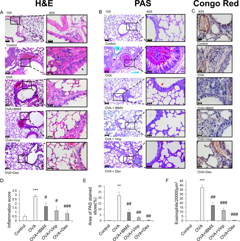
The histological changes in each group were monitored by H&E staining (Fig 2A and 2D). The alveoli and bronchi were thicker in the OVA group than in the control group. The OVA group also showed the infiltration of inflammatory cells into the lungs (P<0.001). IBMX ameliorated these effects slightly (P<0.05). Vinp also improved the morphological abnormality induced by OVA in the lungs (P<0.05). Mucus secretion (Fig 2B and 2E) and eosinophil recruitment in lung tissues (Fig 2C and 2F) were increased in the OVA group (P<0.01). IBMX and Vinp treatment reduced the mucus secretion compared with the OVA group (P<0.01) and decreased the infiltration of eosinophils significantly (IBMX; P<0.01, Vinp; P<0.001). Dex (positive control) significantly decreased these histological changes (H&E staining; P<0.001, PAS staining; P<0.01, Congo red staining; P<0.001).

Fig 2. Inhibition of ovalbumin-induced histological changes by IBMX and Vinp.

Fig 2

(A) H&E, (B) PAS, and (C) Congo red staining were performed to investigate the histological changes. H&E and PAS staining (left-hand side: magnification 10×; scale bar, 100 μm; right-hand side: magnification 40×; scale bar, 30 μm) show infiltration of inflammatory cells (yellow arrows), damage to epithelial cells (black arrows), and mucus stained magenta (red arrows). Congo red staining (magnification 63×, scale bar, 20 μm) shows the eosinophils (white arrows). (D) The inflammation scores were determined based on criteria. (E) PAS-stained areas were analyzed using ImageJ software. (F) Congo red-stained eosinophils were counted in a 20,000-μm2 area. Data are expressed as mean ± SEM. Statistical analysis was performed using one-way ANOVA (Data were considered significant at **P<0.01 and ***P<0.001 compared with the control group and #P<0.05, ##P<0.01, and ###P<0.001 compared with the OVA group). IBMX, 3-isobutyl-1-methylxanthine; H&E, hematoxylin–eosin; PAS, periodic acid–Schiff; OVA, ovalbumin; Vinp, vinpocetine; Dex, dexamethasone.

Down-regulation of allergic inflammation by IBMX and Vinp through OVA-induced decrease in IgE and IL-13

OVA-specific IgE was analyzed by ELISA to measure the degree of systemic allergic inflammation. OVA-specific IgE was increased significantly in the OVA-exposed group (P<0.01). In the Vinp group, the OVA-specific IgE level in serum was reduced significantly compared with the OVA group (P<0.01). Dex (positive control) also reduced the IgE levels (P<0.01; Fig 3A).

Fig 3. Effects of IBMX and Vinp on OVA-induced increase in allergic inflammatory mediators.

Fig 3

Plasma samples obtained from mice were analyzed by ELISA to investigate systemic allergic inflammation. (A) Anti-OVA IgE in serum and (B) the release of IL-13 in BALF were measured by ELISA. The mRNA expression levels of (C) IL-13 in lung tissues were measured by RT-qPCR. Data are expressed as mean ± SEM. Statistical analysis was performed using one-way ANOVA (Data were considered significant at **P<0.01 compared with the control group and #P<0.05 and ##P<0.01 compared with the OVA group). IBMX, 3-isobutyl-1-methylxanthine; BALF, broncho-alveolar lavage fluid; OVA, ovalbumin; Vinp, vinpocetine; Dex, dexamethasone.

IL-13 in BALF was analyzed to investigate the release of inflammatory cytokines. The OVA group showed a significant increase in the release of IL-13 (P<0.01). Although IBMX did not reduce the release of IL-13, Vinp inhibited the release of IL-13 significantly (P<0.01). The Dex group served as the positive control (P<0.01; Fig 3B).

We performed RT-qPCR using mouse lung tissues to analyze the mRNA expression levels in the lungs associated with allergic inflammation. The IL-13 expression level in the lungs was increased significantly in the OVA group (P<0.01). The increase in IL-13 expression was down-regulated by IBMX, Vinp, and Dex (P<0.05; Fig 3C).

OVA-induced increase in MIP-1β inhibited by both IBMX and Vinp

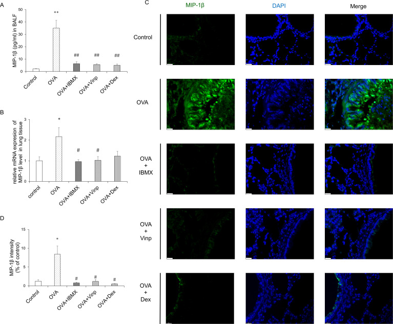
MIP-1β is a chemokine involved in a variety of inflammatory responses. To investigate the changes in MIP-1β, we performed ELISA, RT-qPCR, and immunofluorescence staining. OVA significantly induced MIP-1β release in BALF (P<0.01) and mRNA expression in the lungs (P<0.05). IBMX and Vinp both down-regulated the release (P<0.01) and mRNA expression of MIP-1β (P<0.05). Dex significantly reduced the MIP-1β release in BALF (P<0.01), but the MIP-1β mRNA expression level did not reduce significantly compared with the OVA group (Fig 4A and 4B). Immunofluorescence staining supported the MIP-1β expression results in the lungs (Fig 4C and 4D).

Fig 4. Effects of IBMX and Vinp on MIP-1β expression and release in eosinophilic lung inflammation.

Fig 4

(A) MIP-1β in BALF was measured by ELISA. (B) The mRNA expression level of MIP-1β in lung tissues was measured by RT-qPCR. (C) MIP-1β expression in lung tissue was detected by immunofluorescence staining (magnification, 63×; scale bar, 20 μm). (D) In immunofluorescence-stained tissue, the green fluorescence intensity of stained images was measured using ImageJ. Data are expressed as mean ± SEM. Statistical analysis was performed using one-way ANOVA (Data were considered significant at **P<0.01 compared with the control group and #P<0.05 compared with the OVA group). IBMX, 3-isobutyl-1-methylxanthine; BALF, broncho-alveolar lavage fluid; Vinp, vinpocetine; MIP-1β, macrophage inflammatory protein-1β; OVA, ovalbumin; Dex, dexamethasone.

Up-regulation of PDE 1 expression in the lungs of OVA-induced asthma mice model

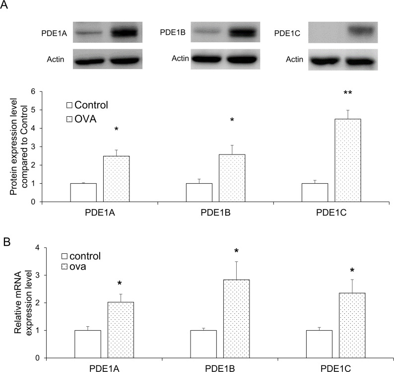
To investigate the changes in the expression of PDE1A, 1B, and 1C levels in the lungs following OVA exposure, we performed western blot and RT-qPCR analysis. For all three proteins, the expression levels increased significantly in the OVA group relative to the control (P<0.05 for all, except PDE1C, where P<0.01; Fig 5A). The mRNA expression levels of PDE1A, 1B, and 1C were also significantly up-regulated in the OVA group (P<0.05 for all; Fig 5B).

Fig 5. PDE1 expression levels in lung tissues were selectively increased by OVA exposure.

Fig 5

(A) PDE1A, PDE1B, and PDE1C protein expression levels in the lung tissues were analyzed by western blot. (B) PDE1A, PDE1B, and PDE1C mRNA expression levels in the lung tissues were measured by RT-qPCR. Data are expressed as mean ± SEM. Statistical analysis was performed using the Student’s t-test (*P<0.05 and **P<0.01 were compared with the control). OVA; ovalbumin.

Discussion

It was known that PDE inhibitors have effect on respiratory disease such as asthma and chronic obstruction pulmonary disease [22, 39]. In particular, PDE 3, 4, and 7 inhibitors are known to be effective in mouse models of asthma from previous studies [22]. However, studies on asthma using the PDE 1 inhibitor in allergic asthma mice model have not been studied yet. In this study, we investigated whether Vinp, a known PDE1 inhibitor, affects the OVA-induced asthma model. This study also focused on the contribution of PDE1 to asthma and its potential as a therapeutic target. To investigate the relationship with asthma, the appropriate asthma mice model and treatment schedule were determined from previous research and a pilot study [5, 10, 4042]. In the asthma model, OVA exposure led to an increase in airway sensitivity. However, both IBMX and Vinp treatment, respectively, significantly inhibited the increased airway resistance. Furthermore, Vinp treatment alleviated the increased airway resistance more than the IBMX treatment. Considering that IBMX is a non-selective PDE inhibitor and that Vinp is a selective PDE1 inhibitor, these results indicate that PDE, especially PDE1, is associated with airway hypersensitivity. Previous research reported that PDE4 inhibition was shown to have an effect on the attenuation of airway resistance in asthma mice model [43]. PDE3 inhibitor and PDE1/4 dual inhibitor, has been reported to cause the inhibition of the ovalbumin‐induced bronchoconstriction [44]. These previous studies supported that PDE inhibition can alleviated the increased airway resistance. Through this study, we showed that PDE 1 inhibitor can ameliorated the airway resistance increased in asthma mice model. In tidal volume, there were no significant differences among the groups (S1 Fig). The mouse model of asthma induced in this study does not appear to be associated with a decrease in tidal volume.

Patients with asthma have hypersensitive airways because the inflammatory cells infiltrate the airway and lungs [45]. The infiltrating inflammatory cells release cytokines, a major factor in airway hyper-responsiveness [10]. Among the inflammatory cells, eosinophils are well-known to increase airway hyper-responsiveness [46]. It was reported that PDE 4 inhibitor reduced the number of eosinophils, neutrophils, and lymphocytes in BALF [47]. In this study, OVA exposure increased the total inflammatory cells in BALF. In particular, the number of eosinophils increased significantly. This increase was significantly inhibited by the administration of both IBMX and Vinp, individually. As a result, it showed that PDE inhibition ameliorated eosinophilia in BALF, and suppression of PDE1 significantly reduced inflammatory cells, including eosinophils. The effect on inflammatory cells was similar to previous reports using PDE 4 inhibitors.

The histological analysis of lung tissues also supported the hypothesis that OVA caused the infiltration of inflammatory cells, especially eosinophils, into lung tissue. Structural remodeling and mucus hypersecretion were also caused by OVA exposure through inflammation. Both IBMX and Vinp treatment, respectively, relieved the increased inflammatory cell infiltration and structural remodeling. PAS staining and Congo red staining also showed mucus hypersecretion in bronchioles and eosinophil infiltration into the lungs. Although the absolute quantification of PAS and Congo red staining was not possible because of the difficulty of quantifying the total volume or total cell numbers, these results support the pathophysiological changes. As mentioned above, this increase in inflammatory cells was related to PDE, especially PDE1, and the inhibition of PDE reduced inflammation and structural remodeling in the lungs.

In allergic inflammation, the reactions to allergens, including inflammatory cell infiltration, mucin secretion, and increased IgE levels in serum, result from various inflammatory cytokines secreted by activated Th2 cells [48]. In previous research, among the PDE inhibitors, PDE 4 inhibitors were reported to affect the inhibition of inflammatory cytokines of BALF (IL-4, 5 and 13) [43]. In our study, the release of IL-13 in BALF was increased significantly by OVA exposure, and the mRNA expression levels in lung tissues increased significantly. Vinp treatment significantly decreased the IL-13 release in BALF and mRNA expression in the lungs compared with the OVA group. These results suggest that IL-13 secretion and expression are closely related to the PDE1 subtype. IL-4 and IL-5 were also measured, but they did not show significant differences between groups (S2 Fig). In the asthma mouse model, the changes in Th2 cytokines depend on the induction method and schedule of the model [49]. In previous work, IL-13 alone induced lung inflammation, mucus hypersecretion, and chemokine production [50]. In the current study, IL-13 predominated in its contribution to the pathophysiology in asthma, and the regulation of IL-13 expression and release by Vinp contributed to alleviating the pathophysiological changes.

In patients with asthma, the IgE level in the blood is increased systemically, which activates mast cells that release histamine and other cytokines [51]. The released histamine and cytokines then induce allergic inflammation in the lungs [52]. B cells play a critical role in the Th2-type immune response and eosinophilic airway inflammation [53]. It is also known that IL-13 contributes to IgE switching and production from B cells [54]. In the current study, Vinp inhibited OVA-induced IL-13 expression and release. The OVA-specific IgE level in the blood increased significantly in the OVA-exposed group, and Vinp significantly decreased the increased IgE level. These results suggested that the down-regulation of IL-13 production by Vinp reduced IgE production from B cells. It can be assumed that through this process, Vinp can reduce allergic inflammation in the lungs.

MIP-1β is a well-known chemokine produced by various cells, such as neutrophils, epithelial cells, B cells, T cells, and eosinophils [55]. In a previous study, MIP-1β was found to have chemo-attractant activity for eosinophils [56]. These chemo-attractant activities of MIP-1β can cause the infiltration of eosinophils into the lungs and bronchi, which results in allergic lung inflammation [8]. In the current study, both IBMX and Vinp, respectively, inhibited the increase in MIP-1β expression level in lung tissues and its release in BALF. This result indicates that PDE1 is mainly involved in the regulation of MIP-1β expression and release. Therefore, the reduction in eosinophil infiltration into the lungs is likely caused by decreased production of MIP-1β via PDE inhibition.

The present study found similar effects between IBMX and Vinp on allergic lung inflammation, such as increased inflammatory cells, histological changes, and MIP-1β expression level. In light of the similarity between the effects of PDE1-specific inhibitor and non-specific PDE inhibitor, PDE1 is likely a substantial contributor to allergic lung inflammation.

To further support the correlation between PDE1 and allergic lung inflammation, we also demonstrated that the increase in PDE1 expression in the lungs was caused by OVA exposure. Through previous studies, it was already demonstrated that PDE4A, 4C, and 4D expression level was increased in allergic pulmonary inflammatory conditions [57, 58]. These research showed the PDE4 was associated with asthmatic conditions. In this study, PDE1A, 1B, and 1C mRNA expression levels and protein expression levels in the lungs were all increased. These results show that PDE1 expression is also associated with asthma. PDE1 is distributed in pulmonary arterial smooth muscle cells, epithelial cells, fibroblasts, macrophages, and lymphocytes [21, 25]. PDE1 inhibitors have been associated with reactive oxygen species-mediated lung inflammation via the effect on bronchial epithelial cells and macrophages, besides transforming growth factor-beta (TGF-β)-induced myofibroblastic conversion of fibroblasts in the lungs [59, 60]. Such evidence and our study suggested that PDE1 is associated with allergic lung inflammation. Thus, Vinp, a PDE1 inhibitor, can affect asthma through PDE1A, 1B, and 1C inhibition.

In conclusion, the PDE1 inhibitor alleviated asthma symptoms, such as airway hyper-responsiveness, lung inflammation, eosinophil recruitment, and mucin secretion, by reducing Th2 cytokines and MIP-1β. OVA exposure also induced PDE1A, 1B, and 1C expression. Although this study investigated the relationship between PDE1 and allergic lung inflammation, the specific mechanism remains unclear. Through further experiments, such as using PDE1 knockout mice, the relationship between PDE1 and allergic lung inflammation should be fully investigated. Despite this limitation, the hypothesis that PDE1 contributes to allergic lung inflammation and is a potential therapeutic target for asthma treatment was supported by this study.

Supporting information

S1 Fig. Measurement of tidal volume of lungs.

The methacholine test was performed to measure tidal volume. Mice in the OVA, OVA + IBMX, OVA + Vinp, and OVA + Dex groups were exposed to methacholine (4, 8, 16 mg/ml). After methacholine exposure, the tidal volume of the lungs was measured by plethysmography. Data are expressed as mean ± SEM. Statistical analysis was performed using the Student’s t-test, one-way ANOVA, and two-way ANOVA. OVA, ovalbumin; IBMX, 3-isobutyl-1-methylxanthine; Vinp, vinpocetine; Dex, dexamethasone.

(TIF)

S2 Fig. The release of IL-4 and IL-5 in BALF.

(A) and (B) The release of IL-4 and IL-5 in BALF was measured by ELISA. Data are expressed as mean ± SEM. Statistical analysis was performed using one-way ANOVA. BALF, broncho-alveolar lavage fluid; OVA, ovalbumin; Vinp, vinpocetine; Dex, dexamethasone.

(TIF)

S1 Raw images. Uncropped western blot data.

(PDF)

Data Availability

All relevant data are within the paper and its Supporting Information files.

Funding Statement

This research was supported by the National Research Foundation of Korea (NRF) grant funded by the Korea government (Ministry of Science & ICT) (grant number NRF-2018R1C1B6008326).

References

  • 1.Lambrecht BN, Hammad H. The airway epithelium in asthma. Nat Med. 2012;18(5):684–92. 10.1038/nm.2737 [DOI] [PubMed] [Google Scholar]
  • 2.Camargo LDN, Righetti RF, Aristoteles L, Dos Santos TM, de Souza FCR, Fukuzaki S, et al. Effects of anti-IL-17 on inflammation, remodeling, and oxidative stress in an experimental model of asthma exacerbated by LPS. Front Immunol. 2017;8:1835. 10.3389/fimmu.2017.01835 [DOI] [PMC free article] [PubMed] [Google Scholar]
  • 3.Hogan AD, Bernstein JA. GINA updated 2019: Landmark changes recommended for asthma management. Ann Allergy Asthma Immunol. 2020;124(4):311–3. 10.1016/j.anai.2019.11.005 [DOI] [PubMed] [Google Scholar]
  • 4.Lommatzsch M, Buhl R, Korn S. The treatment of mild and moderate asthma in adults. Dtsch Arztebl Int. 2020;117(25):434–44. 10.3238/arztebl.2020.0434 [DOI] [PMC free article] [PubMed] [Google Scholar]
  • 5.Qian J, Ma X, Xun Y, Pan L. Protective effect of forsythiaside A on OVA-induced asthma in mice. Eur J Pharmacol. 2017;812:250–5. 10.1016/j.ejphar.2017.07.033 [DOI] [PubMed] [Google Scholar]
  • 6.Bagnasco D, Ferrando M, Varricchi G, Passalacqua G, Canonica GW. A critical evaluation of anti-IL-13 and anti-IL-4 strategies in severe asthma. Int Arch Allergy Immunol. 2016;170(2):122–31. 10.1159/000447692 [DOI] [PubMed] [Google Scholar]
  • 7.Lambrecht BN, Hammad H, Fahy JV. The cytokines of asthma. Immunity. 2019;50(4):975–91. 10.1016/j.immuni.2019.03.018 [DOI] [PubMed] [Google Scholar]
  • 8.Mattes J, Yang M, Mahalingam S, Kuehr J, Webb DC, Simson L, et al. Intrinsic defect in T cell production of interleukin (IL)-13 in the absence of both IL-5 and eotaxin precludes the development of eosinophilia and airways hyperreactivity in experimental asthma. J Exp Med. 2002;195(11):1433–44. 10.1084/jem.20020009 [DOI] [PMC free article] [PubMed] [Google Scholar]
  • 9.Elias JA, Lee CG, Zheng T, Ma B, Homer RJ, Zhu Z. New insights into the pathogenesis of asthma. J Clin Invest. 2003;111(3):291–7. 10.1172/JCI17748 [DOI] [PMC free article] [PubMed] [Google Scholar]
  • 10.Park HJ, Lee JH, Park YH, Han H, Sim da W, Park KH, et al. Roflumilast ameliorates airway hyperresponsiveness caused by diet-induced obesity in a murine model. Am J Respir Cell Mol Biol. 2016;55(1):82–91. 10.1165/rcmb.2015-0345OC [DOI] [PubMed] [Google Scholar]
  • 11.Kita H. Eosinophils: multifaceted biological properties and roles in health and disease. Immunol Rev. 2011;242(1):161–77. 10.1111/j.1600-065X.2011.01026.x [DOI] [PMC free article] [PubMed] [Google Scholar]
  • 12.Kay AB. The role of eosinophils in the pathogenesis of asthma. Trends Mol Med. 2005;11(4):148–52. 10.1016/j.molmed.2005.02.002 [DOI] [PubMed] [Google Scholar]
  • 13.Kim S-H, Kim B-K, Lee Y-C. Effects of Corni fructus on ovalbumin-induced airway inflammation and airway hyper-responsiveness in a mouse model of allergic asthma. J Inflamm. 2012;9(1):9. 10.1186/1476-9255-9-9 [DOI] [PMC free article] [PubMed] [Google Scholar]
  • 14.Matucci A, Vultaggio A, Maggi E, Kasujee I. Is IgE or eosinophils the key player in allergic asthma pathogenesis? Are we asking the right question? Respir Res. 2018;19(1):113. 10.1186/s12931-018-0813-0 [DOI] [PMC free article] [PubMed] [Google Scholar]
  • 15.Oliveira SHP, Lira S, Martinez-A C, Wiekowski M, Sullivan L, Lukacs NW. Increased responsiveness of murine eosinophils to MIP-1β (CCL4) and TCA-3 (CCL1) is mediated by their specific receptors, CCR5 and CCR8. J Leukoc Biol. 2002;71(6):1019–25. 10.1189/jlb.71.6.1019 [DOI] [PubMed] [Google Scholar]
  • 16.Zhang J, Zeng X, Li Y, Zhao W, Chen Z, Du Q, et al. Exposure to ambient particles alters the evolution of macrophage phenotype and amplifies the inducible release of eotaxin-1 in allergen-sensitized mice. J Biomed Nanotechnol. 2019;15(2):382–95. 10.1166/jbn.2019.2692 [DOI] [PubMed] [Google Scholar]
  • 17.Kobayashi Y, Konno Y, Kanda A, Yamada Y, Yasuba H, Sakata Y, et al. Critical role of CCL4 in eosinophil recruitment into the airway. Clin Exp Allergy. 2019;49(6):853–60. 10.1111/cea.13382 [DOI] [PubMed] [Google Scholar]
  • 18.Provost V, Larose MC, Langlois A, Rola-Pleszczynski M, Flamand N, Laviolette M. CCL26/eotaxin-3 is more effective to induce the migration of eosinophils of asthmatics than CCL11/eotaxin-1 and CCL24/eotaxin-2. J Leukoc Biol. 2013;94(2):213–22. 10.1189/jlb.0212074 [DOI] [PubMed] [Google Scholar]
  • 19.Pleasants RA. Clinical pharmacology of oral maintenance therapies for obstructive lung diseases. Respir Care. 2018;63(6):671. 10.4187/respcare.06068 [DOI] [PubMed] [Google Scholar]
  • 20.Chen Y-f, Huang G, Wang Y-m, Cheng M, Zhu F-f, Zhong J-n, et al. Exchange protein directly activated by cAMP (Epac) protects against airway inflammation and airway remodeling in asthmatic mice. Respir Res. 2019;20(1):285. 10.1186/s12931-019-1260-2 [DOI] [PMC free article] [PubMed] [Google Scholar]
  • 21.Zuo H, Cattani-Cavalieri I, Musheshe N, Nikolaev VO, Schmidt M. Phosphodiesterases as therapeutic targets for respiratory diseases. Pharmacol Ther. 2019;197:225–42. 10.1016/j.pharmthera.2019.02.002 [DOI] [PubMed] [Google Scholar]
  • 22.Page CP. Phosphodiesterase inhibitors for the treatment of asthma and chronic obstructive pulmonary disease. Int Arch Allergy Immunol. 2014;165(3):152–64. 10.1159/000368800 [DOI] [PubMed] [Google Scholar]
  • 23.Wedzicha JA, Calverley PM, Rabe KF. Roflumilast: a review of its use in the treatment of COPD. Int J Chron Obstruct Pulmon Dis. 2016;11:81–90. 10.2147/COPD.S89849 [DOI] [PMC free article] [PubMed] [Google Scholar]
  • 24.Giembycz MA. Phosphodiesterase 4 inhibitors and the treatment of asthma: where are we now and where do we go from here? Drugs. 2000;59(2):193–212. 10.2165/00003495-200059020-00004 [DOI] [PubMed] [Google Scholar]
  • 25.Francis SH, Corbin JD. Cyclic GMP phosphodiesterases. In: Lennarz WJ, Lane MD, editors. Encyclopedia of Biological Chemistry (Second Edition). Waltham: Academic Press; 2013. p. 567–73. [Google Scholar]
  • 26.O’Brien JJ, O’Callaghan JP, Miller DB, Chalgeri S, Wennogle LP, Davis RE, et al. Inhibition of calcium-calmodulin-dependent phosphodiesterase (PDE1) suppresses inflammatory responses. Mol Cell Neurosci. 2020;102:103449. 10.1016/j.mcn.2019.103449 [DOI] [PMC free article] [PubMed] [Google Scholar]
  • 27.Kogiso H, Hosogi S, Ikeuchi Y, Tanaka S, Shimamoto C, Matsumura H, et al. A low [Ca(2+)]i-induced enhancement of cAMP-activated ciliary beating by PDE1A inhibition in mouse airway cilia. Pflugers Arch. 2017;469(9):1215–27. 10.1007/s00424-017-1988-9 [DOI] [PubMed] [Google Scholar]
  • 28.Rajagopal S, Nalli AD, Kumar DP, Bhattacharya S, Hu W, Mahavadi S, et al. Cytokine-induced S-nitrosylation of soluble guanylyl cyclase and expression of phosphodiesterase 1A contribute to dysfunction of longitudinal smooth muscle relaxation. J Pharmacol Exp Ther. 2015;352(3):509–18. 10.1124/jpet.114.221929 [DOI] [PMC free article] [PubMed] [Google Scholar]
  • 29.Ren L, Yang C, Dou Y, Zhan R, Sun Y, Yu Y. MiR-541-5p regulates lung fibrosis by targeting cyclic nucleotide phosphodiesterase 1A. Exp Lung Res. 2017;43(6–7):249–58. 10.1080/01902148.2017.1349210 [DOI] [PubMed] [Google Scholar]
  • 30.Azevedo MF, Faucz FR, Bimpaki E, Horvath A, Levy I, de Alexandre RB, et al. Clinical and molecular genetics of the phosphodiesterases (PDEs). Endocr Rev. 2014;35(2):195–233. 10.1210/er.2013-1053 [DOI] [PMC free article] [PubMed] [Google Scholar]
  • 31.Stenson L, Degerman E, Manganiello VC. Chapter 175—cAMP/cGMP dual-specificity phosphodiesterases. In: Bradshaw RA, Dennis EA, editors. Handbook of Cell Signaling (Second Edition). San Diego: Academic Press; 2010. p. 1425–37. [Google Scholar]
  • 32.Kanda N, Watanabe S. Regulatory roles of adenylate cyclase and cyclic nucleotide phosphodiesterases 1 and 4 in interleukin-13 production by activated human T cells. Biochem Pharmacol. 2001;62(4):495–507. 10.1016/s0006-2952(01)00688-8 [DOI] [PubMed] [Google Scholar]
  • 33.Kim NJ, Baek JH, Lee J, Kim H, Song JK, Chun KH. A PDE1 inhibitor reduces adipogenesis in mice via regulation of lipolysis and adipogenic cell signaling. Exp Mol Med. 2019;51(1):1–15. 10.1038/s12276-018-0198-7 [DOI] [PMC free article] [PubMed] [Google Scholar]
  • 34.Heckman PRA, Blokland A, Bollen EPP, Prickaerts J. Phosphodiesterase inhibition and modulation of corticostriatal and hippocampal circuits: Clinical overview and translational considerations. Neurosci Biobehav Rev. 2018;87:233–54. 10.1016/j.neubiorev.2018.02.007 [DOI] [PubMed] [Google Scholar]
  • 35.Ruiz-Miyazawa KW, Pinho-Ribeiro FA, Zarpelon AC, Staurengo-Ferrari L, Silva RL, Alves-Filho JC, et al. Vinpocetine reduces lipopolysaccharide-induced inflammatory pain and neutrophil recruitment in mice by targeting oxidative stress, cytokines and NF-kappaB. Chem Biol Interact. 2015;237:9–17. 10.1016/j.cbi.2015.05.007 [DOI] [PubMed] [Google Scholar]
  • 36.Essayan DM. Cyclic nucleotide phosphodiesterases. J Allergy Clin Immunol. 2001;108(5):671–80. 10.1067/mai.2001.119555 [DOI] [PubMed] [Google Scholar]
  • 37.Oliveira TL, Candeia-Medeiros N, Cavalcante-Araújo PM, Melo IS, Fávaro-Pípi E, Fátima LA, et al. SGLT1 activity in lung alveolar cells of diabetic rats modulates airway surface liquid glucose concentration and bacterial proliferation. Scientific Reports. 2016;6(1):21752. 10.1038/srep21752 [DOI] [PMC free article] [PubMed] [Google Scholar]
  • 38.Albert EJ, Duplisea J, Dawicki W, Haidl ID, Marshall JS. Tissue eosinophilia in a mouse model of colitis is highly dependent on TLR2 and independent of mast cells. Am J Pathol. 2011;178(1):150–60. 10.1016/j.ajpath.2010.11.041 [DOI] [PMC free article] [PubMed] [Google Scholar]
  • 39.Kwak HJ, Nam JY, Song JS, No Z, Yang SD, Cheon HG. Discovery of a novel orally active PDE-4 inhibitor effective in an ovalbumin-induced asthma murine model. Eur J Pharmacol. 2012;685(1):141–8. 10.1016/j.ejphar.2012.04.016 [DOI] [PubMed] [Google Scholar]
  • 40.Højen JF, Kristensen MLV, McKee AS, Wade MT, Azam T, Lunding LP, et al. IL-1R3 blockade broadly attenuates the functions of six members of the IL-1 family, revealing their contribution to models of disease. Nat Immunol. 2019;20(9):1138–49. 10.1038/s41590-019-0467-1 [DOI] [PMC free article] [PubMed] [Google Scholar]
  • 41.Bao A, Li F, Zhang M, Chen Y, Zhang P, Zhou X. Impact of ozone exposure on the response to glucocorticoid in a mouse model of asthma: involvements of p38 MAPK and MKP-1. Respir Res. 2014;15:126. 10.1186/s12931-014-0126-x [DOI] [PMC free article] [PubMed] [Google Scholar]
  • 42.Ye P, Yang XL, Chen X, Shi C. Hyperoside attenuates OVA-induced allergic airway inflammation by activating Nrf2. Int Immunopharmacol. 2017;44:168–73. 10.1016/j.intimp.2017.01.003 [DOI] [PubMed] [Google Scholar]
  • 43.Kim SW, Kim JH, Park CK, Kim TJ, Lee SY, Kim YK, et al. Effect of roflumilast on airway remodelling in a murine model of chronic asthma. Clin Exp Allergy. 2016;46(5):754–63. 10.1111/cea.12670 [DOI] [PubMed] [Google Scholar]
  • 44.Myou Fujimura, Kurashima Tachibana, Hirose Nakao. Effect of aerosolized administration of KF19514, a phosphodiesterase 4 inhibitor, on bronchial hyperresponsiveness and airway inflammation induced by antigen inhalation in guinea-pigs. Clin Exp Allergy. 2000;30(5):713–8. 10.1046/j.1365-2222.2000.00782.x [DOI] [PubMed] [Google Scholar]
  • 45.Ding F, Liu B, Zou W, Tian D, Li Q, Dai J, et al. LPS exposure in early life protects against mucus hypersecretion in ovalbumin-induced asthma by down-regulation of the IL-13 and JAK-STAT6 pathways. Cell Physiol Biochem. 2018;46(3):1263–74. 10.1159/000489109 [DOI] [PubMed] [Google Scholar]
  • 46.Pavord ID. Eosinophilic phenotypes of airway disease. Ann Am Thorac Soc. 2013;10 Suppl:S143–S9. 10.1513/AnnalsATS.201306-168AW [DOI] [PubMed] [Google Scholar]
  • 47.Sun J-g Deng Y-m, Wu X Tang H-f, Deng J-f Chen J-q, et al. Inhibition of phosphodiesterase activity, airway inflammation and hyperresponsiveness by PDE4 inhibitor and glucocorticoid in a murine model of allergic asthma. Life Sci. 2006;79(22):2077–85. 10.1016/j.lfs.2006.07.001 [DOI] [PubMed] [Google Scholar]
  • 48.Rudolph AK, Walter T, Erkel G. The fungal metabolite cyclonerodiol inhibits IL-4/IL-13 induced Stat6-signaling through blocking the association of Stat6 with p38, ERK1/2 and p300. Int Immunopharmacol. 2018;65:392–401. 10.1016/j.intimp.2018.10.033 [DOI] [PubMed] [Google Scholar]
  • 49.Epstein MM, Tilp C, Erb KJ. The use of mouse asthma models to successfully discover and develop novel drugs. Int Arch Allergy Immunol. 2017;173(2):61–70. 10.1159/000473699 [DOI] [PubMed] [Google Scholar]
  • 50.Zhu Z, Homer RJ, Wang Z, Chen Q, Geba GP, Wang J, et al. Pulmonary expression of interleukin-13 causes inflammation, mucus hypersecretion, subepithelial fibrosis, physiologic abnormalities, and eotaxin production. J Clin Invest. 1999;103(6):779–88. 10.1172/JCI5909 [DOI] [PMC free article] [PubMed] [Google Scholar]
  • 51.Oettgen HC, Geha RS. IgE regulation and roles in asthma pathogenesis. J Allergy Clin Immunol. 2001;107(3):429–41. 10.1067/mai.2001.113759 [DOI] [PubMed] [Google Scholar]
  • 52.Wernersson S, Pejler G. Mast cell secretory granules: armed for battle. Nat Rev Immunol. 2014;14(7):478–94. 10.1038/nri3690 [DOI] [PubMed] [Google Scholar]
  • 53.Drake LY, Iijima K, Hara K, Kobayashi T, Kephart GM, Kita H. B cells play key roles in th2-type airway immune responses in mice exposed to natural airborne allergens. PLoS One. 2015;10(3):e0121660. 10.1371/journal.pone.0121660 [DOI] [PMC free article] [PubMed] [Google Scholar]
  • 54.Stone KD, Prussin C, Metcalfe DD. IgE, mast cells, basophils, and eosinophils. J Allergy Clin Immunol. 2010;125(2 Suppl 2):S73–80. 10.1016/j.jaci.2009.11.017 [DOI] [PMC free article] [PubMed] [Google Scholar]
  • 55.Lillard JW Jr., Singh UP, Boyaka PN, Singh S, Taub DD, McGhee JR. MIP-1alpha and MIP-1beta differentially mediate mucosal and systemic adaptive immunity. Blood. 2003;101(3):807–14. 10.1182/blood-2002-07-2305 [DOI] [PubMed] [Google Scholar]
  • 56.Nakagome K, Nagata M. Possible mechanisms of eosinophil accumulation in eosinophilic pneumonia. Biomolecules. 2020;10(4):1–12. 10.3390/biom10040638 [DOI] [PMC free article] [PubMed] [Google Scholar]
  • 57.Tang H-F, Song Y-H, Chen J-C, Chen J-Q, Wang P. Upregulation of phosphodiesterase-4 in the lung of allergic rats. Am J Respir Crit Care Med. 2005;171(8):823–8. 10.1164/rccm.200406-771OC [DOI] [PubMed] [Google Scholar]
  • 58.Trian T, Burgess JK, Niimi K, Moir LM, Ge Q, Berger P, et al. β2-agonist induced cAMP is decreased in asthmatic airway smooth muscle due to increased PDE4D. PLoS One. 2011;6(5):e20000. 10.1371/journal.pone.0020000 [DOI] [PMC free article] [PubMed] [Google Scholar]
  • 59.Brown DM, Hutchison L, Donaldson K, MacKenzie SJ, Dick CA, Stone V. The effect of oxidative stress on macrophages and lung epithelial cells: the role of phosphodiesterases 1 and 4. Toxicol Lett. 2007;168(1):1–6. 10.1016/j.toxlet.2006.10.016 [DOI] [PubMed] [Google Scholar]
  • 60.Dunkern TR, Feurstein D, Rossi GA, Sabatini F, Hatzelmann A. Inhibition of TGF-beta induced lung fibroblast to myofibroblast conversion by phosphodiesterase inhibiting drugs and activators of soluble guanylyl cyclase. Eur J Pharmacol. 2007;572(1):12–22. 10.1016/j.ejphar.2007.06.036 [DOI] [PubMed] [Google Scholar]

Decision Letter 0

Heinz Fehrenbach

11 Nov 2020

PONE-D-20-32932

Vinpocetine alleviates lung inflammation via macrophage inflammatory protein-1β inhibition in an ovalbumin-induced allergic asthma model

PLOS ONE

Dear Dr. Lee,

Thank you for submitting your manuscript to PLOS ONE. After careful consideration, we feel that it has merit but does not fully meet PLOS ONE’s publication criteria as it currently stands. Therefore, we invite you to submit a revised version of the manuscript that addresses the points raised during the review process.

Two expert reviewers from the field raised a number of points that should be addressed when revising the manuscript. In particular, the role of the other Th2 cytokines should be addressed throughout the manuscript, i.e. including the Introduction and Discussion section. Moreover, since you analysed the levels of IL-4, IL-5, IL-13, MIP-1β, and interferon-gamma (IFN-γ) in BALF, all the data should be presented - at least in the supplement. Further, I ask you to expand your discussion to include additional references (see suggestions by reviewer #2) allowing you to discuss the potential molecular mechanism. It will be mandatory that you submit a detailed point-by-point response to all the comments raused by the reviewers and me.

Please submit your revised manuscript by Dec 26 2020 11:59PM. If you will need more time than this to complete your revisions, please reply to this message or contact the journal office at plosone@plos.org. When you're ready to submit your revision, log on to https://www.editorialmanager.com/pone/ and select the 'Submissions Needing Revision' folder to locate your manuscript file.

Please include the following items when submitting your revised manuscript:

  • A rebuttal letter that responds to each point raised by the academic editor and reviewer(s). You should upload this letter as a separate file labeled 'Response to Reviewers'.

  • A marked-up copy of your manuscript that highlights changes made to the original version. You should upload this as a separate file labeled 'Revised Manuscript with Track Changes'.

  • An unmarked version of your revised paper without tracked changes. You should upload this as a separate file labeled 'Manuscript'.

If you would like to make changes to your financial disclosure, please include your updated statement in your cover letter. Guidelines for resubmitting your figure files are available below the reviewer comments at the end of this letter.

If applicable, we recommend that you deposit your laboratory protocols in protocols.io to enhance the reproducibility of your results. Protocols.io assigns your protocol its own identifier (DOI) so that it can be cited independently in the future. For instructions see: http://journals.plos.org/plosone/s/submission-guidelines#loc-laboratory-protocols

We look forward to receiving your revised manuscript.

Kind regards,

Heinz Fehrenbach

Academic Editor

PLOS ONE

Additional Editor Comments:

In addition to the comments of our reviewers, I have some comments to the quantitative analysis of histopathological changes.

1) regarding the analysis of mucus, which was done as follows "After periodic acid–Schiff (PAS) staining of the lung tissues using the PAS Stain Kit (Abcam, Cambridge, UK), mucus secretion was evaluated by measuring the bronchoalveolar red-stained regions using ImageJ software program (NIH Image, MD, USA)": in any tissue section (2-dimensional), the area occupied by a specific tissue component depends on the total volume of this component. Consequently, you cannot distinguish between an increase in mucus due to an increase in the mucus volume stored in the cells and an increase the number of mucus producing / storing goblet cells. Please take into account this limitation of your methodological approach when discussing the results derived from this analysis.

2) regarding the analysis of eosinophils, which was done as follows "Eosinophils were counted in an area of 20,000 µm2 of lung tissues after staining with Congo red using the Congo Red Stain Kit (Abcam, Cambridge, UK).": Doing the analysis in a two-dimensional section, you in fact counted the number of cell profiles, but clearly not the cell numbers. The cell profile counts again depend on the volume of the cells analysed. Therefore, you cannot distinguish between an increase in cell numbers (due to proliferation or infiltration) and an increase in cell volume (due to hypertrophy). I therefore suggest, that you clearly indicate this limitation, or that you use a similar scoring system as you had implemented for estimating the inflammation in the tissues done in the H & E stainings.

Journal Requirements:

When submitting your revision, we need you to address these additional requirements.

1. Please ensure that your manuscript meets PLOS ONE's style requirements, including those for file naming. The PLOS ONE style templates can be found at

https://journals.plos.org/plosone/s/file?id=wjVg/PLOSOne_formatting_sample_main_body.pdf and

https://journals.plos.org/plosone/s/file?id=ba62/PLOSOne_formatting_sample_title_authors_affiliations.pdf

2. Thank you for including your ethics statement: ' All experimental procedures were in accordance with the guidelines established by the Institutional Animal Care and Use Committee of Chung-Ang University (IACUC 2019-00031).'

(a) Please amend your current ethics statement to include the full name of the ethics committee/institutional review board(s) that approved your specific study.  

(b) Once you have amended this/these statement(s) in the Methods section of the manuscript, please add the same text to the “Ethics Statement” field of the submission form (via “Edit Submission”).

For additional information about PLOS ONE ethical requirements for human subjects research, please refer to https://journals.plos.org/plosone/s/animal-research.

3. To comply with PLOS ONE submissions requirements, please provide the method of euthanasia in the Methods section of your manuscript.

4. At this time, we request that you  please report additional details in your Methods section regarding animal care, as per our editorial guidelines:

(1) Please describe the post-operative care received by the animals, including the frequency of monitoring and the specific clinical, physiological and behavioural criteria used to assess animal health and well-being.

Thank you for your attention to these requests.

5. In the Methods section, please provide the product number and any lot numbers of the primary antibodies purchased from chemical companies for your study.

6. To comply with PLOS ONE submission guidelines, in your Methods section, please provide additional information regarding your statistical analyses, including the name and version of the software used for the statistical analyses. For more information on PLOS ONE's expectations for statistical reporting, please see https://journals.plos.org/plosone/s/submission-guidelines.#loc-statistical-reporting.

7. At this time, we ask that you please provide scale bars on the microscopy images presented in Figure 1 and 2 and refer to the scale bar in the corresponding Figure legend.

8. PLOS ONE now requires that authors provide the original uncropped and unadjusted images underlying all blot or gel results reported in a submission’s figures or Supporting Information files. This policy and the journal’s other requirements for blot/gel reporting and figure preparation are described in detail at https://journals.plos.org/plosone/s/figures#loc-blot-and-gel-reporting-requirements and https://journals.plos.org/plosone/s/figures#loc-preparing-figures-from-image-files. When you submit your revised manuscript, please ensure that your figures adhere fully to these guidelines and provide the original underlying images for all blot or gel data reported in your submission. See the following link for instructions on providing the original image data: https://journals.plos.org/plosone/s/figures#loc-original-images-for-blots-and-gels.

In your cover letter, please note whether your blot/gel image data are in Supporting Information or posted at a public data repository, provide the repository URL if relevant, and provide specific details as to which raw blot/gel images, if any, are not available. Email us at plosone@plos.org if you have any questions.

[Note: HTML markup is below. Please do not edit.]

Reviewers' comments:

Reviewer's Responses to Questions

Comments to the Author

1. Is the manuscript technically sound, and do the data support the conclusions?

The manuscript must describe a technically sound piece of scientific research with data that supports the conclusions. Experiments must have been conducted rigorously, with appropriate controls, replication, and sample sizes. The conclusions must be drawn appropriately based on the data presented.

Reviewer #1: Partly

Reviewer #2: Yes

**********

2. Has the statistical analysis been performed appropriately and rigorously?

Reviewer #1: Yes

Reviewer #2: Yes

**********

3. Have the authors made all data underlying the findings in their manuscript fully available?

The PLOS Data policy requires authors to make all data underlying the findings described in their manuscript fully available without restriction, with rare exception (please refer to the Data Availability Statement in the manuscript PDF file). The data should be provided as part of the manuscript or its supporting information, or deposited to a public repository. For example, in addition to summary statistics, the data points behind means, medians and variance measures should be available. If there are restrictions on publicly sharing data—e.g. participant privacy or use of data from a third party—those must be specified.

Reviewer #1: Yes

Reviewer #2: Yes

**********

4. Is the manuscript presented in an intelligible fashion and written in standard English?

PLOS ONE does not copyedit accepted manuscripts, so the language in submitted articles must be clear, correct, and unambiguous. Any typographical or grammatical errors should be corrected at revision, so please note any specific errors here.

Reviewer #1: Yes

Reviewer #2: Yes

**********

5. Review Comments to the Author

Please use the space provided to explain your answers to the questions above. You may also include additional comments for the author, including concerns about dual publication, research ethics, or publication ethics. (Please upload your review as an attachment if it exceeds 20,000 characters)

Reviewer #1: The manuscript number PONE-D-20-32932, entitled "Vinpocetine alleviates lung inflammation via macrophage inflammatory protein-1β inhibition in an ovalbumin-induced allergic asthma model"generally is well written and describes an interesting relationship between PDE 1 inhibition and reduction of lung inflammation in mice. Nevertheless, in my opinion manuscript requires minor corrections before further processing. Please find some comments that should be considered in the revision version of the manuscript.

Introduction:

1. In the introduction part, the Authors write that the treatment of asthma is mainly based on the use of glucocorticosteroids. I believe that this is too simplified and requires supplementing with current pharmacotherapy.

2. Also in the introduction, the authors mention the role of IL-13 as the leading one. However, the role of the remaining TH2 cytokines and their role in driving chronic asthma inflammation cannot be overlooked.

3. Third paragraph of the introduction: Please add citations confirming the role of eosinophils in inflammatory diseases. It is also worth supplementing the information on eosinophilia in asthma - focus on this disease entity.

4. Phosphodiesterases paragraph, second sentence, this is unclear. Please explain and provide the reason why PDE inhibitors are being tested as a potential therapy.

5. What kind of relationship between PDE1 and allergic lung inflammation?

Materials and methods:

1. Lack of DEX origin in materials part, how the all compounds were prepared/dissolved, what was the stock solution/concentration used in the study?

2. Does treatment with methacholine affect the parameters of inflammation? Was airway resistance measured within the same groups as the inflammatory parameters?

3. Did you try to compare resistance of the lungs also with untreated group?

4. General: Please supplement the material and method parts with the number of repeats in each experiment: eg. WB, PCR repeats, number of cells counted, fields of view, number of biological and technical repeats.

5. Please add antibodies catalog numbers used in the Immunofluorescence and Western Blot. How much protein was applied for electrophoresis. How much of RNA and cDNA was used in the qPCR experiments ?

Results and discussion

1. It would be useful to add comparisons between the VIN and IBMX groups in the description of the results. This would help to draw conclusions about the significance between these groups and conclude on superiority / or no superiority of the PDE1 inhibitor over the non-specific IBMX inhibitor. Data available in the literature show that the activity of vinpocetin and IBMX on PDE1 may be similar. The authors checked the expression of PDE1 in the lung tissue of mice with induced allergic asthma. Did the Authors also check the expression of other PDE isoforms? Are there any changes in the other isoforms? It would be interesting to see if the expression of other PDEs is also increasing in the asthmatic model. To clearly state that PDE1 is mainly responsible for the described changes, it is worth considering the implementation of knockout experiments in the future.

2. Third result: Have the other TH2 cytokines been studied? If only IL-13 has been tested then this title is too general, it is known that the response may be varied and heterogeneous.

3. Results description and MIP-1beta – the lack of DEX group description/comparison. Were the protein levels also tested?

4. The discussion needs improvement. Do not duplicate the results, but rather relate them to the current literature. Regarding the results for MIP-1beta, the differences between VIN and IBMX are poorly marked, so can we conclude that the effects depend only on PDE1 inhibition? It is also worth adding directions for further research and work limitations.

Reviewer #2: In the present manuscript Lee et. al. analyzed the therapeutic effectiveness of the PDE1 inhibitor vinpocetine in a murine model of allergic asthma.

Animals were systemically sensitized with OVA/Alum, to induce the allergic airway disease the animals were challenged by 5 times performed nebulization with OVA.

1 hr prior each challenge the animals were treated with the PDE1 inhibitor vinpocetine (Vinp), a general non specific PDE inhibitor (IBMX) or with dexamethasone (Dex).

Animals treated with Vinp demonstrated attenuated asthmatic phenotype. Asthma hallmarks like, lung function, cellular infiltrates in BAL and lung tissue and goblet cell metaplasia were improved upon treatment with Vinp in comparison to untreated animals.

Moreover, the group demonstrated reduced levels of OVA specific IgE in serum and reduced protein concentrations of IL-13 in BAL and reduced mRNA signals of the Th2 cytokine in lung tissue.

Finally the authors demonstrate that MIP-1ß was downregulated in BAL and lung tissue.

The authors conclude that the PDE1 inhibitor vinpocetine suppress the asthma phenotype by reducing Th2 cytokines and MIP-1ß.

The paper is well and comprehensibly written. It demonstrates that PDE1 proteins are upregulated in asthma and that specific blocking of PDE1 by vinpocetine is more effective as unspecific blocking of PDEs with IBMX.

Analyses in the OVA model sound solid unfortunately information upon the mode of action of vinpocetine are missing to the greatest extant.

Major:

1. The discussion exclusively focuses on the own observation of the PDE1 inhibitor vinpocetine in the murine model of allergic airway diseases without cross referencing other studies which analyzed the mode of action of vinpocetine or studies analyzing the effect of other PDE inhibitors in asthma.

This cross references will contribute to understand the possible mechanisms of the PDE1 inhibitor.

In the lung PDE1 inhibitors are associated with smooth muscle cells, oxidative stress of bronchial epithelial cells and macrophages [Brown 2007], TGFb induced fibroblast conversion [Dunkern 2007] and other mechanisms.

PDE inhibitors affect immune cells please cross reference.

2. Please mention shortly downsides of the chosen model:

a. Treatment was performed during primary challenge phase; here the lung disease is developing for the first time. It is more an acute model than a therapeutic model.

3. How do you explain the strong effect on OVA specific IgE? Are B cells affected, is it more a systemic effect? How are other immunoglobulins affected?

4. Are other immune cells affected by the inhibition of PDE1.

5. Are other side effects observable upon the systemic application of the inhibitors?

Minor:

1. Please provide information of the solvent of IBMX, Vinp, or Dex and the total volume injected.

2. Please add treatments in the schedule for inducing asthma (Fig. 1A)

3. Please specify the “airway resistance” measured with a double chamber plethysmograph.

4. Please specify how total protein concentrations for OVA specific IgE and Cytokines were calculated.

5. Please specify how relative mRNA expression was determined.

6. Please enlarge y-axis labeling of almost all graphs.

**********

6. PLOS authors have the option to publish the peer review history of their article (what does this mean?). If published, this will include your full peer review and any attached files.

If you choose “no”, your identity will remain anonymous but your review may still be made public.

Do you want your identity to be public for this peer review? For information about this choice, including consent withdrawal, please see our Privacy Policy.

Reviewer #1: No

Reviewer #2: No

[NOTE: If reviewer comments were submitted as an attachment file, they will be attached to this email and accessible via the submission site. Please log into your account, locate the manuscript record, and check for the action link "View Attachments". If this link does not appear, there are no attachment files.]

While revising your submission, please upload your figure files to the Preflight Analysis and Conversion Engine (PACE) digital diagnostic tool, https://pacev2.apexcovantage.com/. PACE helps ensure that figures meet PLOS requirements. To use PACE, you must first register as a user. Registration is free. Then, login and navigate to the UPLOAD tab, where you will find detailed instructions on how to use the tool. If you encounter any issues or have any questions when using PACE, please email PLOS at figures@plos.org. Please note that Supporting Information files do not need this step.

PLoS One. 2021 Apr 29;16(4):e0251012. doi: 10.1371/journal.pone.0251012.r002

Author response to Decision Letter 0


15 Dec 2020

PLOS ONE

Manuscript number: PONE-D-20-32932

Title: Vinpocetine alleviates lung inflammation via macrophage inflammatory protein-1β inhibition in an ovalbumin-induced allergic asthma model

Revision date: December 12, 2020

We would like to thank you and the reviewers for the comments on our manuscript, which is entitled as “Vinpocetine alleviates lung inflammation via macrophage inflammatory protein-1β inhibition in an ovalbumin-induced allergic asthma model” We revised the manuscript according to the reviewer’s helpful comments. We made red color revised part. For the convenience, line number was added in left side in the manuscript. This revised manuscript was received English proofreading by ESSAY REVIEW, and an editorial certificate was attached. We hope that this revised manuscript can be suitable for PLoS ONE’s style requirements.

Additional Editor Comments:

In addition to the comments of our reviewers, I have some comments to the quantitative analysis of histopathological changes.

1) regarding the analysis of mucus, which was done as follows "After periodic acid–Schiff (PAS) staining of the lung tissues using the PAS Stain Kit (Abcam, Cambridge, UK), mucus secretion was evaluated by measuring the bronchoalveolar red-stained regions using ImageJ software program (NIH Image, MD, USA)": in any tissue section (2-dimensional), the area occupied by a specific tissue component depends on the total volume of this component. Consequently, you cannot distinguish between an increase in mucus due to an increase in the mucus volume stored in the cells and an increase the number of mucus producing / storing goblet cells. Please take into account this limitation of your methodological approach when discussing the results derived from this analysis.

→ Thank you for your comment. To quantify mucus production, mucus secretion was evaluated by measuring the broncho-alveolar red-stained regions using ImageJ software based on previous research. However, we agree with and discuss the limitation in the Discussion section. We also revised the relevant method to clarify the method of quantification.

Page 9, Lines 177-179

To quantify mucus production, mucus secretion was evaluated by measuring the broncho-alveolar red-stained regions using the ImageJ software program (NIH Image, Bethesda, MD, USA) [37].

Page 16, Lines 356–360

PAS staining and Congo red staining also showed mucus hypersecretion in bronchioles and eosinophil infiltration into the lungs. Although the absolute quantification of PAS and Congo red staining was not possible because of the difficulty of quantifying the total volume or total cell numbers, these results support the pathophysiological changes.

References

37. Oliveira TL, Candeia-Medeiros N, Cavalcante-Araújo PM, Melo IS, Fávaro-Pípi E, Fátima LA, et al. SGLT1 activity in lung alveolar cells of diabetic rats modulates airway surface liquid glucose concentration and bacterial proliferation. Scientific Reports. 2016;6(1):21752.http://doi.org/10.1038/srep21752 PMID: 26902517

2) regarding the analysis of eosinophils, which was done as follows "Eosinophils were counted in an area of 20,000 µm2 of lung tissues after staining with Congo red using the Congo Red Stain Kit (Abcam, Cambridge, UK).": Doing the analysis in a two-dimensional section, you in fact counted the number of cell profiles, but clearly not the cell numbers. The cell profile counts again depend on the volume of the cells analysed. Therefore, you cannot distinguish between an increase in cell numbers (due to proliferation or infiltration) and an increase in cell volume (due to hypertrophy). I therefore suggest, that you clearly indicate this limitation, or that you use a similar scoring system as you had implemented for estimating the inflammation in the tissues done in the H & E stainings.

→ Thank you for your comment. To quantify the number of eosinophils in Congo red staining, we referred to previous research and counted the eosinophils in an area of 20,000 µm2. We also agree with and discuss the limitation in the Discussion and revised the relevant Methods section to clarify the quantification procedure.

Page 9, Lines 179–182

Congo red staining of the lung tissues was performed using the Congo Red Stain Kit (Abcam). Eosinophils were counted in an area of 20,000 µm2 of lung tissues after staining with Congo red [38]. Six random fields of each stained tissue section……

Page 16, Lines 356-360

PAS staining and Congo red staining also showed mucus hypersecretion in bronchioles and eosinophil infiltration into the lungs. Although the absolute quantification of PAS and Congo red staining was not possible because of the difficulty of quantifying the total volume or total cell numbers, these results support the pathophysiological changes.

References

38. Albert EJ, Duplisea J, Dawicki W, Haidl ID, Marshall JS. Tissue eosinophilia in a mouse model of colitis is highly dependent on TLR2 and independent of mast cells. Am J Pathol. 2011;178(1):150-60.http://doi.org/10.1016/j.ajpath.2010.11.041 PMID: 21224053

Journal Requirements:

When submitting your revision, we need you to address these additional requirements.

1. Please ensure that your manuscript meets PLOS ONE's style requirements, including those for file naming. The PLOS ONE style templates can be found at

https://journals.plos.org/plosone/s/file?id=wjVg/PLOSOne_formatting_sample_main_body.pdf and

https://journals.plos.org/plosone/s/file?id=ba62/PLOSOne_formatting_sample_title_authors_affiliations.pdf

→ Thank you for your comment. Accordingly, we revised all parts of the manuscript to comply with PLoS ONE’s style requirements.

2. Thank you for including your ethics statement: ' All experimental procedures were in accordance with the guidelines established by the Institutional Animal Care and Use Committee of Chung-Ang University (IACUC 2019-00031).'

(a) Please amend your current ethics statement to include the full name of the ethics committee/institutional review board(s) that approved your specific study.

→ Thank you for your comment. We already mentioned the full name of the ethics committee that approved our study (the Institutional Animal Care and Use Committee of Chung-Ang University, Seoul, South Korea). However, we wrote the incorrect IACUC number. We amended the IACUC number. The IACUC Protocol Approval document is presented below.

Page 6 and 7, Lines 123-130

All experimental procedures were in accordance with the guidelines established by the Institutional Animal Care and Use Committee of Chung-Ang University (IACUC 2018-00124).

(b) Once you have amended this/these statement(s) in the Methods section of the manuscript, please add the same text to the “Ethics Statement” field of the submission form (via “Edit Submission”).

For additional information about PLOS ONE ethical requirements for human subjects research, please refer to https://journals.plos.org/plosone/s/animal-research.

→ Thank you for your comment. We included the ethics statement in the Methods section and added the same text to the “Ethics Statement” field of the submission form.

3. To comply with PLOS ONE submissions requirements, please provide the method of euthanasia in the Methods section of your manuscript.

→ Thank you for your helpful comment. Accordingly, we included the method of euthanasia in the Methods section, as follows.

Pages 6, Lines 126-128

After the experiment, mice were euthanized by intramuscular injection with 40 mg/kg Zoletil 50 (125 mg tiletamine and 125 mg zolazepam; Virbac, Carros, France)–10 mg/kg xylazine (Sigma�Aldrich).

4. At this time, we request that you please report additional details in your Methods section regarding animal care, as per our editorial guidelines:

→ Thank you for your helpful comment. Accordingly, we included additional details regarding animal care in the Methods section.

Pages 6, Lines 119-128

Balb/c male mice (5 weeks old, weighing 20–30 g) were obtained from Samtako Bio Korea, Gyeonggi-do, Republic of Korea, and housed in the mouse facility in the R&D Center of Chung-Ang University (Seoul, Republic of Korea) with sterilized bedding. The air condition was maintained at 24±2 ℃ with 50±5% humidity, and the light/dark cycle was synchronized to 12:12 h. Pathogen-free food and water were provided. The mice were acclimated for 1 week, and 10 mice per group were randomized to five treatment groups (control, OVA, OVA + IBMX, OVA + Vinp, and OVA + Dex). Overall health was monitored twice a week. After the experiment, mice were euthanized by intramuscular injection with 40 mg/kg Zoletil 50 (125 mg tiletamine and 125 mg zolazepam; Virbac, Carros, France)–10 mg/kg xylazine (Sigma�Aldrich).

(1) Please describe the post-operative care received by the animals, including the frequency of monitoring and the specific clinical, physiological and behavioural criteria used to assess animal health and well-being.

→ Thank you for your comment. In our study, we did not perform any surgery. Instead, we monitored twice a week to check the overall health of the mice following the drug injection or OVA nebulization.

Page 6, Line 125–126

Overall health was monitored twice a week.

5. In the Methods section, please provide the product number and any lot numbers of the primary antibodies purchased from chemical companies for your study.

→ Thank you for your comment. Accordingly, we added the product number and lot numbers of the antibodies we used in this study.

Page 9, Lines 187-190

MIP-1β protein was detected using 20 µg/ml rabbit anti-CCL4 polyclonal antibody (Invitrogen, Carlsbad, CA, USA, catalog number PA5-34509, lot #UL2898185) and 24 µg/ml goat anti-rabbit IgG FITC secondary antibody (Invitrogen, catalog number 65-6111, lot #UG285467) in the 4-µm paraffin section of mouse lung tissue (n = 6).

Pages 11 and 12, Lines 244-251

The primary antibodies used were all purchased from Santa Cruz Biotechnology, Inc., and included the following: mouse anti-PDE1A antibody (catalog number sc-374602, lot #H1219, dilution ratio 1:200), mouse anti-PDE1B antibody (catalog number sc-393112, lot #E2218, dilution ratio 1:200), mouse anti-PDE1C antibody (catalog number sc-376474, lot #F2717, dilution ratio 1:200), and mouse anti-β-actin antibody (catalog number sc-47778, lot #A2317, dilution ratio 1:1000). HRP-linked horse anti-mouse IgG antibody was used as the secondary antibody (Cell Signaling Technology, Inc., Danvers, MA, USA, catalog number 7076S, lot #33, dilution ratio 1:3000 ).

6. To comply with PLOS ONE submission guidelines, in your Methods section, please provide additional information regarding your statistical analyses, including the name and version of the software used for the statistical analyses. For more information on PLOS ONE's expectations for statistical reporting, please see https://journals.plos.org/plosone/s/submission-guidelines.#loc-statistical-reporting.

→ Thank you for your comment. Accordingly, we reported the details of the software used to perform the statistical analysis in this study.

Page 12, Line 258

Data were statistically analyzed by the Student's t-test, one-way ANOVA, and two-way ANOVA using GraphPad Prism 7 (GraphPad Software, Inc., San Diego, CA, USA).

7. At this time, we ask that you please provide scale bars on the microscopy images presented in Figure 1 and 2 and refer to the scale bar in the corresponding Figure legend.

→ Thank you for your comment. Accordingly, we revised all microscopy images to include scale bars. We also mentioned the scale bar in the legends of Figs 1E (below) and 2A�2C (overleaf).

Page 29, Lines 627–629 (Fig 1, legend)

(E) Stained cells were observed using a microscope) (red arrows, eosinophils; black arrows, macrophages; magnification, 63×; scale bar, 10 µm).

Pages 29, Lines 638-641 (Fig 2, legend)

H&E and PAS staining (left-hand side: magnification 10×; scale bar, 100 μm; right-hand side: magnification 40×; scale bar, 30 μm)…… Congo red staining (magnification 63×, scale bar, 20 μm)……

In Fig 4C, the white line in each image indicates the scale bar.

Page 30, Lines 662 and 663

(C) MIP-1β expression in lung tissue was detected by immunofluorescence staining (magnification, 63×; scale bar, 20 μm).

8. PLOS ONE now requires that authors provide the original uncropped and unadjusted images underlying all blot or gel results reported in a submission’s figures or Supporting Information files. This policy and the journal’s other requirements for blot/gel reporting and figure preparation are described in detail at https://journals.plos.org/plosone/s/figures#loc-blot-and-gel-reporting-requirements and https://journals.plos.org/plosone/s/figures#loc-preparing-figures-from-image-files. When you submit your revised manuscript, please ensure that your figures adhere fully to these guidelines and provide the original underlying images for all blot or gel data reported in your submission. See the following link for instructions on providing the original image data: https://journals.plos.org/plosone/s/figures#loc-original-images-for-blots-and-gels.

In your cover letter, please note whether your blot/gel image data are in Supporting Information or posted at a public data repository, provide the repository URL if relevant, and provide specific details as to which raw blot/gel images, if any, are not available. Email us at plosone@plos.org if you have any questions.

→ Thank you for your comment. We already submitted the original uncropped and unadjusted images of the blot data. We attached the images and provided the data as Supporting information files, as follows:

Reviewers' comments:

Reviewer #1: The manuscript number PONE-D-20-32932, entitled "Vinpocetine alleviates lung inflammation via macrophage inflammatory protein-1β inhibition in an ovalbumin-induced allergic asthma model"generally is well written and describes an interesting relationship between PDE 1 inhibition and reduction of lung inflammation in mice. Nevertheless, in my opinion manuscript requires minor corrections before further processing. Please find some comments that should be considered in the revision version of the manuscript.

Introduction:

1. In the introduction part, the Authors write that the treatment of asthma is mainly based on the use of glucocorticosteroids. I believe that this is too simplified and requires supplementing with current pharmacotherapy.

→ Thank you for your helpful comment. Accordingly, the current pharmacotherapy of asthma was reported in the Introduction section

Page 3, Lines 39-47

Treatment of asthma is usually dependent on corticosteroids and beta2-agonist as a bronchodilator [3]. Some biologic agents, including anti-IgE and anti-interleukin-5 (IL-5), have been developed recently and used in the treatment of severe asthma [4]. However, the specific mechanisms of asthma remain unclear. Although inhaled corticosteroids are the gold-standard therapy used to treat patients with asthma, long-term use of high-dose inhaled corticosteroids can cause adverse effects, such as hypothalamic–pituitary–adrenal axis suppression, reduced bone growth, and increased risk of opportunistic infections [5]. Therefore, there is still a need for the development of new asthma treatment.

References

3. Hogan AD, Bernstein JA. GINA updated 2019: Landmark changes recommended for asthma management. Ann Allergy Asthma Immunol. 2020;124(4):311-3.https://doi.org/10.1016/j.anai.2019.11.005 PMID: 31734328

4. Lommatzsch M, Buhl R, Korn S. The treatment of mild and moderate asthma in adults. Dtsch Arztebl Int. 2020;117(25):434-44.http://doi.org/10.3238/arztebl.2020.0434 PMID: 32885783

5. Qian J, Ma X, Xun Y, Pan L. Protective effect of forsythiaside A on OVA-induced asthma in mice. Eur J Pharmacol. 2017;812:250-5.http://doi.org/10.1016/j.ejphar.2017.07.033 PMID: 28733217

2. Also in the introduction, the authors mention the role of IL-13 as the leading one. However, the role of the remaining TH2 cytokines and their role in driving chronic asthma inflammation cannot be overlooked.

→ Thank you for your helpful comment. Accordingly, besides IL-13, we reported the role of other Th2 cytokines, such as IL-4 and IL-5, in driving chronic asthma inflammation in the Introduction.

Page 3, Lines 48-54

Asthma has been associated with T helper cell 2 (Th2)-mediated immunity due to aberrant production of IL-4, IL5, and IL13. In one study, 50% of asthmatic patients showed Th2-related inflammation [6]. Atopic asthma and the genetic predisposition to produce immunoglobulin E (IgE) to common allergens is driven by IL-4-dependent Ig class switching in B cells [7]. Airway eosinophilia depends on both IL-5 and Stat6 signaling [8]. Each cytokine has distinct functional effects in the induction of disease, but IL-13 predominates in its contribution to the pathophysiology in asthma [9].

References

7. Lambrecht BN, Hammad H, Fahy JV. The cytokines of asthma. Immunity. 2019;50(4):975-91.http://doi.org/10.1016/j.immuni.2019.03.018 PMID: 30995510

8. Mattes J, Yang M, Mahalingam S, Kuehr J, Webb DC, Simson L, et al. Intrinsic defect in T cell production of interleukin (IL)-13 in the absence of both IL-5 and eotaxin precludes the development of eosinophilia and airways hyperreactivity in experimental asthma. J Exp Med. 2002;195(11):1433-44.http://doi.org/10.1084/jem.20020009 PMID: 12045241

9. Elias JA, Lee CG, Zheng T, Ma B, Homer RJ, Zhu Z. New insights into the pathogenesis of asthma. J Clin Invest. 2003;111(3):291-7.http://doi.org/10.1172/jci17748 PMID: 12569150

3. Third paragraph of the introduction: Please add citations confirming the role of eosinophils in inflammatory diseases. It is also worth supplementing the information on eosinophilia in asthma - focus on this disease entity.

→ Thank you for your helpful comment. Accordingly, we added citations to confirm the role of eosinophils and supplement the information on eosinophilia in asthma.

Pages 3 and 4, Lines 58-60

In most asthma phenotypes, there are increases in eosinophils in the tissues, blood, and bone marrow and in general, the numbers increase with disease severity [12]. The eosinophil is the central effector cell responsible for ongoing airway inflammation [12, 13].

References

12. Kay AB. The role of eosinophils in the pathogenesis of asthma. Trends Mol Med. 2005;11(4):148-52.https://doi.org/10.1016/j.molmed.2005.02.002 PMID: 15823751

13. Kim S-H, Kim B-K, Lee Y-C. Effects of Corni fructus on ovalbumin-induced airway inflammation and airway hyper-responsiveness in a mouse model of allergic asthma. J Inflamm. 2012;9(1):9.http://doi.org/10.1186/1476-9255-9-9 PMID: 22439901

4. Phosphodiesterases paragraph, second sentence, this is unclear. Please explain and provide the reason why PDE inhibitors are being tested as a potential therapy.

→ Thank you for your helpful comment. To clarify why PDE inhibitors are being tested as a potential therapy, we revised the PDE paragraph in the Introduction. In brief, PDE inhibitors prevent the inactivation of intracellular cAMP and cGMP. Studies have demonstrated that cAMP and cGMP have a role in airway smooth muscle relaxation and down-regulate the airway inflammation and airway remodeling. In addition, most PDEs are expressed in lung and immune cells. In this context, we hypothesized that targeting PDEs has potential application in asthma treatment.

Page 4, Lines 71–82

Phosphodiesterase (PDE) inhibitors prevent the inactivation of intracellular cyclic adenosine monophosphate (cAMP) and cyclic guanosine monophosphate (cGMP) [19]. Studies have demonstrated that cAMP and cGMP have a role in airway smooth muscle relaxation and down-regulate the airway inflammation and airway remodeling [20, 21]. Consequently, most PDEs are expressed in lung and immune cells [21]. The cAMP-specific PDE family negatively regulates the function of almost all pro-inflammatory and immune cells and exerts widespread anti-inflammatory activity in animal models of asthma. Some PDE inhibitors have been implicated in the treatment of chronic obstructive pulmonary disease and asthma [22]. For example, Roflumilast, a PDE4 inhibitor, is currently being used to treat chronic obstructive pulmonary disease and effectively improves asthma symptoms [10, 23]. Based on this evidence, we hypothesized that targeting PDEs has potential application in asthma treatment.

References

20. Chen Y-f, Huang G, Wang Y-m, Cheng M, Zhu F-f, Zhong J-n, et al. Exchange protein directly activated by cAMP (Epac) protects against airway inflammation and airway remodeling in asthmatic mice. Respir Res. 2019;20(1):285.http://doi.org/10.1186/s12931-019-1260-2 PMID: 31852500

21. Zuo H, Cattani-Cavalieri I, Musheshe N, Nikolaev VO, Schmidt M. Phosphodiesterases as therapeutic targets for respiratory diseases. Pharmacol Ther. 2019;197:225-42.https://doi.org/10.1016/j.pharmthera.2019.02.002 PMID: 30759374

22. Page CP. Phosphodiesterase inhibitors for the treatment of asthma and chronic obstructive pulmonary disease. Int Arch Allergy Immunol. 2014;165(3):152-64.http://doi.org/10.1159/000368800 PMID: 25532037

5. What kind of relationship between PDE1 and allergic lung inflammation?

→ Thank you for your helpful comment. PDE1 is expressed in lung and immune cells, such as pulmonary arterial smooth muscle cells, airway smooth muscles, epithelial cells, fibroblasts, macrophages, and T cells. PDE1 inhibition can regulate cAMP and cGMP degradation, associated with smooth muscle contraction, airway inflammation, and airway remodeling. Furthermore, PDE1 inhibitors activate T cells and suppress IL-13 production, implicated in allergic lung inflammation. Based on this knowledge, we hypothesize that PDE1 inhibition can regulate allergic lung inflammation. To clarify the relationship between PDE1 and allergic lung inflammation, we revised the Introduction section as follows:

Page 5, Lines 83-96

PDE1, as one of the subtypes in the PDE superfamily, is expressed in pulmonary arterial smooth muscle cells, epithelial cells, fibroblasts, macrophages, and lymphocytes [21, 24, 25]. It is well-known that PDE1 degrades both cAMP and cGMP [26]. Inhibition of cAMP and cGMP degradation can regulate airway inflammation and airway smooth muscle contraction [20]. Although the direct association between PDE1 and asthma remains unclear, recent studies have provided clues to the relationship between allergic lung inflammation and PDE1. PDE1A and PDE1C protein expression was detected in the isolated lung cells of mice, and it was increased in the inflammation state [27, 28]. A previous study also reported that PDE1A inhibition prevented lung fibrosis [29]. Other work demonstrated that PDE1 inhibition may dampen inflammatory responses of microglia in the disease state [26]. Moreover, PDE1B has been associated with the activation or differentiation of immune cells [30, 31]. It was found to be expressed in T lymphocytes and modulate the allergic response by regulating IL-13 production, which was closely related to allergic lung inflammation [30-32]. In this context, we hypothesized that PDE1 inhibition could down-regulate allergic inflammation.

Materials and methods:

1. Lack of DEX origin in materials part, how the all compounds were prepared/dissolved, what was the stock solution/concentration used in the study?

→ Thank you for your helpful comment. Accordingly, we reported the DEX origin in the Materials section. We also detailed the methods used to prepare all compounds in the Materials and Methods. We prepared stock solutions of 3.5 mg/ml DEX, IBMX, and Vinp in DMSO. Before the intraperitoneal injection, DEX, IBMX, and Vinp were diluted to 1 mg/ml with normal saline.

Page 6, Lines 110–115

OVA, grade V from hen egg white (lyophilized powder, �98%) and dexamethasone (Dex; catalog number D1756) were purchased from Sigma–Aldrich (St Louis, MO, USA). Aluminum hydroxide (Imject® Alum) was purchased from Thermo Scientific, Rockford, IL, USA. IBMX and Vinp were obtained from Tocris Bioscience, Bristol, UK. Stock solutions of IBMX, Vinp, and Dex were prepared at 3.5 mg/ml in DMSO. Before the intraperitoneal (i.p.) injection to the mice, IBMX, Vinp, and Dex were diluted to 1 mg/ml with normal saline.

2. Does treatment with methacholine affect the parameters of inflammation? Was airway resistance measured within the same groups as the inflammatory parameters?

→ Thank you for your helpful comment. We performed the methacholine test to measure airway resistance. After measuring airway resistance, mice were sacrificed to obtain blood, BALF, and lung tissues. Using these samples, we measured the inflammatory parameters, such as OVA-specific IgE, IL-13, and MIP-1β. All groups, including the control group, underwent the methacholine test; therefore, we believed that comparing the inflammatory parameters between groups was possible. We have also used this protocol in previous experiments and confirmed that the methacholine test did not affect allergic lung inflammation.

Page 7, Lines 146 and 147

We also confirmed that the methacholine test did not affect the inflammatory parameter in this study.

3. Did you try to compare resistance of the lungs also with untreated group?

→ Thank you for your comment. We measured airway resistance in the control group as an untreated group. In this group, the methacholine test did not interfere with further analysis (e.g., inflammatory parameters).

→ In the present study, we did not measure the direct resistance of lung tissue. Instead, we measured tidal volume. Tidal volume did not show significant differences between groups. We added the tidal volume data as Supplementary data and discussed this result.

Page 16, Lines 340-342

In tidal volume, there were no significant differences among the groups (S1 Fig). The mouse model of asthma induced in this study does not appear to be associated with a decrease in tidal volume.

Page 33, Lines 1131-1137

S1 Fig. Measurement of tidal volume of lungs. The methacholine test was performed to measure tidal volume. Mice in the OVA, OVA + IBMX, OVA + Vinp, and OVA + Dex groups were exposed to methacholine (4, 8, 16 mg/ml). After methacholine exposure, the tidal volume of the lungs was measured by plethysmography. Data are expressed as mean ± SEM. Statistical analysis was performed using the Student's t-test, one-way ANOVA, and two-way ANOVA. OVA, ovalbumin; IBMX, 3-isobutyl-1-methylxanthine; Vinp, vinpocetine; Dex, dexamethasone.

4. General: Please supplement the material and method parts with the number of repeats in each experiment: eg. WB, PCR repeats, number of cells counted, fields of view, number of biological and technical repeats.

→ Thank you for your helpful comment. We reported the number of repeats in each experiment of the Methods section.

Page 8, Line 171 and 172

The left lung was removed from each mouse, fixed with 10% formalin solution, embedded using Tissue-Tek® (Sakura Finetek®, Torrance, CA, USA), then sectioned using a Leica microtome 820 (Leica Microsystems, Wetzlar, Germany) at 4 µm (n = 6). The sectioned tissues were stained with hematoxylin–eosin (H&E).

Page 9, Line 182

Six random fields of each stained tissue section were observed under a microscope (Leica Microsystems), and images were captured using a Leica DM 480 camera (Leica Microsystems).

Page 9, Lines 190-192

…… in the 4-µm paraffin section of mouse lung tissue (n=6). The nucleus was stained using UltraCruz® Aqueous Mounting Medium with DAPI (Santa Cruz Biotechnology, Inc., Dallas, TX, USA). Six random field of each ……

Page 9, Line 198

Blood samples were obtained from the inferior vena cava for biochemical assay (n = 10). Obtained blood samples were centrifuged at 1,500g for 10 min, and the serum was separated.

Page 10, Line 208 and 209

The levels of IL-4, IL-5, IL-13, and MIP-1β in BALF were measured using an ELISA kit (R&D Systems, Inc., Minneapolis, MN, USA) according to the manufacturer's instructions (n = 6�8 per group). Absorbance was measured as described above.

Page 11, Lines 228 and 229

The RT-qPCR analysis was performed in three independent experiments (n = 6�8 per group)

Page 12, Lines 252 and 253

Western blot analysis was performed in three independent experiments (n = 4�5 per group).

5. Please add antibodies catalog numbers used in the Immunofluorescence and Western Blot. How much protein was applied for electrophoresis. How much of RNA and cDNA was used in the qPCR experiments?

→ Thank you for your helpful comment. Accordingly, we added the catalog numbers of the antibodies used in the immunofluorescence and western blot. We also revised the Methods section to clarify the western blot and qPCR procedures.

Page 9, Lines 187–190

MIP-1β protein was detected using 20 µg/ml rabbit anti-CCL4 polyclonal antibody (Invitrogen, Carlsbad, CA, USA, catalog number PA5-34509, lot #UL2898185) and 24 µg/ml goat anti-rabbit IgG FITC secondary antibody (Invitrogen, catalog number 65-6111, lot #UG285467) in the 4-µm paraffin section of mouse lung tissue (n = 6).

Pages 11 and 12, Lines 232-251

Protein expression in the lungs was measured by western blot analysis. Proteins were extracted using RIPA buffer (Thermo Scientific) containing protease inhibitor cocktail (Roche, Basel, Switzerland) and phosphatase inhibitor cocktail (Roche), as per the manufacturer's instructions. Extracted protein concentrations were quantified by the BCA protein assay reagent (Thermo Scientific) using bovine serum albumin (BSA) as a standard. For electrophoresis, 20 μg of protein was loaded into each well. The protein was separated on a 10% sodium dodecyl sulfate-polyacrylamide gel electrophoresis (SDS-PAGE) gel at 60 V for 30 min, followed by 90 V for 2 h, and then transferred to polyvinylidene fluoride (PVDF) membranes (Merck, Darmstadt, Germany). The membranes were blocked by incubation with 5% BSA in PBS-T buffer (0.1% Tween in PBS, pH 7.6) at room temperature for 1 h to prevent non-specific binding. The membranes were incubated overnight with primary antibodies at 4 °C. Then, the membranes were incubated with secondary antibodies at room temperature for 2 h. The primary antibodies used were all purchased from Santa Cruz Biotechnology, Inc., and included the following: mouse anti-PDE1A antibody (catalog number sc-374602, lot #H1219, dilution ratio 1:200), mouse anti-PDE1B antibody (catalog number sc-393112, lot #E2218, dilution ratio 1:200), mouse anti-PDE1C antibody (catalog number sc-376474, lot #F2717, dilution ratio 1:200), and mouse anti-β-actin antibody (catalog number sc-47778, lot #A2317, dilution ratio 1:1000). HRP-linked horse anti-mouse IgG antibody was used as the secondary antibody (Cell Signaling Technology, Inc., Danvers, MA, USA, catalog number 7076S, lot #33, dilution ratio 1:3000 ).

Pages 10 and 11, Lines 214-228

The RNA was spectrophotometrically quantified using a NanoDrop ND-1000 (Thermo Fisher Scientific). One microgram of total RNA was used to synthesize cDNA using the iScript™ cDNA Synthesis Kit (Bio-Rad, Hercules, CA, USA). After synthesis, 100 ng cDNA was used for quantitative PCR (qPCR). qPCR was performed using the iQ™ SYBR® Green Supermix (Bio-Rad). The primer sequences used in this study are shown in Table 1. The CFX96 Real-Time PCR Detection System (Bio-Rad) was used to monitor fluorescent intensity during amplification. cDNA was initially denatured at 95 °C for 3 min, then cycled 40 times through the following cycle: denaturation (95 °C for 10 s), annealing (55 °C for 30 s), plate read. A melting curve was generated after cycling by increasing the temperature from 55 to 95 °C at 0.5 °C for 5 s and performing a plate read at each increment. CFX Manager software was used to automatically calculate the cycle quantification value at which samples amplified at a high enough value to be detected (ΔCt values). Each ΔCt value was normalized against GAPDH, used as a housekeeping gene. Transcript expression was determined relative to the control group.

Results and discussion

1. It would be useful to add comparisons between the VIN and IBMX groups in the description of the results. This would help to draw conclusions about the significance between these groups and conclude on superiority / or no superiority of the PDE1 inhibitor over the non-specific IBMX inhibitor. Data available in the literature show that the activity of vinpocetin and IBMX on PDE1 may be similar. The authors checked the expression of PDE1 in the lung tissue of mice with induced allergic asthma. Did the Authors also check the expression of other PDE isoforms? Are there any changes in the other isoforms? It would be interesting to see if the expression of other PDEs is also increasing in the asthmatic model. To clearly state that PDE1 is mainly responsible for the described changes, it is worth considering the implementation of knockout experiments in the future.

→ Thank you for your helpful comment. Accordingly, we compared the Vinp and IBMX groups and reviewed this result in the Discussion section. Interestingly, the effects on allergic lung inflammation were similar between Vinp and IBMX. IBMX is a non-specific PDE inhibitor, and Vinp is a PDE1-specific inhibitor. The fact that the PDE1 inhibitor had a similar level of effect to the non-specific PDE inhibitor suggests that the effect of the PDE inhibitor on allergic lung inflammation is due to PDE1. Therefore, we considered that PDE1 contributes to allergic lung inflammation and is a potential therapeutic target in asthma treatment. We also mentioned further research using PDE1 knockout mice.

Pages 18, Lines 396-404

The present study found similar effects between IBMX and Vinp on allergic lung inflammation, such as increased inflammatory cells, histological changes, and MIP-1β expression level. In light of the similarity between the effects of PDE1-specific inhibitor and non-specific PDE inhibitor, PDE1 is likely a substantial contributor to allergic lung inflammation.

To further support the correlation between PDE1 and allergic lung inflammation, we also demonstrated that the increase in PDE1 expression in the lungs was caused by OVA exposure. PDE1A, 1B, and 1C mRNA expression levels and protein expression levels in the lungs were all increased.

Page 19, Lines 415–418

Although this study investigated the relationship between PDE1 and allergic lung inflammation, the specific mechanism remains unclear. Through further experiments, such as using PDE1 knockout mice, the relationship between PDE1 and allergic lung inflammation should be fully investigated.

2. Third result: Have the other TH2 cytokines been studied? If only IL-13 has been tested then this title is too general, it is known that the response may be varied and heterogeneous.

→ Thank you for your helpful comment. We also studied other Th2 cytokines, such as IL-4 and IL-5, that are also known to contribute to allergic lung inflammation. In this study, we measured the levels of IL-4 and IL-5 released in BALF. However, we did not find any significant differences between the groups. We added the IL-4 and IL-5 data as Supplementary data. We also revised the third subtitle of the Results and Discussion sections.

Page 14, Lines 294 and 295

Down-regulation of allergic inflammation by IBMX and Vinp through OVA-induced decrease in IgE and IL-13

Page 17, Lines 370–376

IL-4 and IL-5 were also measured, but they did not show significant differences between groups (S2 Fig). In the asthma mouse model, the changes in Th2 cytokines depend on the induction method and schedule of the model [45]. In previous work, IL-13 alone induced lung inflammation, mucus hypersecretion, and chemokine production [46]. In the current study, IL-13 predominated in its contribution to the pathophysiology in asthma, and the regulation of IL-13 expression and release by Vinp contributed to alleviating the pathophysiological changes.

Page 33, Lines 1139–1142

S2 Fig. The release of IL-4 and IL-5 in BALF. (A) and (B) The release of IL-4 and IL-5 in BALF was measured by ELISA. Data are expressed as mean ± SEM. Statistical analysis was performed using one-way ANOVA. BALF, broncho-alveolar lavage fluid; OVA, ovalbumin; Vinp, vinpocetine; Dex, dexamethasone.

References

45. Epstein MM, Tilp C, Erb KJ. The use of mouse asthma models to successfully discover and develop novel drugs. Int Arch Allergy Immunol. 2017;173(2):61-70.http://doi.org/10.1159/000473699 PMID: 28586774

46. Zhu Z, Homer RJ, Wang Z, Chen Q, Geba GP, Wang J, et al. Pulmonary expression of interleukin-13 causes inflammation, mucus hypersecretion, subepithelial fibrosis, physiologic abnormalities, and eotaxin production. J Clin Invest. 1999;103(6):779-88.http://doi.org/10.1172/jci5909 PMID: 10079098

3. Results description and MIP-1beta – the lack of DEX group description/comparison. Were the protein levels also tested?

→ Thank you for your comment. We included the results of MIP-1β release in BALF and mRNA expression for the Dex group. Our data indicated that Dex treatment decreased the protein level (release in BALF). However, the mRNA expression level of MIP-1β was not significantly different between groups OVA and Dex. We thought that the mRNA expression level might differ from the protein level during post-transcriptional regulation, translational regulation, and regulation of protein degradation. Although we do not know the exact reason for the dissimilarity between the mRNA expression level and protein level in this study, we will investigate this in more detail through further study.

Page 14 and 15, Lines 316–318

Dex significantly reduced the MIP-1β release in BALF (P<0.01), but the MIP-1β mRNA expression level did not reduce significantly compared with the OVA group (Fig 4A and 4B).

4. The discussion needs improvement. Do not duplicate the results, but rather relate them to the current literature. Regarding the results for MIP-1beta, the differences between VIN and IBMX are poorly marked, so can we conclude that the effects depend only on PDE1 inhibition? It is also worth adding directions for further research and work limitations.

→ Thank you for your helpful comment. We reviewed the entire Discussion section for improvement. Especially, we explained the similarity between the effects of Vinp and IBMX on allergic lung inflammation, particularly regarding MIP-1β. We also described the study limitations in the Conclusion section.

Pages 18 and 19, Lines 396-401

The present study found similar effects between IBMX and Vinp on allergic lung inflammation, such as increased inflammatory cells, histological changes, and MIP-1β expression level. In light of the similarity between the effects of PDE1-specific inhibitor and non-specific PDE inhibitor, PDE1 is likely a substantial contributor to allergic lung inflammation.

To further support the correlation between PDE1 and allergic lung inflammation, ……

Page 19, Lines 414-420

OVA exposure also induced PDE1A, 1B, and 1C expression. Although this study investigated the relationship between PDE1 and allergic lung inflammation, the specific mechanism remains unclear. Through further experiments, such as using PDE1 knockout mice, the relationship between PDE1 and allergic lung inflammation should be fully investigated. Despite this limitation, the hypothesis that PDE1 contributes to allergic lung inflammation and is a potential therapeutic target for asthma treatment was supported by this study.

Reviewer #2: In the present manuscript Lee et. al. analyzed the therapeutic effectiveness of the PDE1 inhibitor vinpocetine in a murine model of allergic asthma.

Animals were systemically sensitized with OVA/Alum, to induce the allergic airway disease the animals were challenged by 5 times performed nebulization with OVA.

1 hr prior each challenge the animals were treated with the PDE1 inhibitor vinpocetine (Vinp), a general non specific PDE inhibitor (IBMX) or with dexamethasone (Dex).

Animals treated with Vinp demonstrated attenuated asthmatic phenotype. Asthma hallmarks like, lung function, cellular infiltrates in BAL and lung tissue and goblet cell metaplasia were improved upon treatment with Vinp in comparison to untreated animals.

Moreover, the group demonstrated reduced levels of OVA specific IgE in serum and reduced protein concentrations of IL-13 in BAL and reduced mRNA signals of the Th2 cytokine in lung tissue.

Finally the authors demonstrate that MIP-1ß was downregulated in BAL and lung tissue.

The authors conclude that the PDE1 inhibitor vinpocetine suppress the asthma phenotype by reducing Th2 cytokines and MIP-1ß.

The paper is well and comprehensibly written. It demonstrates that PDE1 proteins are upregulated in asthma and that specific blocking of PDE1 by vinpocetine is more effective as unspecific blocking of PDEs with IBMX.

Analyses in the OVA model sound solid unfortunately information upon the mode of action of vinpocetine are missing to the greatest extant.

Major:

1. The discussion exclusively focuses on the own observation of the PDE1 inhibitor vinpocetine in the murine model of allergic airway diseases without cross referencing other studies which analyzed the mode of action of vinpocetine or studies analyzing the effect of other PDE inhibitors in asthma.

This cross references will contribute to understand the possible mechanisms of the PDE1 inhibitor.

In the lung PDE1 inhibitors are associated with smooth muscle cells, oxidative stress of bronchial epithelial cells and macrophages [Brown 2007], TGFb induced fibroblast conversion [Dunkern 2007] and other mechanisms.

PDE inhibitors affect immune cells please cross reference.

→ Thank you for your helpful comment. Accordingly, we revised the Introduction and Discussion sections to explain the mode of action of PDE1 inhibitors in asthma, with cross-referencing.

Page 5, Lines 83-87

PDE1, as one of the subtypes in the PDE superfamily, is expressed in pulmonary arterial smooth muscle cells, epithelial cells, fibroblasts, macrophages, and lymphocytes [21, 24, 25]. It is well-known that PDE1 degrades both cAMP and cGMP [26]. Inhibition of cAMP and cGMP degradation can regulate airway inflammation and airway smooth muscle contraction [20].

Page 18 and 19, Lines 404–410

These results show that PDE1 expression is associated with asthma. PDE1 is distributed in pulmonary arterial smooth muscle cells, epithelial cells, fibroblasts, macrophages, and lymphocytes [21, 25]. PDE1 inhibitors have been associated with reactive oxygen species-mediated lung inflammation via the effect on bronchial epithelial cells and macrophages, besides transforming growth factor-beta (TGF-β)-induced myofibroblastic conversion of fibroblasts in the lungs [53, 54]. Such evidence and our study suggested that PDE1 is associated with allergic lung inflammation.

References

20. Chen Y-f, Huang G, Wang Y-m, Cheng M, Zhu F-f, Zhong J-n, et al. Exchange protein directly activated by cAMP (Epac) protects against airway inflammation and airway remodeling in asthmatic mice. Respir Res. 2019;20(1):285.http://doi.org/10.1186/s12931-019-1260-2 PMID: 31852500

21. Zuo H, Cattani-Cavalieri I, Musheshe N, Nikolaev VO, Schmidt M. Phosphodiesterases as therapeutic targets for respiratory diseases. Pharmacol Ther. 2019;197:225-42.https://doi.org/10.1016/j.pharmthera.2019.02.002 PMID: 30759374

24. Giembycz MA. Phosphodiesterase 4 inhibitors and the treatment of asthma: where are we now and where do we go from here? Drugs. 2000;59(2):193-212.http://doi.org/10.2165/00003495-200059020-00004 PMID: 10730545

25. Francis SH, Corbin JD. Cyclic GMP phosphodiesterases. In: Lennarz WJ, Lane MD, editors. Encyclopedia of Biological Chemistry (Second Edition). Waltham: Academic Press; 2013. p. 567-73.

26. O'Brien JJ, O'Callaghan JP, Miller DB, Chalgeri S, Wennogle LP, Davis RE, et al. Inhibition of calcium-calmodulin-dependent phosphodiesterase (PDE1) suppresses inflammatory responses. Mol Cell Neurosci. 2020;102:103449.http://doi.org/10.1016/j.mcn.2019.103449 PMID: 31770590

53. Brown DM, Hutchison L, Donaldson K, MacKenzie SJ, Dick CA, Stone V. The effect of oxidative stress on macrophages and lung epithelial cells: the role of phosphodiesterases 1 and 4. Toxicol Lett. 2007;168(1):1-6.http://doi.org/10.1016/j.toxlet.2006.10.016 PMID: 17129690

54. Dunkern TR, Feurstein D, Rossi GA, Sabatini F, Hatzelmann A. Inhibition of TGF-beta induced lung fibroblast to myofibroblast conversion by phosphodiesterase inhibiting drugs and activators of soluble guanylyl cyclase. Eur J Pharmacol. 2007;572(1):12-22.http://doi.org/10.1016/j.ejphar.2007.06.036 PMID: 17659276

2. Please mention shortly downsides of the chosen model:

a. Treatment was performed during primary challenge phase; here the lung disease is developing for the first time. It is more an acute model than a therapeutic model.

→ Thank you for your helpful comment. The schedule for sensitization and challenge in a murine OVA-induced asthma mouse model is very important because its affects the inflammation pattern. Through the pilot study, we decided to time-schedule the sensitization and challenge.

→ Furthermore, we also focused on the contribution of PDE1 in asthma and its potential as a therapeutic target. In previous research, treatment began on or before the 1st challenge to investigate the relationship between the target and asthma [5, 10, 37�39]. Based on our findings, we hypothesize that PDE1 contributes to allergic lung inflammation and is a potential therapeutic target for asthma treatment. To explain why this model was chosen, we revised the Discussion.

Page 15, Lines 332–335

This study also focused on the contribution of PDE1 to asthma and its potential as a therapeutic target. To investigate the relationship with asthma, the appropriate asthma mice model and treatment schedule were determined from previous research and a pilot study [5, 10, 39-41].

References

5. Qian J, Ma X, Xun Y, Pan L. Protective effect of forsythiaside A on OVA-induced asthma in mice. Eur J Pharmacol. 2017;812:250-5.http://doi.org/10.1016/j.ejphar.2017.07.033 PMID: 28733217

10. Park HJ, Lee JH, Park YH, Han H, Sim da W, Park KH, et al. Roflumilast ameliorates airway hyperresponsiveness caused by diet-induced obesity in a murine model. Am J Respir Cell Mol Biol. 2016;55(1):82-91.http://doi.org/10.1165/rcmb.2015-0345OC PMID: 26756251

39. Højen JF, Kristensen MLV, McKee AS, Wade MT, Azam T, Lunding LP, et al. IL-1R3 blockade broadly attenuates the functions of six members of the IL-1 family, revealing their contribution to models of disease. Nat Immunol. 2019;20(9):1138-49.http://doi.org/10.1038/s41590-019-0467-1 PMID: 31427775

40. Bao A, Li F, Zhang M, Chen Y, Zhang P, Zhou X. Impact of ozone exposure on the response to glucocorticoid in a mouse model of asthma: involvements of p38 MAPK and MKP-1. Respir Res. 2014;15:126.http://doi.org/10.1186/s12931-014-0126-x PMID: 25287866

41. Ye P, Yang XL, Chen X, Shi C. Hyperoside attenuates OVA-induced allergic airway inflammation by activating Nrf2. Int Immunopharmacol. 2017;44:168-73.http://doi.org/10.1016/j.intimp.2017.01.003 PMID: 28107754

3. How do you explain the strong effect on OVA specific IgE? Are B cells affected, is it more a systemic effect? How are other immunoglobulins affected?

→ Thank you for your helpful comment. B cells play a critical role in Th2-type immune responses and eosinophilic airway inflammation. It is also known that IL-13 contributes to IgE switching and production. In our study, Vinp inhibited OVA-induced IL-13 expression and release. Therefore, we thought that the regulation of IL-13 production reduced IgE production. We revised the Discussion to explain the relationship between IL-13 and IgE.

Page 17, Lines 379–386

B cells play a critical role in the Th2-type immune response and eosinophilic airway inflammation [49]. It is also known that IL-13 contributes to IgE switching and production from B cells [50]. In the current study, Vinp inhibited OVA-induced IL-13 expression and release. The OVA-specific IgE level in the blood increased significantly in the OVA-exposed group, and Vinp significantly decreased the increased IgE level. These results suggested that the down-regulation of IL-13 production by Vinp reduced IgE production from B cells. It can be assumed that through this process, Vinp can reduce allergic inflammation in the lungs.

In this study, we did not examine the other immunoglobulins. We will investigate the effect of Vinp on the stimulation of other immunoglobulins in the OVA-induced asthma mice model through further study.

References

49. Drake LY, Iijima K, Hara K, Kobayashi T, Kephart GM, Kita H. B cells play key roles in th2-type airway immune responses in mice exposed to natural airborne allergens. PLoS One. 2015;10(3):e0121660.http://doi.org/10.1371/journal.pone.0121660 PMID: 25803300

50. Stone KD, Prussin C, Metcalfe DD. IgE, mast cells, basophils, and eosinophils. J Allergy Clin Immunol. 2010;125(2 Suppl 2):S73-80.http://doi.org/10.1016/j.jaci.2009.11.017 PMID: 20176269

4. Are other immune cells affected by the inhibition of PDE1.

→ Thank you for your helpful comment. This study showed that macrophages and eosinophils were decreased by PDE1 inhibition. It is known that cytokines, such as IL-13, are also affected by the inhibition of PDE1, suggesting that it directly or indirectly affects T lymphocytes. PDE1 is distributed in epithelial cells, macrophages, and lymphocytes, and all of these cells can affect allergic lung inflammation, although the exact mechanism is currently unknown. In further studies, we want to decipher the relationship between PDE1 and immune cells through research at the primary cell level.

5. Are other side effects observable upon the systemic application of the inhibitors?

→ Thank you for your helpful comment. According to a previous reference, the systemic application of PDE inhibitors is not known to cause toxicity at the dose used in the present study [1]. In other previous studies, a minimum of 10 mg/kg dose of Vinp was typically used in mice [2, 3]. Thus, we decided to use 10 mg/kg of Vinp, and this dose did not show any side effects in the study.

References

1. Chemical Information Review Document for Vinpocetine [CAS No. 42971-09-5]. National Toxicology Program. U.S. Department of Health and Human Services 2013. https://ntp.niehs.nih.gov/ntp/htdocs/chem_background/exsumpdf/vinpocetine091613_508.pdf

2. Kim NJ, Baek JH, Lee J, Kim H, Song JK, Chun KH. A PDE1 inhibitor reduces adipogenesis in mice via regulation of lipolysis and adipogenic cell signaling. Exp Mol Med. 2019;51(1):1-15.http://doi.org/10.1038/s12276-018-0198-7

3. Ruiz-Miyazawa KW, Pinho-Ribeiro FA, Zarpelon AC, Staurengo-Ferrari L, Silva RL, Alves-Filho JC, et al. Vinpocetine reduces lipopolysaccharide-induced inflammatory pain and neutrophil recruitment in mice by targeting oxidative stress, cytokines and NF-kappaB. Chem Biol Interact. 2015;237:9-17.http://doi.org/10.1016/j.cbi.2015.05.007

Minor:

1. Please provide information of the solvent of IBMX, Vinp, or Dex and the total volume injected.

→ Thank you for your helpful comment. We included the information on the solvent of IBMX, Vinp, or Dex and the total volume injected.

Page 6, Lines 113-115

Stock solutions of IBMX, Vinp, and Dex were prepared at 3.5 mg/ml in DMSO. Before the intraperitoneal (i.p.) injection to the mice, IBMX, Vinp, and Dex were diluted to 1 mg/ml with normal saline.

Page 7, Line 138-140

The administration dosage was 10 mg/kg (the injection volume was 0.2 ml per mouse). In the OVA group, 0.2 ml of the vehicle was injected (i.p.) 1 h before 5% OVA exposure (Fig 1A).

2. Please add treatments in the schedule for inducing asthma (Fig. 1A)

→ Thank you for your helpful comment. Accordingly, we revised Fig 1A to add treatments in the schedule for inducing asthma.

3. Please specify the “airway resistance” measured with a double chamber plethysmograph.

→ Thank you for your helpful comment. Accordingly, we specified the meanings of “airway resistance” and “tidal volume” and described how they were measured.

Pages 7 and 8, Lines 143-154

Airway resistance is the resistance of the respiratory tract to airflow during inhalation and exhalation. Tidal volume is the lung volume representing the normal volume of air displaced between normal inhalation and exhalation when extra effort is not applied. Airway resistance and tidal volume can be measured by plethysmography. ~~~~~ Measured airway resistance and tidal volume were automatically calculated by FinePointe software (Data Sciences International) and expressed as a ratio from the value at 0 mg/ml (n = 9).

4. Please specify how total protein concentrations for OVA specific IgE and Cytokines were calculated.

→ Thank you for your helpful comment. The protein concentration was calculated using a standard curve of manufacturer’s IgE standard solution that was measured at 450 nm. We described the procedure for calculating the protein concentration of OVA-specific IgE and cytokines in the Methods.

Page 10, Lines 201-204

Absorbance was measured using a FlexStation3 Microplate Reader (Molecular Devices, Sunnyvale, CA, USA) at 450 nm according to the manufacturer's protocol. The concentration was determined using a standard curve of manufacturer’s IgE standard solution.

Page 10, Lines 209

Absorbance was measured as described above.

5. Please specify how relative mRNA expression was determined.

→ Thank you for your helpful comment. We detailed the procedure for calculating the relative mRNA expression in the Methods.

Page 11, Lines 2�6

CFX Manager software was used to automatically calculate the cycle quantification value at which samples amplified at a high enough value to be detected (ΔCt values). Each ΔCt value was normalized against GAPDH, used as a housekeeping gene. Transcript expression was determined relative to the control group. The RT-qPCR analysis was performed in three independent experiments (n = 6�8 per group)

6. Please enlarge y-axis labeling of almost all graphs.

→ Thank you for your helpful comment. Accordingly, we enlarged the y-axis labeling of all figures.

Attachment

Submitted filename: Rebuttal letter_JYL_PLOS ONE.docx

Decision Letter 1

Heinz Fehrenbach

17 Feb 2021

PONE-D-20-32932R1

Vinpocetine alleviates lung inflammation via macrophage inflammatory protein-1β inhibition in an ovalbumin-induced allergic asthma model

PLOS ONE

Dear Dr. Lee,

Thank you for submitting your manuscript to PLOS ONE. After careful consideration, we feel that it has merit but does not fully meet PLOS ONE’s publication criteria as it currently stands. Therefore, we invite you to submit a revised version of the manuscript that addresses the points raised during the review process.

Many thanks for having adressed almost all of the comments raised by the two reviewers. However, we feel that in view of your statement that your study "focused on the contribution of PDE1 to asthma and its potential as a therapeutic target", the impact of your paper could be clearly improved if you not only discuss your data on Vinp but also in view of other studies using different PDE inhibitors in asthma models. Please revise the discussion accordingly. I will assess your revised manuscript and come to a decision without involving the reviewers anymore.

Please submit your revised manuscript by Apr 03 2021 11:59PM. If you will need more time than this to complete your revisions, please reply to this message or contact the journal office at plosone@plos.org. When you're ready to submit your revision, log on to https://www.editorialmanager.com/pone/ and select the 'Submissions Needing Revision' folder to locate your manuscript file.

Please include the following items when submitting your revised manuscript:

  • A rebuttal letter that responds to each point raised by the academic editor and reviewer(s). You should upload this letter as a separate file labeled 'Response to Reviewers'.

  • A marked-up copy of your manuscript that highlights changes made to the original version. You should upload this as a separate file labeled 'Revised Manuscript with Track Changes'.

  • An unmarked version of your revised paper without tracked changes. You should upload this as a separate file labeled 'Manuscript'.

If you would like to make changes to your financial disclosure, please include your updated statement in your cover letter. Guidelines for resubmitting your figure files are available below the reviewer comments at the end of this letter.

If applicable, we recommend that you deposit your laboratory protocols in protocols.io to enhance the reproducibility of your results. Protocols.io assigns your protocol its own identifier (DOI) so that it can be cited independently in the future. For instructions see: http://journals.plos.org/plosone/s/submission-guidelines#loc-laboratory-protocols

We look forward to receiving your revised manuscript.

Kind regards,

Heinz Fehrenbach

Academic Editor

PLOS ONE

[Note: HTML markup is below. Please do not edit.]

Reviewers' comments:

Reviewer's Responses to Questions

Comments to the Author

1. If the authors have adequately addressed your comments raised in a previous round of review and you feel that this manuscript is now acceptable for publication, you may indicate that here to bypass the “Comments to the Author” section, enter your conflict of interest statement in the “Confidential to Editor” section, and submit your "Accept" recommendation.

Reviewer #1: All comments have been addressed

Reviewer #2: (No Response)

**********

2. Is the manuscript technically sound, and do the data support the conclusions?

The manuscript must describe a technically sound piece of scientific research with data that supports the conclusions. Experiments must have been conducted rigorously, with appropriate controls, replication, and sample sizes. The conclusions must be drawn appropriately based on the data presented.

Reviewer #1: Yes

Reviewer #2: Yes

**********

3. Has the statistical analysis been performed appropriately and rigorously?

Reviewer #1: Yes

Reviewer #2: Yes

**********

4. Have the authors made all data underlying the findings in their manuscript fully available?

The PLOS Data policy requires authors to make all data underlying the findings described in their manuscript fully available without restriction, with rare exception (please refer to the Data Availability Statement in the manuscript PDF file). The data should be provided as part of the manuscript or its supporting information, or deposited to a public repository. For example, in addition to summary statistics, the data points behind means, medians and variance measures should be available. If there are restrictions on publicly sharing data—e.g. participant privacy or use of data from a third party—those must be specified.

Reviewer #1: Yes

Reviewer #2: Yes

**********

5. Is the manuscript presented in an intelligible fashion and written in standard English?

PLOS ONE does not copyedit accepted manuscripts, so the language in submitted articles must be clear, correct, and unambiguous. Any typographical or grammatical errors should be corrected at revision, so please note any specific errors here.

Reviewer #1: Yes

Reviewer #2: Yes

**********

6. Review Comments to the Author

Please use the space provided to explain your answers to the questions above. You may also include additional comments for the author, including concerns about dual publication, research ethics, or publication ethics. (Please upload your review as an attachment if it exceeds 20,000 characters)

Reviewer #1: NA

Reviewer #2: The present manuscript adressed all my comments. Especially the material and method part is clearly improved.

However, the discussion part still strongly focuses on the own results, here a comparison to other PDE inhibitors would be desirable.

**********

7. PLOS authors have the option to publish the peer review history of their article (what does this mean?). If published, this will include your full peer review and any attached files.

If you choose “no”, your identity will remain anonymous but your review may still be made public.

Do you want your identity to be public for this peer review? For information about this choice, including consent withdrawal, please see our Privacy Policy.

Reviewer #1: No

Reviewer #2: No

[NOTE: If reviewer comments were submitted as an attachment file, they will be attached to this email and accessible via the submission site. Please log into your account, locate the manuscript record, and check for the action link "View Attachments". If this link does not appear, there are no attachment files.]

While revising your submission, please upload your figure files to the Preflight Analysis and Conversion Engine (PACE) digital diagnostic tool, https://pacev2.apexcovantage.com/. PACE helps ensure that figures meet PLOS requirements. To use PACE, you must first register as a user. Registration is free. Then, login and navigate to the UPLOAD tab, where you will find detailed instructions on how to use the tool. If you encounter any issues or have any questions when using PACE, please email PLOS at figures@plos.org. Please note that Supporting Information files do not need this step.

PLoS One. 2021 Apr 29;16(4):e0251012. doi: 10.1371/journal.pone.0251012.r004

Author response to Decision Letter 1


11 Mar 2021

PLOS ONE

Manuscript number: PONE-D-20-32932

Title: Vinpocetine alleviates lung inflammation via macrophage inflammatory protein-1β inhibition in an ovalbumin-induced allergic asthma model

Revision date: March 10, 2021

We would like to thank you and the reviewers for the comments on our manuscript, which is entitled as “Vinpocetine alleviates lung inflammation via macrophage inflammatory protein-1β inhibition in an ovalbumin-induced allergic asthma model” We revised the manuscript according to the reviewer’s helpful comments. We made red color revised part. We hope that this revised manuscript can be suitable for PLOS ONE’s publication criteria.

Review Comments to the Author

Reviewer #1: NA

Reviewer #2: The present manuscript adressed all my comments. Especially the material and method part is clearly improved. However, the discussion part still strongly focuses on the own results, here a comparison to other PDE inhibitors would be desirable.

Author’s response

Thank you for your helpful comment. We revised the discussion part to discuss not only PDE 1 based on our data but also another PDE subtype using other previous studies using different PDE inhibitors in asthma models

[Line 332-336]

It was known that PDE inhibitors have effect on respiratory disease such as asthma and chronic obstruction pulmonary disease [22, 39]. In particular, PDE 3, 4, and 7 inhibitors are known to be effective in mouse models of asthma from previous studies [22]. However, studies on asthma using the PDE 1 inhibitor in allergic asthma mice model have not been studied yet.

[Line 346-351]

Previous research reported that PDE4 inhibition was shown to have an effect on the attenuation of airway resistance in asthma mice model [43]. PDE3 inhibitor and PDE1/4 dual inhibitor, has been reported to cause the inhibition of the ovalbumin‐induced bronchoconstriction [44]. These previous studies supported that PDE inhibition can alleviated the increased airway resistance. Through this study, we showed that PDE 1 inhibitor can ameliorated the airway resistance increased in asthma mice model.

[Line 357-364]

It was reported that PDE 4 inhibitor reduced the number of eosinophils, neutrophils, and lymphocytes in BALF [47]. …………….………..The effect on inflammatory cells was similar to previous reports using PDE 4 inhibitors.

[Line 378-380]

In previous research, among the PDE inhibitors, PDE 4 inhibitors were reported to affect the inhibition of inflammatory cytokines of BALF (IL-4, 5 and 13) [43].

[Line 417-421]

Through previous studies, it was already demonstrated that PDE4A, 4C, and 4D expression level was increased in allergic pulmonary inflammatory conditions [57, 58]. These research showed the PDE4 was associated with asthmatic conditions. In this study, PDE1A, 1B, and 1C mRNA expression levels and protein expression levels in the lungs were all increased. These results show that PDE1 expression is also associated with asthma.

[References]

22. Page CP. Phosphodiesterase inhibitors for the treatment of asthma and chronic obstructive pulmonary disease. Int Arch Allergy Immunol. 2014;165(3):152-64.http://doi.org/10.1159/000368800 PMID: 25532037

39. Kwak HJ, Nam JY, Song JS, No Z, Yang SD, Cheon HG. Discovery of a novel orally active PDE-4 inhibitor effective in an ovalbumin-induced asthma murine model. Eur J Pharmacol. 2012;685(1):141-8.https://doi.org/10.1016/j.ejphar.2012.04.016 PMID: 22554769

43. Kim SW, Kim JH, Park CK, Kim TJ, Lee SY, Kim YK, et al. Effect of roflumilast on airway remodelling in a murine model of chronic asthma. Clin Exp Allergy. 2016;46(5):754-63.https://doi.org/10.1111/cea.12670 PMID: 26542330

44. Myou, Fujimura, Kurashima, Tachibana, Hirose, Nakao. Effect of aerosolized administration of KF19514, a phosphodiesterase 4 inhibitor, on bronchial hyperresponsiveness and airway inflammation induced by antigen inhalation in guinea-pigs. Clin Exp Allergy. 2000;30(5):713-8.https://doi.org/10.1046/j.1365-2222.2000.00782.x PMID: 10792364

47. Sun J-g, Deng Y-m, Wu X, Tang H-f, Deng J-f, Chen J-q, et al. Inhibition of phosphodiesterase activity, airway inflammation and hyperresponsiveness by PDE4 inhibitor and glucocorticoid in a murine model of allergic asthma. Life Sci. 2006;79(22):2077-85.https://doi.org/10.1016/j.lfs.2006.07.001 PMID: 16875702

57. Tang H-F, Song Y-H, Chen J-C, Chen J-Q, Wang P. Upregulation of phosphodiesterase-4 in the lung of allergic rats. Am J Respir Crit Care Med. 2005;171(8):823-8.http://doi.org/10.1164/rccm.200406-771OC PMID: 15665325

58. Trian T, Burgess JK, Niimi K, Moir LM, Ge Q, Berger P, et al. β2-agonist induced cAMP is decreased in asthmatic airway smooth muscle due to increased PDE4D. PLoS One. 2011;6(5):e20000.http://doi.org/10.1371/journal.pone.0020000 PMID: 21611147

Attachment

Submitted filename: Authors response_PLOS ONE.docx

Decision Letter 2

Saba Al Heialy

19 Apr 2021

Vinpocetine alleviates lung inflammation via macrophage inflammatory protein-1β inhibition in an ovalbumin-induced allergic asthma model

PONE-D-20-32932R2

Dear Dr. Lee,

We’re pleased to inform you that your manuscript has been judged scientifically suitable for publication and will be formally accepted for publication once it meets all outstanding technical requirements.

Within one week, you’ll receive an e-mail detailing the required amendments. When these have been addressed, you’ll receive a formal acceptance letter and your manuscript will be scheduled for publication.

An invoice for payment will follow shortly after the formal acceptance. To ensure an efficient process, please log into Editorial Manager at http://www.editorialmanager.com/pone/, click the 'Update My Information' link at the top of the page, and double check that your user information is up-to-date. If you have any billing related questions, please contact our Author Billing department directly at authorbilling@plos.org.

If your institution or institutions have a press office, please notify them about your upcoming paper to help maximize its impact. If they’ll be preparing press materials, please inform our press team as soon as possible -- no later than 48 hours after receiving the formal acceptance. Your manuscript will remain under strict press embargo until 2 pm Eastern Time on the date of publication. For more information, please contact onepress@plos.org.

Kind regards,

Saba Al Heialy

Academic Editor

PLOS ONE

Additional Editor Comments (optional):

Reviewers' comments:

Reviewer's Responses to Questions

Comments to the Author

1. If the authors have adequately addressed your comments raised in a previous round of review and you feel that this manuscript is now acceptable for publication, you may indicate that here to bypass the “Comments to the Author” section, enter your conflict of interest statement in the “Confidential to Editor” section, and submit your "Accept" recommendation.

Reviewer #2: All comments have been addressed

Reviewer #3: All comments have been addressed

**********

2. Is the manuscript technically sound, and do the data support the conclusions?

The manuscript must describe a technically sound piece of scientific research with data that supports the conclusions. Experiments must have been conducted rigorously, with appropriate controls, replication, and sample sizes. The conclusions must be drawn appropriately based on the data presented.

Reviewer #2: Yes

Reviewer #3: Yes

**********

3. Has the statistical analysis been performed appropriately and rigorously?

Reviewer #2: N/A

Reviewer #3: Yes

**********

4. Have the authors made all data underlying the findings in their manuscript fully available?

The PLOS Data policy requires authors to make all data underlying the findings described in their manuscript fully available without restriction, with rare exception (please refer to the Data Availability Statement in the manuscript PDF file). The data should be provided as part of the manuscript or its supporting information, or deposited to a public repository. For example, in addition to summary statistics, the data points behind means, medians and variance measures should be available. If there are restrictions on publicly sharing data—e.g. participant privacy or use of data from a third party—those must be specified.

Reviewer #2: Yes

Reviewer #3: Yes

**********

5. Is the manuscript presented in an intelligible fashion and written in standard English?

PLOS ONE does not copyedit accepted manuscripts, so the language in submitted articles must be clear, correct, and unambiguous. Any typographical or grammatical errors should be corrected at revision, so please note any specific errors here.

Reviewer #2: Yes

Reviewer #3: Yes

**********

6. Review Comments to the Author

Please use the space provided to explain your answers to the questions above. You may also include additional comments for the author, including concerns about dual publication, research ethics, or publication ethics. (Please upload your review as an attachment if it exceeds 20,000 characters)

Reviewer #2: The authors adressed all my recommendations and improved the discussion. I appreaciate the efforts the authors have done.

Reviewer #3: After analyzing both rounds of the manuscript reviews , I can see that the authors addresses most of the reviewers as well as the editors concerns in a fair way.

Results

Regarding the quantitative analysis of histopathological changes, I think the editor made an important point regarding the specificity of the staining. I have 2 suggestions to improve the quality of Figure 2 and I wish the authors will consider those suggestions before publication:

Figure 2A, OVI+ IBMX , (10X) might need to be changes as there is some aggregate of cells that might mimic inflammatory cells and affect the interpretation.

Figure 2C, the last picture also might need to be replaced by better picture as I can see strong background that might give wrong impression about positive cells.

Discussion

In review of the reviews comments as well as the author responses , the discussion was fairly improved to address the literature in the field in addition to the authors own results

**********

7. PLOS authors have the option to publish the peer review history of their article (what does this mean?). If published, this will include your full peer review and any attached files.

If you choose “no”, your identity will remain anonymous but your review may still be made public.

Do you want your identity to be public for this peer review? For information about this choice, including consent withdrawal, please see our Privacy Policy.

Reviewer #2: No

Reviewer #3: Yes: Ibrahim Yaseen Hachim

Acceptance letter

Saba Al Heialy

21 Apr 2021

PONE-D-20-32932R2

Vinpocetine alleviates lung inflammation via macrophage inflammatory protein-1β inhibition in an ovalbumin-induced allergic asthma model

Dear Dr. Lee:

I'm pleased to inform you that your manuscript has been deemed suitable for publication in PLOS ONE. Congratulations! Your manuscript is now with our production department.

If your institution or institutions have a press office, please let them know about your upcoming paper now to help maximize its impact. If they'll be preparing press materials, please inform our press team within the next 48 hours. Your manuscript will remain under strict press embargo until 2 pm Eastern Time on the date of publication. For more information please contact onepress@plos.org.

If we can help with anything else, please email us at plosone@plos.org.

Thank you for submitting your work to PLOS ONE and supporting open access.

Kind regards,

PLOS ONE Editorial Office Staff

on behalf of

Dr. Saba Al Heialy

Academic Editor

PLOS ONE

Associated Data

    This section collects any data citations, data availability statements, or supplementary materials included in this article.

    Supplementary Materials

    S1 Fig. Measurement of tidal volume of lungs.

    The methacholine test was performed to measure tidal volume. Mice in the OVA, OVA + IBMX, OVA + Vinp, and OVA + Dex groups were exposed to methacholine (4, 8, 16 mg/ml). After methacholine exposure, the tidal volume of the lungs was measured by plethysmography. Data are expressed as mean ± SEM. Statistical analysis was performed using the Student’s t-test, one-way ANOVA, and two-way ANOVA. OVA, ovalbumin; IBMX, 3-isobutyl-1-methylxanthine; Vinp, vinpocetine; Dex, dexamethasone.

    (TIF)

    S2 Fig. The release of IL-4 and IL-5 in BALF.

    (A) and (B) The release of IL-4 and IL-5 in BALF was measured by ELISA. Data are expressed as mean ± SEM. Statistical analysis was performed using one-way ANOVA. BALF, broncho-alveolar lavage fluid; OVA, ovalbumin; Vinp, vinpocetine; Dex, dexamethasone.

    (TIF)

    S1 Raw images. Uncropped western blot data.

    (PDF)

    Attachment

    Submitted filename: Rebuttal letter_JYL_PLOS ONE.docx

    Attachment

    Submitted filename: Authors response_PLOS ONE.docx

    Data Availability Statement

    All relevant data are within the paper and its Supporting Information files.


    Articles from PLoS ONE are provided here courtesy of PLOS

    RESOURCES