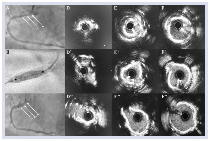
A 64-year-old symptomatic man with Canadian Cardiovascular Society (CCS) class III angina was referred for percutaneous coronary intervention of subtotal occlusion of the ostial right coronary artery (RCA; Fig. 1A). Following intubation with a 7F AL 0.75 guiding catheter, and sequential high-pressure predilatation (1.2 mm semi-compliant balloon, and 2.0 mm to 3.0 mm non-compliant balloons), intravascular ultrasound (IVUS) revealed extensive three-to-four-quadrant (270° to 360°) calcification within proximal RCA along with persistent ostial stenosis of the vessel (Fig. 1D–F). To modify plaque within proximal RCA, a 4.0 × 12 mm intravascular lithotripsy (IVL) balloon was inflated to 4 atm, and 8 cycles of 10 pulses each were delivered, followed by further dilatation to nominal pressure (Fig. 1B). IVUS after IVL confirmed multiple calcium disruptions (Fig. 1D’–F’) allowing for guideliner-facilitated delivery and deployment of 2 drug eluting stents (4.0 mm each), and further high-pressure postdilatation (at 22 atm) using a 4.5 non-compliant balloon. Optimal angiographic result (Fig. 1C) was subsequently verified with both IVUS (Fig. 1D”–F”; Suppl. Video 1) and instantaneous wave-free ratio.
Figure 1.
A. Subtotal occlusion of the ostial right coronary artery (RCA) with tortuous uptake from the aorta; B. Angiographic appearance of the Shockwave intravascular lithotripsy (IVL) balloon; C. Final angiographic result after stent implantation; D–F. Intravascular ultrasound (IVUS) before IVL revealing three-to-four-quadrant (270° to 360°) calcification of the proximal RCA along with severe ostial stenosis; D’–F’. IVUS after IVL demonstrating successful fracture of the calcified lesion within proximal RCA; D”–F”. Final IVUS showing optimal stent expansion.
Coronary IVL is a novel catheter-based technique that utilizes sonic pressure waves to disrupt calcified lesions. Herein we present a case of IVL for treatment of subtotal ostial coronary occlusion with severe calcification resulting in successful delivery and optimal expansion of coronary stents. Whether IVL may supplement available percutaneous techniques in coronary total occlusions is to be elucidated in future trials.
Supplementary Information
Footnotes
Conflict of interest: None declared
Associated Data
This section collects any data citations, data availability statements, or supplementary materials included in this article.


