Abstract
Background
Myocardial infarction (MI) is a leading cause of disease with high morbidity and mortality worldwide. Recent studies have revealed that long non-coding RNAs (lncRNAs) are involved in heart disease pathogenesis. This study aimed to investigate the effect and the molecular basis of THRIL on hypoxia-injured H9C2 cells.
Methods
THRIL, miR-99a and Brahma-related gene 1 (Brg1) expressions in H9C2 cells were altered by transient transfections. The cells were subjected to hypoxia for 4 h, and then the levels of THRIL, miR-99a and Brg1 were investigated. Cell viability, migration and invasion, and apoptotic cells were respectively measured by trypan blue exclusion assay, transwell migration assay and flow cytometry assay. Dual luciferase reporter assay was conducted to verify the interaction between miR-99a and THRIL. Furthermore, levels of apoptosis-, PI3K/AKT and mTOR pathways-related factors were measured by western blotting.
Results
Hypoxia induced an increase of THRIL but a reduction of miR-99a and Brg1. THRIL inhibition significantly attenuated hypoxia-induced cell injuries, as increased cell viability, migration and invasion, and decreased cell apoptosis. THRIL negatively regulated miR-99a expression through sponging with miR-99a binding site, and miR-99a inhibition abolished the protective effects of THRIL knockdown against hypoxia-induced injury in H9C2 cells. Furthermore, miR-99a positively regulated the expression of Brg1. Brg1 inhibition promoted hypoxia-induced cell injuries, while Brg1 overexpression alleviated hypoxia-induced cell injuries. Moreover, Brg1 overexpression activated PI3K/AKT and mTOR pathways.
Conclusions
This study demonstrated that THRIL inhibition represented a protective effect against hypoxia-induced injuries in H9C2 cells by up-regulating miR-99a expression.
Keywords: THRIL, miR-99a, Brg1, myocardial infarction, hypoxia
Introduction
Ischemic heart disease, such as myocardial infarction (MI), poses a major threat to human health and is one of the leading causes of diseases with high morbidity and mortality worldwide [1]. MI is mainly caused by a life-threatening interruption of blood supply to part of the heart [2]. Despite rapid progress obtained in targeted therapy strategies and improvement of life quality of patients, there still remain limitations in the clinical treatment and prevention of MI. Thus, it is of great significance to understand further mechanisms of physiological and pathological processes in MI.
Non-coding RNAs (ncRNAs) are classified into small ncRNAs and long ncRNAs (lncRNAs) according to their size. Small ncRNAs, such as short interfering RNAs (siRNAs), piwi-interactingRNA (piRNAs), and microRNAs (miRNAs) that have a length of less than 200 nucleotides (nt) [3]. LncRNAs are nonprotein coding transcripts with length longer than 200 nucleotides, and many of which are emerged as an important class of regulatory molecules in governing fundamental biological processes [4]. The mechanism of gene regulation by lncRNAs are involved in activation or inhibition of gene expression and modulation of chromatin architecture [5]. Recent studies have revealed that a number of lncRNAs have significant roles in a diverse range of cellular functions including development, differentiation, and cell fate as well as disease pathogenesis [3, 6, 7]. In the cardiovascular system, it has been reported that several lncRNAs, such as Novlnc6 and Mhrt, are involved in acute MI and heart failure; whereas others (such as CARL, CHRF, Novlnc6 and et al.) control hypertrophy, mitochondrial function and apoptosis of cardiomyocytes [8]. THRIL (TNFα and hn-RNPL related immunoregulatory lincRNA) is first reported to regulate lipopolysaccharide-induced tumor necrosis factor alpha (TNFα) by interacting with heterogenous nuclear ribonucleoprotein L (hnRNPL), and plays a crucial role in the regulation of physiological and pathological inflammatory immune responses [9]. However, its biological role and regulatory mechanism in hypoxia-induced injury of H9C2 cells are poorly defined.
In the present study, the role of THRIL in the survival and metastasis of H9C2 cells against hypoxia stimulus as well as the underlying mechanism is investigated. It was found that THRIL knockdown attenuated hypoxia-induced cell injuries in H9C2 cells. THRIL negatively regulated miR-99a expression through sponging with its banding site. In addition, we also proved that Brahma-related gene 1 (Brg1) expression was positively regulated by miR-99a and Brg1 was involved in the hypoxia-induced injuries in H9C2 cells. This study will provide new insight into the fundamental information and novel pharmacological target for therapeutic approaches of cardiovascular disease.
Methods
Cell culture and hypoxia treatment
H9C2 cells, obtained from the American Type Culture Collection (ATCC; Rockville, MD, USA), were cultured in Dulbecco’s modified Eagle’s medium (DMEM; HyClone, Logan, UT, USA) supplemented with 10% fetal bovine serum (FBS; Gibco, Gaithersburg, MD, USA), 100 U/mL penicillin and 100 μg/mL streptomycin (Life Technologies Corporation, Carlsbad, CA, USA) at 37°C in an atmosphere of 95% air and 5% CO2. When cells reached about 80% of confluence in appropriate culture dishes, cells were pre-starved using DMEM supplemented with 0.5% FBS for 1 h and then were incubated in hypoxic incubator containing 94% N2, 5% CO2, and 1% O2 for 4 h to simulate hypoxia injury.
Transfection and generation of stably transfected cell lines
Short-hairpin RNA (shRNA) directed against human lncRNA THRIL was ligated into the U6/ GFP/Neo plasmid (GenePharma, Shanghai, China) and was referred as to sh-THRIL. The plasmid PGPU6/GFP/neo-shControl (GenePharma) was used as the negative control (NC) and encoded with a nonsense sequence, and was referred as to sh-NC. For the analysis of the Brg1 functions, the full-length Brg1 sequences and shRNA directed against Brg1 were constructed in pEX-2 and U6/GFP/Neo plasmids (GenePharma), respectively. And they were referred as to pEX-Brg1 and sh-Brg1. The plasmid carrying a non-targeting sequence was used as a NC of pEX-Brg1 and sh-Brg1, which were referred as to pEX and sh-NC. The lipofectamine 3000 reagent (Life Technologies Corporation) was used for cell transfection according to the manufacturer instructions. The stably transfected cells were selected by the culture medium containing 0.5 mg/mL G418 (Sigma-Aldrich, St. Louis, MO, USA). After approximately 4 weeks, G418-resistant cell clones were established. miR-99a mimic, inhibitor and their respective NC were synthesized (Life Technologies Corporation) and transfected into cells in the study. Since the highest transfection efficiency occurred at 48 h, thus 72 h post-transfection was considered as the harvest time in the subsequent experiments.
Quantitative real-time PCR
Total RNA was extracted from cells using TRIzol reagent (Life Technologies Corporation) according to the manufacturer instructions. RNA was reverse transcribed to cDNA using a Reverse Transcription Kit (Takara, Dalian, China). THRIL and Brg1 expression levels were determined by quantitative real time polymerase chain reaction (qRT-PCR) using the SYBR Green Master Mix (Takara). The expression of miR-99a was measured using Taqman MicroRNA Assay (Applied Biosystems, Foster City, CA, USA) according to manufacturer instructions. GAPDH and U6 were used for the normalization of mRNA, lncRNA and miRNA. The results were presented as fold changes relative to U6 or GAPDH and were calculated using the 2−ΔΔCT method. The primer sequences used in qRT-PCR analysis were shown as follows: lncRNA THRIL (Forward: 5′-GAG TGC AGT GGC GTG ATC TC-3′, Reverse: 5′-AAA ATT AGT CAG GCA TGG TGG TG-3′); miR-99a (Forward: 5′-CGG AAC CCG TAG ATC CGA T-3′, Reverse: 5′-CAG TGC AGG GTC CGA GGT-3′); Brg1 (Forward: 5′-CCT CTC TCA ACG CTG TCC AAC TG-3′, Reverse: 5′-ATC TTG GCG AGG ATG TGC TTG TCT T-3′).
Cell viability assay
For cell viability assay, 1 × 10⁵ cells were seeded in duplicate in 60-mm dishes. After 4 h for hypoxia followed by 72 h transfection, cells were washed with phosphate buffered saline (PBS) and living cell numbers were determined by trypan blue exclusion (Beyotime Biotechnology, Shanghai, China) as previously described [10].
Apoptosis assay
For apoptosis assay, cells were stained with propidium iodide (PI) and fluorescein-isothiocyanate (FITC) — conjugated Annexin V using an Annexin V-FITC/PI Apoptosis Detection Kit (Beyotime Biotechnology). Briefly, the cells were harvested after transfection in 72 h and 4 h for hypoxia incubation and then centrifuged at 800 g for 5 min at 4°C. Cells were washed with PBS 3 times and resuspended with 500 μL 1 × binding buffer. Then cells were mixed with 5 μL Annexin V-FITC and 5 μL PI for 15 min in the dark at 37°C. Flow cytometry analysis was done using a FACS can (Beckman Coulter, Fullerton, CA, USA). The data were analyzed using FlowJo software (Tree Star Inc., Ashland, OR).
Migration and invasion assay
Cell migration and invasion was measured using a transwell chamber (8 μm, 24-well format; Corning, Lowell, MA, USA). For migration assay, 1 × 10⁵ cells in 0.2 mL of serum-free medium were plated on the upper compartment of 24-well transwell culture chamber, and 0.6 mL of medium containing 10% FBS was added to the lower chamber. For invasion assay, 1.5 × 10⁵ cells were planted on the upper chamber which is pre-coated with 20 μg of Matrigel (BD Biosciences, Bedford, MA, USA). After 48 h of incubation, the migrated and invaded cells in the lower chamber were fixed in 100% methanol and stained with 0.1% crystal violet (Sigma-Aldrich) and cells that did not migrate or invade through the pores were removed by cotton swabs. The migrated and invaded cells were counted in 5 random fields and expressed as the average number of cells per field. These experiments were done in triplicate and performed a minimum of three times.
Reporter vectors constructs and luciferase reporter assay
The segment from the 3′-UTR of the THRIL containing the predicted miR-99a binding site was amplified by PCR and then cloned into a pmirGlO Dual-luciferase miRNA Target Expression Vector (Promega, Madison, WI, USA) to form the reporter vector THRIL-wild-type (THRIL-wt). To mutate the putative binding site of miR-99a in the THRIL, the sequence of putative binding site was replaced and was named as THRIL-mutated-type (THRIL-mt). Then the vector and miR-99a mimic were co-transfected into H9C2 cells with lipofectamine 3000 (Life Technologies Corporation), and the Dual-Luciferase Reporter Assay System (Promega) were used for detecting the luciferase activity.
Western blot
Cells were homogenized in RIPA protein lysis buffer (Beyotime Biotechnology) supplemented with protease inhibitors (Roche, Basel, Switzerland) at 4°C for 30 min before centrifugation at 12,000 g for 15 min at 4°C. The protein concentration was quantified using the BCA Protein Assay Kit (Pierce, Rockford, IL, USA). Denatured samples (20–40 μg of protein) were loaded into each well, separated by 10–12% sodium dodecyl sulfate-polyacrylamide gel (SDS-PAGE), and transferred to a polyvinylidene difluoride (PVDF) membrane (Millipore, Billerica, MA). Subsequently, the membrane was blocked in 5% BSA (Roche) in TBST at room temperature for 1 h. Primary antibodies: anti-Bcl-2 (#4223), anti-Bax (#5023), anti-caspase-3 (#9662), anti-caspase-9 (#9502), anti-GAPDH (#2118), anti-Brg1 (#49360), anti-PI3K (#4249), anti-p-PI3K(#4228), anti-AKT (#4685), anti-p-AKT (#4060), anti-mTOR (#2983), anti-p-mTOR (#5536), anti-p70S6K (#2708), and anti-p-p70S6K (#9204, Cell Signaling Technology, Beverly, MA, USA) were prepared in 5% BSA at a dilution of 1:1000. The blot was incubated respectively with primary antibodies overnight at 4°C. After washing with TBST for 3 times, the membranes were further incubated with horseradish peroxidase-conjugated goat anti-rabbit IgG or anti-mouse IgG (Sigma-Aldrich) at a 1:5000 dilution for 2 h at room temperature. The blots were developed with ECL solution (Pierce) and visualized by using Image Lab™ Software (Bio-Rad, Hercules, CA, USA).
Statistical analysis
All experiments were repeated 3 times. The results of multiple experiments are presented as the mean ± standard deviation (SD). Statistical analyses were performed using Graphpad 6.0 statistical software (GraphPad Software, San Diego, CA, USA). The p-values were calculated using a one-way analysis of variance (ANOVA). A p-value of < 0.05 was considered to indicate a statistically significant result.
Results
Hypoxia induced the increase of THRIL but the reduction of miR-99a and Brg1
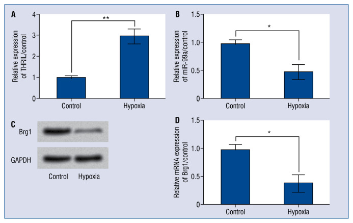
H9C2 cells were exposed to hypoxia incubator for 4 h to stimulate hypoxia injury. As shown in Figure 1A, the expression of THRIL was significantly up-regulated by hypoxia treatment in H9C2 cells (p < 0.01). However, hypoxia treatment induced a significant reduction of miR-99a expression (p < < 0.05; Fig. 1B). In addition, the protein and mRNA expression of Brg1 was also down-regulated by hypoxia injury in H9C2 cells (p < 0.05; Fig. 1C, D).
Figure 1.
Hypoxia induced an increase of THRIL but the reduction of miR-99a and Brahma-related gene 1 (Brg1). H9C2 cells were incubated in hypoxia incubator for 4 h to induced hypoxia injury. Then, the expression levels of (A) THRIL and (B) miR-99a were measured by quantitative real time polymerase chain reaction (qRT-PCR). The protein (C) and mRNA expression of Brg1 (D) was respectively determined by Western blotting and qRT-PCR; *p < 0.05; **p < 0.01.
THRIL knockdown attenuated hypoxia-induced cell injuries in H9C2 cells
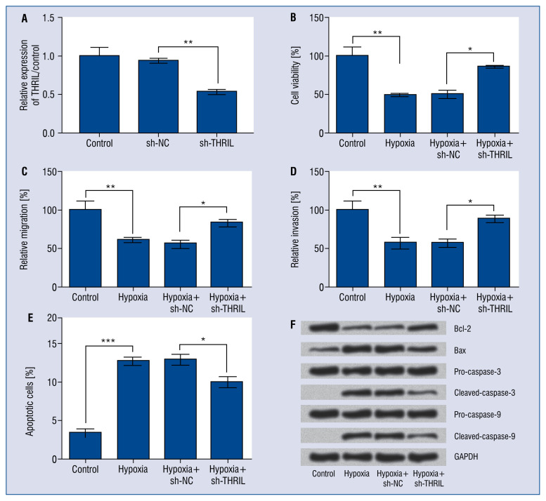
To analyze the potential role of THRIL knockdown in hypoxia-induced injuries in H9C2 cells, we transfected THRIL targeted shRNA (sh-THRIL) into H9C2 cells to induce silence of THRIL and the efficiency of transfection was performed by qRT-PCR analysis (Fig. 2A). As expected, the expression level of THRIL was significantly reduced after transfection with sh-THRIL in H9C2 cells (p < 0.01). As the migration of cardiomyocytes into the injury site has been reported to be regulated independently of proliferation, and that coordination of both processes is necessary for heart regeneration [11], thus not only the effect of THRIL knockdown on cell viability and apoptosis was investigated, but also the migration and invasion of H9C2 cells were examined. As shown in Figure 2B–E, there was a significant reduction of cell viability, relative migration and invasion (all p < 0.01), while an increase of cell apoptotic rate in H9C2 cells after hypoxia treatment (p < 0.001). Consistently, hypoxia treatment induced the upregulation of pro-apoptotic proteins including Bax, Cleaved capase-3 and Cleaved caspase-9, and the down-regulation of Bcl-2 (Fig. 2F). Interestingly, THRIL knockdown significantly increased the cell viability, migration and invasion, and declined apoptotic cell rate after hypoxia treatment (all p < 0.05) (Fig. 2B–E). Besides, THRIL inhibition decreased pro-apoptotic proteins levels and increased Bcl-2 levels in hypoxia-exposed H9C2 cells (Fig. 2F). Taken together, these results indicated that THRIL inhibition exerted a protective effect on hypoxiainduced cell injuries in H9C2 cells.
Figure 2.
THRIL knockdown attenuated hypoxia-induced cell injuries in H9C2 cells. H9C2 cells were transfected with sh-THRIL or sh-NC and then were incubated in hypoxic incubator for 4 h to simulate hypoxia. A. The transfection efficiency was verified by quantitative real time polymerase chain reaction (qRT-PCR). Cell viability (B), relative migration (C) and invasion (D), apoptotic cells rate (E), and the expression of apoptosis-related factors (F) were respectively assessed by trypan blue exclusion assay, transwell analysis, flow cytometry, and Western blotting; *p < 0.05; **p < 0.01; ***p < 0.001.
miR-99a was negatively regulated by THRIL
As it was found that hypoxia treatment upregulated expression of THRIL but down-regulated the expression of miR-99a, it was hypothesized as to whether there was a relationship between those two genes. By seeking bioinformatics online databases including TargetScan (http://www.targetscan.org/vert_71/) and NCBI (https://www.ncbi.nlm.nih.gov/), and using the bioinformatics method, lncRNA THRIL was predicted to bind to miR-99a. The predicted banding sequences were shown in Figure 3A. Thus, it was speculated that there might be an interaction between THRIL and miR-99a in H9C2 cells. To verify whether THRIL has a direct interaction with miR-99a, a dual-luciferase reporter system was employed by co-transfection of miR-99a and luciferase reporter plasmids containing 3’-UTR of THRIL, or mutated THRIL (bearing deletions of the putative miR-99a target sites). The dual-luciferase reporter assay revealed that co-transfection of miR-99a mimic and THRIL-wt specifically decreased the luciferase activity (p < 0.05), and there was no significant difference between co-transfection with miR-99a mimic and THRIL-mt in H9C2 cells (p > 0.05) (Fig. 3B), which suggested that THRIL might function as endogenous sponge RNA to interact with miR-99a. As shown in Figure 3C, THRIL inhibition up-regulated the expression of miR-99a in H9C2 cells (p < 0.001). These findings indicated that THRIL negatively regulated the expression of miR-99a through sponging with miR-99a in H9C2 cells.
Figure 3.
miR-99a was negatively regulated by THRIL. A. The predicted banding sequences of THRIL and miR-99a; B. H9C2 cells were co-transfected with the luciferase reporter plasmid containing wild-type or mutant THRIL and miR-99a mimic or mimic control. Dual luciferase activity assay was performed to detect the interaction between miR-99a and THRIL; C. The expressions of miR-99a in H9C2 cells were assessed by quantitative real time polymerase chain reaction after transfection with sh-THRIL or sh-NC; *p < 0.05; **p < 0.01.
THRIL knockdown alleviated hypoxia-induced cell injuries through up-regulation of miR-99a in H9C2 cells
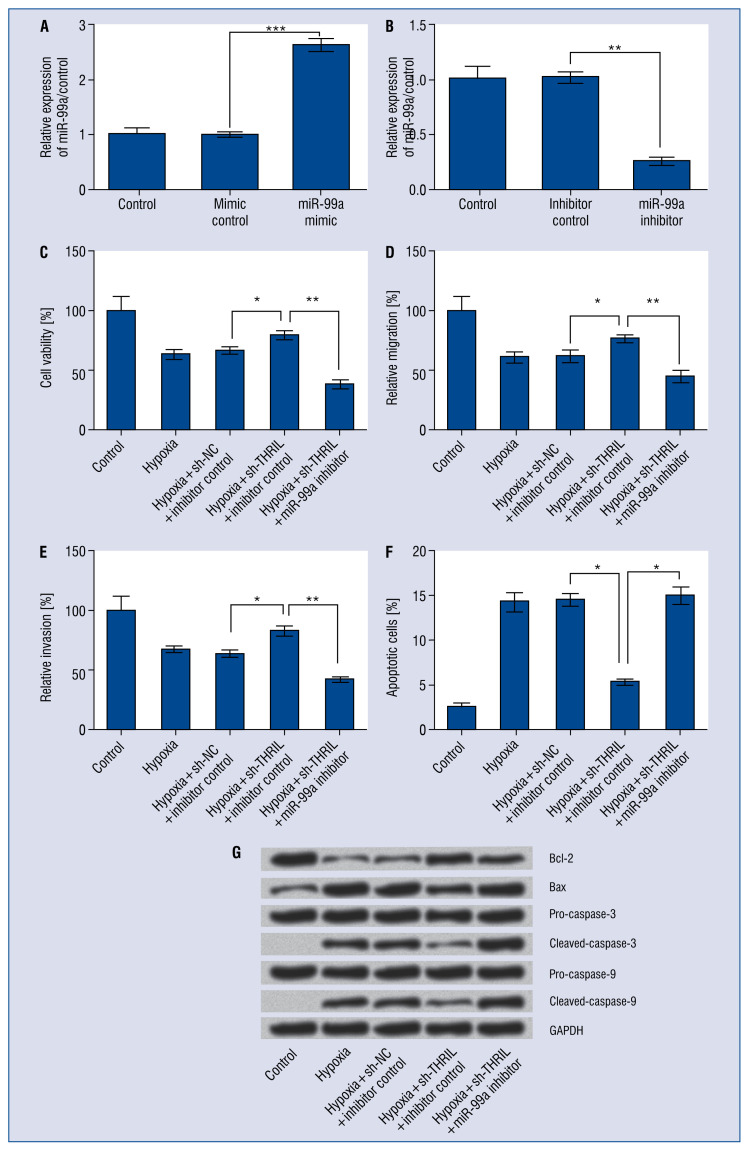
Further, exploring whether THRIL regulated hypoxia-induced cell injuries through targeting miR-99a. miR-99a expression in H9C2 cells were altered by transfection with miR-99a mimic or miR-99a inhibitor. As shown in Figure 4A, B, the expression miR-99a was dramatically up-regulated after transfection with miR-99a mimic, but was down-regulated with miR-99a inhibitor (p < 0.01, or p < 0.001). miR-99a inhibitor blocked the protective effects of THRIL on hypoxia-induced cell injuries, as miR-99a inhibition significantly decreased cell viability (p < 0.01; Fig. 4C), suppressed cell migration and invasion (p < 0.01; Fig. 4D, E), and promoted apoptotic cell rates (p < 0.05; Fig. 4F). Furthermore, the effect of THRIL suppression on apoptosis related proteins was reversed by miR-99a inhibition in H9C2 cells (Fig. 4G). Overall, these results indicate that THRIL knockdown alleviated hypoxia-induced cell injuries through up-regulation of miR-99a in H9C2 cells.
Figure 4.
THRIL knockdown alleviated hypoxia-induced cell injuries through up-regulation of miR-99a in H9C2 cells. H9C2 cells were transfected with miR-99a mimic, miR-99a inhibitor or their corresponding controls, i.e., mimic control and inhibitor control, or co-transfected with 99a inhibitor and sh-THRIL. Cells were incubated in hypoxic incubator for 4 h to simulate hypoxia. A, B. The expression of miR-99a was assessed by quantitative real time polymerase chain reaction. Cell viability (C), relative migration (D) and invasion (E), apoptotic cells rate (F), and the expression of apoptosis-related factors (G) were respectively assessed by trypan blue exclusion assay, transwell analysis, flow cytometry, and Western blotting; *p < 0.05; **p < 0.01, ***p < 0.001.
Brg1 was positively regulated by miR-99a
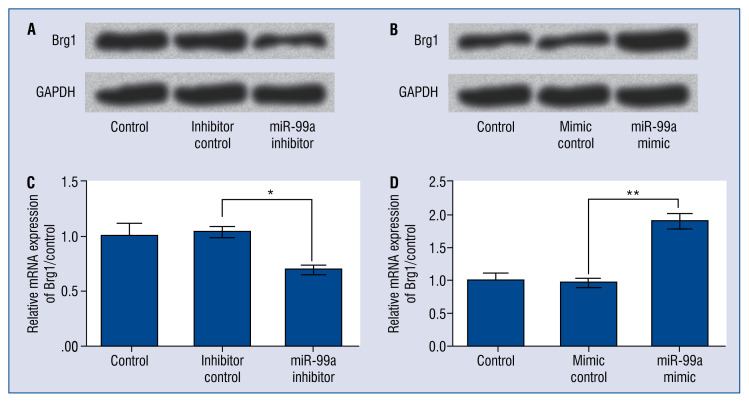
Given that, Brg1 is required for cell proliferation to form the compact and septal myocardium [12]. Cross-regulation was detected between miR-99 and Brg1, to further reveal the mechanism(s) via which miR-99a modulated H9C2 cells. It was found that miR-99a inhibition declined both the protein and mRNA levels of Brg1 (p < 0.05; Fig. 5A, C). Of contrast, miR-99a mimic elevated Brg1 levels in H9C2 cells (p < 0.01; Fig. 5B, D). Taken together, these results indicated that Brg1 was positively regulated by miR-99a in H9C2 cells.
Figure 5.
Brahma-related gene 1 (Brg1) was positively regulated by miR-99a. H9C2 cells were transfected with miR-99a mimic, miR-99a inhibitor or their corresponding controls, i.e., mimic control and inhibitor control; then the (A, B) mRNA and (C, D) protein expressions of Brg1 were assessed by quantitative real time polymerase chain reaction and Western blotting; *p < 0.05, **p < 0.01.
Brg1 was involved in hypoxia-induced cell injuries in H9C2 cells
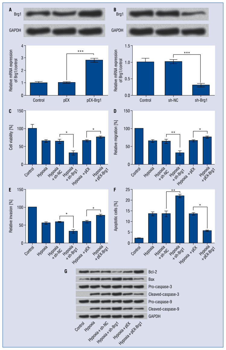
To investigate whether Brg1 was required for hypoxia-induced cell injuries in H9C2 cells, H9C2 cells were transfected with overexpressing-vector and shRNA specific targeted Brg1. As shown in Figure 6A, B, the protein and mRNA levels of Brg1 were significantly up-regulated in H9C2 cells after transfection with pEX-Brg1 (p < 0.001), while were down-regulated in cells which were transfected with sh-Brg1 (p < 0.001). Results revealed that Brg1 inhibition promoted hypoxia-induced cell injuries, as decreased cell viability (p < 0.05; Fig. 6C), cell migration and invasion (p < 0.05, or p < 0.01; Fig. 6D, E), and induced apoptosis (p < 0.01; Fig. 6F, G). Of contrast, inverse regulations were found in cells which were transfected with pEX-Brg1 (all p < 0.05; Fig. 6C, G). These results suggested that Brg1 was involved in hypoxia-induced cell injuries in H9C2 cells.
Figure 6.
Brahma-related gene 1 (Brg1) was involved in hypoxia-induced cell injuries in H9C2 cells. H9C2 cells were transfected with pEX-Brg1, or sh-Brg1 or their corresponding controls, i.e., pEX and sh-NC and then were incubated in hypoxic incubator for 4 h to simulate hypoxia. A, B. The mRNA and protein expression of Brg1 were detected by quantitative real time polymerase chain reaction (qRT-PCR) and Western blotting. Cell viability (C), relative migration (D) and invasion (E), apoptotic cells rate (F), and the expression of apoptosis-related factors (G) were respectively assessed by trypan blue exclusion assay, transwell analysis, flow cytometry, and Western blotting; *p < 0.05; **p < 0.01; ***p < 0.001.
Brg1 regulated PI3K/AKT and mTOR signaling pathways
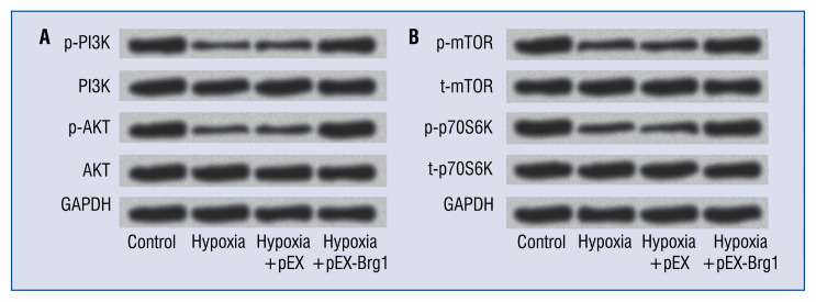
miR-99a has been identified to regulate PI3K/AKT and mTOR signaling pathways [13, 14]. Here, the effect of Brg1 overexpression on PI3K/AKT and mTOR signaling pathways in H9C2 cells was explored. As shown in Figure 7A, hypoxia suppressed the activation of PI3K and AKT. However, Brg1 overexpression increased the levels of p-PI3K and p-AKT, indicating the activated effect of PI3K/AKT signaling pathway. Furthermore, it was found that Brg1 also activated mTOR signaling pathway, as increased expression of p-mTOR and p-p70S6K (Fig. 7B).
Figure 7.
Brahma-related gene 1 (Brg1) regulated PI3K/AKT and mTOR signaling pathways. H9C2 cells were transfected with pEX-Brg1 or pEX and then were incubated in hypoxic incubator for 4 h to simulate hypoxia. The protein expressions of main factors related with PI3K/AKT (A) and mTOR (B) signaling pathways were detected by Western blotting.
Discussion
Myocardial hypoxia triggers cell injuries, which is associated with the pathogenesis of many cardiovascular diseases including heart failure, MI, myocardial ischemia as well as reperfusion injury [15]. The present data indicated that miR-99a was negatively regulated by THRIL, and THRIL knockdown alleviated hypoxia-induced cell injuries through up-regulation of miR-99a in H9C2 cells. Moreover, it was shown that Brg1 was positively regulated by miR-99a, and was involved in hypoxia-induced cell injuries in H9C2 cells. These data suggested that THRIL may play a crucial role in hypoxia-induced cell injuries via regulating miR-99a in H9C2 cells.
LncRNAs have been defined to have important functions in various cellular processes such as genomic imprinting, cell fate determination, RNA processing, chromatin modification, modulation of apoptosis and invasion, and is important for development, differentiation and metabolism [16–19]. Emerging evidences have reported that lncRNAs are essential for the development of cardiomyocytes [20–22]. For example, the lateral mesoderm-specific lncRNA Fendrr is an essential regulator of the fate of lateral mesoderm derivatives, specifically the heart and the body wall in mice [23]. APF regulates autophagy and MI by targeting miR-188-3p [18]. Recent evidence suggests that THRIL is associated with TNFα regulation and may contribute to other common inflammatory diseases [9]. Another report showed that THRIL regulates helicobacter pylori cagA induced-inflammation in gastric cancer cells via inhibition of NF-κB translocation [24]. However, the potential role of THRIL in regulating hypoxia-induced injuries in H9C2 cells have not been elucidated. The present results revealed that THRIL knockdown functioned as a protector against hypoxia-induced injuries in H9C2 cells, as increased cell viability, cell migration and invasion, and reduced cell apoptosis.
Recently, lncRNAs have been identified as competing endogenous RNAs to sponge miRNAs thus modulating the depression of miRNA targets and imposing an additional level of post-transcriptional regulation [25, 26]. miR-99a has been demonstrated to play an important role in cardiomyogenesis. Recent evidence showed that miR-99a expression significantly declined in patients with MI, and overexpression of miR-99a attenuated ventricular remodeling, cardiac hypertrophy and improved cardiac performance after MI [27–29]. In the present study, it was found that THRIL negatively regulated the expression of miR-99a, and THRIL acted as a sponge of miR-99a in H9C2 cells. Furthermore, we also found that THRIL inhibition alleviated hypoxia-induced cell injuries through up-regulation of miR-99a in H9C2 cells.
Brg1 is one of the central ATPase catalytic subunits, which is essential for zygote genome activation, erythropoiesis, cardiac development and neuronal development [30]. In embryos, Brg1 promotes cardiomyocyte proliferation by maintaining Bmp10 and suppressing p57kip2 expression. However, in adults, Brg1 is turned off in cardiomyocytes and it is reactivated by cardiac stresses, and preventing Brg1 re-expression decreases hypertrophy [31]. Zebrafish experiment also showed that Brg1 promotes heart regeneration by repressing cyclin-dependent kinase inhibitors partly through Dnmt3ab-dependent DNA methylation [30]. Results herein showed that Brg1 was positively regulated by miR-99a in H9C2 cells, and overexpression of Brg1 attenuated hypoxiainduced cell injuries. It was supposed that THRIL may sponge with miR-99a and that miR-99a further regulate Brg1, thus regulating hypoxia-induced injury in H9C2 cells. However, further study is needed to confirm this hypothesis. In addition, it was also found that overexpression of Brg1 activated PI3K/AKT and mTOR signaling pathways. The PI3K/AKT and mTOR signaling pathways are important signal transduction pathways that control cardiomyocyte survival and functions [32, 33]. The activation of PI3K/AKT and mTOR pathways may contribute to inhibiting myocardial cells apoptosis and promoting cell survival in a damaged heart [34, 35]. It has been reported that Brg1 promotes osteogenic differentiation of mesenchymal stem cells (MSCs), and the mechanism might be by regulating Runx2-mediated downstream Wnt and PI3K/AKT pathways. Brg1 overexpression statistically increased the expression of PI3K/AKT pathways key proteins [36]. Present results are consistent with previous reports about regulation of Brg1 on PI3K/AKT and mTOR pathway, and these data suggest that miR-99a overexpression might activate the PI3K/AKT and mTOR signaling pathways through up-regulated Brg1.
Conclusions
In conclusion, the present study demonstrated that THRIL knockdown attenuated hypoxia-induced injuries of H9C2 cells through directly up-regulated expression of miR-99a. Interestingly, miR-99a positively regulated Brg1 expression, and Brg1 overexpression exerted similarly protective effects to THRIL inhibition. According to available research, this is the first study to demonstrate that THRIL is associated with hypoxia-induced injuries of H9C2 cells, which direct sponging with miR-99a. It was also revealed that Brg1 was positively regulated by miR-99a in H9C2 cells for the first time. It may allow a better understanding of the pathological process of MI and ultimately contribute to the development of THRIL-directed diagnostics and therapeutics against myocardial infarction.
Footnotes
Conflict of interest: None declared
References
- 1.Hoffmann R. The potential for reduction of educational differences in ischaemic heart disease mortality in Europe: The role of three lifestyle risk factors. Eur J Public Health. 2016:ckw104. [Google Scholar]
- 2.Ren Li, Liu W, Wang Y, et al. Investigation of hypoxia-induced myocardial injury dynamics in a tissue interface mimicking microfluidic device. Anal Chem. 2013;85(1):235–244. doi: 10.1021/ac3025812. [DOI] [PubMed] [Google Scholar]
- 3.Peng W, Si S, Zhang Q, et al. Long non-coding RNA MEG3 functions as a competing endogenous RNA to regulate gastric cancer progression. J Exp Clin Cancer Res. 2015;34:79. doi: 10.1186/s13046-015-0197-7. [DOI] [PMC free article] [PubMed] [Google Scholar]
- 4.Zhou RM, Wang XQ, Yao J, et al. Identification and characterization of proliferative retinopathy-related long noncoding RNAs. Biochem Biophys Res Commun. 2015;465(3):324–330. doi: 10.1016/j.bbrc.2015.07.120. [DOI] [PubMed] [Google Scholar]
- 5.Vausort M, Wagner DR, Devaux Y. Long noncoding RNAs in patients with acute myocardial infarction. Circ Res. 2014;115(7):668–677. doi: 10.1161/CIRCRESAHA.115.303836. [DOI] [PubMed] [Google Scholar]
- 6.Haemmerle M, Gutschner T. Long non-coding RNAs in cancer and development: where do we go from here? Int J Mol Sci. 2015;16(1):1395–1405. doi: 10.3390/ijms16011395. [DOI] [PMC free article] [PubMed] [Google Scholar]
- 7.Hung CL, Wang LY, Yu YL, et al. A long noncoding RNA connects c-Myc to tumor metabolism. Proc Natl Acad Sci U S A. 2014;111(52):18697–18702. doi: 10.1073/pnas.1415669112. [DOI] [PMC free article] [PubMed] [Google Scholar]
- 8.Uchida S, Dimmeler S. Long noncoding RNAs in cardiovascular diseases. Circ Res. 2015;116(4):737–750. doi: 10.1161/CIRCRESAHA.116.302521. [DOI] [PubMed] [Google Scholar]
- 9.Li Z, Chao TC, Chang KY, et al. The long noncoding RNA THRIL regulates TNFα expression through its interaction with hn-RNPL. Proc Natl Acad Sci U S A. 2014;111(3):1002–1007. doi: 10.1073/pnas.1313768111. [DOI] [PMC free article] [PubMed] [Google Scholar]
- 10.Hsieh TC, Wijeratne EK, Liang JY, et al. Differential control of growth, cell cycle progression, and expression of NF-kappaB in human breast cancer cells MCF-7, MCF-10A, and MDA-MB-231 by ponicidin and oridonin, diterpenoids from the chinese herb Rabdosia rubescens. Biochem Biophys Res Commun. 2005;337(1):224–231. doi: 10.1016/j.bbrc.2005.09.040. [DOI] [PubMed] [Google Scholar]
- 11.Itou J, Oishi I, Kawakami H, et al. Migration of cardiomyocytes is essential for heart regeneration in zebrafish. Development. 2012;139(22):4133–4142. doi: 10.1242/dev.079756. [DOI] [PubMed] [Google Scholar]
- 12.Hang CT, Yang J, Han P, et al. Chromatin regulation by Brg1 underlies heart muscle development and disease. Nature. 2010;466(7302):62–67. doi: 10.1038/nature09130. [DOI] [PMC free article] [PubMed] [Google Scholar]
- 13.Chakrabarti M, Banik NL, Ray SK. Photofrin based photodynamic therapy and miR-99a transfection inhibited FGFR3 and PI3K/Akt signaling mechanisms to control growth of human glioblastoma In vitro and in vivo. PLoS One. 2013;8(2):e55652. doi: 10.1371/journal.pone.0055652. [DOI] [PMC free article] [PubMed] [Google Scholar]
- 14.Yang Z, Han Y, Cheng K, et al. miR-99a directly targets the mTOR signalling pathway in breast cancer side population cells. Cell Prolif. 2014;47(6):587–595. doi: 10.1111/cpr.12146. [DOI] [PMC free article] [PubMed] [Google Scholar]
- 15.Liu B, Che W, Xue J, et al. SIRT4 prevents hypoxia-induced apoptosis in H9c2 cardiomyoblast cells. Cell Physiol Biochem. 2013;32(3):655–662. doi: 10.1159/000354469. [DOI] [PubMed] [Google Scholar]
- 16.Gong C, Maquat LE. lncRNAs transactivate STAU1-mediated mRNA decay by duplexing with 3’ UTRs via Alu elements. Nature. 2011;470(7333):284–288. doi: 10.1038/nature09701. [DOI] [PMC free article] [PubMed] [Google Scholar]
- 17.Kanduri C. Kcnq1ot1: a chromatin regulatory RNA. Semin Cell Dev Biol. 2011;22(4):343–350. doi: 10.1016/j.semcdb.2011.02.020. [DOI] [PubMed] [Google Scholar]
- 18.Wang K, Liu CY, Zhou LY, et al. APF lncRNA regulates autophagy and myocardial infarction by targeting miR-188-3p. Nat Commun. 2015;6:6779. doi: 10.1038/ncomms7779. [DOI] [PubMed] [Google Scholar]
- 19.Clemson CM, Hutchinson JN, Sara SA, et al. An architectural role for a nuclear noncoding RNA: NEAT1 RNA is essential for the structure of paraspeckles. Mol Cell. 2009;33(6):717–726. doi: 10.1016/j.molcel.2009.01.026. [DOI] [PMC free article] [PubMed] [Google Scholar]
- 20.Liao J, He Q, Li M, et al. LncRNA MIAT. Myocardial infarction associated and more. Gene. 2016;578(2):158–161. doi: 10.1016/j.gene.2015.12.032. [DOI] [PubMed] [Google Scholar]
- 21.Wang P, Fu H, Cui J, et al. Differential lncRNAmRNA coexpression network analysis revealing the potential regulatory roles of lncRNAs in myocardial infarction. Mol Med Rep. 2016;13(2):1195–1203. doi: 10.3892/mmr.2015.4669. [DOI] [PMC free article] [PubMed] [Google Scholar]
- 22.Yang KC, Yamada KA, Patel AY, et al. Deep RNA sequencing reveals dynamic regulation of myocardial noncoding RNAs in failing human heart and remodeling with mechanical circulatory support. Circulation. 2014;129(9):1009–1021. doi: 10.1161/CIRCULATIONAHA.113.003863. [DOI] [PMC free article] [PubMed] [Google Scholar]
- 23.Grote P, Wittler L, Hendrix D, et al. The tissue-specific lncRNA Fendrr is an essential regulator of heart and body wall development in the mouse. Dev Cell. 2013;24(2):206–214. doi: 10.1016/j.devcel.2012.12.012. [DOI] [PMC free article] [PubMed] [Google Scholar]
- 24.Sang KL, Na KL, Yong CL. 150 Long Non-Coding RNA Thril and Hnrnpl Regulates Helicobacter pylori cagA Induced-Inflammation by Inhibition of NF-κ B Translocation. Gastroenterology. 2016;150(4):S37. doi: 10.1016/s0016-5085(16)30251-7. [DOI] [Google Scholar]
- 25.Salmena L, Poliseno L, Tay Y, et al. A ceRNA hypothesis: the Rosetta Stone of a hidden RNA language? Cell. 2011;146(3):353–358. doi: 10.1016/j.cell.2011.07.014. [DOI] [PMC free article] [PubMed] [Google Scholar]
- 26.Liu XH, Sun M, Nie FQ, et al. Lnc RNA HOTAIR functions as a competing endogenous RNA to regulate HER2 expression by sponging miR-331-3p in gastric cancer. Mol Cancer. 2014;13:92. doi: 10.1186/1476-4598-13-92. [DOI] [PMC free article] [PubMed] [Google Scholar]
- 27.Li Q, Xie J, Wang B, et al. Overexpression of microRNA-99a Attenuates Cardiac Hypertrophy. PLoS One. 2016;11(2):e0148480. doi: 10.1371/journal.pone.0148480. [DOI] [PMC free article] [PubMed] [Google Scholar]
- 28.Li Q, Xie J, Li R, et al. Overexpression of microRNA-99a attenuates heart remodelling and improves cardiac performance after myocardial infarction. J Cell Mol Med. 2014;18(5):919–928. doi: 10.1111/jcmm.12242. [DOI] [PMC free article] [PubMed] [Google Scholar]
- 29.Yang SY, Wang YQ, Gao HM, et al. The clinical value of circulating miR-99a in plasma of patients with acute myocardial infarction. Eur Rev Med Pharmacol Sci. 2016;20(24):5193–5197. [PubMed] [Google Scholar]
- 30.Xiao C, Gao Lu, Hou Yu, et al. Chromatin-remodelling factor Brg1 regulates myocardial proliferation and regeneration in zebrafish. Nat Commun. 2016;7:13787. doi: 10.1038/ncomms13787. [DOI] [PMC free article] [PubMed] [Google Scholar]
- 31.Hang CT, Yang J, Han P, et al. Chromatin regulation by Brg1 underlies heart muscle development and disease. Nature. 2010;466(7302):62–67. doi: 10.1038/nature09130. [DOI] [PMC free article] [PubMed] [Google Scholar]
- 32.Bi Y, Wang G, Liu X, et al. Low-after-high glucose down-regulated Cx43 in H9c2 cells by autophagy activation via cross-regulation by the PI3K/Akt/mTOR and MEK/ERK signal pathways. Endocrine. 2017;56(2):336–345. doi: 10.1007/s12020-017-1251-3. [DOI] [PubMed] [Google Scholar]
- 33.Han D, Wan C, Liu F, et al. Jujuboside A Protects H9C2 Cells from Isoproterenol-Induced Injury via Activating PI3K/Akt/mTOR Signaling Pathway. Evid Based Complement Alternat Med. 2016;2016:9593716. doi: 10.1155/2016/9593716. [DOI] [PMC free article] [PubMed] [Google Scholar]
- 34.Wang M, Sun Gb, Sun X, et al. Cardioprotective effect of salvianolic acid B against arsenic trioxide-induced injury in cardiac H9c2 cells via the PI3K/Akt signal pathway. Toxicol Lett. 2013;216(2–3):100–107. doi: 10.1016/j.toxlet.2012.11.023. [DOI] [PubMed] [Google Scholar]
- 35.Zhang ZL, Fan Y, Liu ML. Ginsenoside Rg1 inhibits autophagy in H9c2 cardiomyocytes exposed to hypoxia/reoxygenation. Mol Cell Biochem. 2012;365(1–2):243–250. doi: 10.1007/s11010-012-1265-3. [DOI] [PubMed] [Google Scholar]
- 36.Li J, He Z, Liu R, Ma M, Yang L, Ye C. Brg1 promotes osteogenic differentiation of mesenchymal stem cells by regulating Runx2-mediated Wnt and PI3K/AKT pathways. Int J Clin Exp Pathol. 2016;9(11):11074–11082. [Google Scholar]


