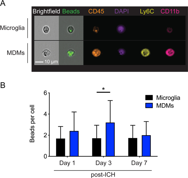
Figure 1. MDMs have higher phagocytic capacity in the ICH brain.
A, Representative images show MDMs and microglia from ICH day 3 brains. MDMs are CD11b+CD45hiLy6C+ and microglia are CD11b+CD45int. Fluorescent beads are visible within these cells. B, MDMs phagocytose higher numbers of fluorescent beads in the perihematomal brain tissues on day 3. Data shows mean ± SD. *p < 0.05 by ANOVA with post-hoc Tukey test, n=10.

