A 2.0 Å resolution cryo-EM structure of a thermostable bacterial nanocompartment is reported, with the high-resolution structure allowing the visualization of key details and new proposed biological functions.
Keywords: cryo-electron microscopy, nanocompartments, encapsulins, thermostability
Abstract
Protein nanocompartments are widespread in bacteria and archaea, but their functions are not yet well understood. Here, the cryo-EM structure of a nanocompartment from the thermophilic bacterium Thermotoga maritima is reported at 2.0 Å resolution. The high resolution of this structure shows that interactions in the E-loop domain may be important for the thermostability of the nanocompartment assembly. Also, the channels at the fivefold axis, threefold axis and dimer interface are assessed for their ability to transport iron. Finally, an unexpected flavin ligand was identified on the exterior of the shell, indicating that this nanocompartment may also play a direct role in iron metabolism.
1. Introduction
Many species of bacteria and archaea have nanocompartments, which are protein assemblies that serve as non-membranous compartments (Nichols et al., 2017 ▸; Giessen & Silver, 2017 ▸). These bacterial nanocompartments, which are also known as encapsulins, strongly resemble icosahedral viral capsids in structure, although there is little sequence conservation between these complexes (Sutter et al., 2008 ▸). Aside from their structural similarities, the size and symmetry of encapsulins can vary to allow different assemblies and volumes (Giessen et al., 2019 ▸). One major difference between encapsulins and viral capsids is that bacterial nanocompartments have binding sites in the interior for a specific peptide tag found on the C-terminus of cargo proteins (Sutter et al., 2008 ▸; Cassidy-Amstutz et al., 2016 ▸). These cargo proteins generally catalyze reactions that remove toxic metabolites or reactions involving toxic substrates, intermediates or products (Giessen & Silver, 2017 ▸). The cargo proteins are diverse and include dye-decolorizing peroxidases (DyPs), ferritin-like proteins, iron-mineralizing encapsulin-associated firmicute (IMEF) proteins, hemerythrins, copper nitrite reductase/hydroxylamine oxidoreductase fusion proteins, cysteine desulfurases and polyprenyl synthetases (Giessen & Silver, 2017 ▸; Nichols et al., 2020 ▸; Tracey et al., 2019 ▸).
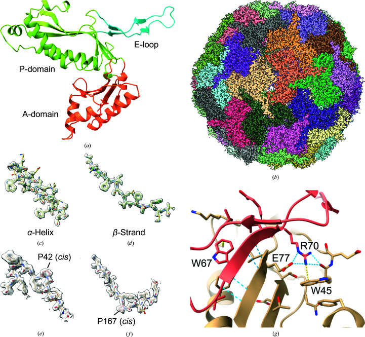
Based on their unique assembly and storage properties, bacterial nanocompartments have been engineered for several different applications, such as catalytic nanoreactors, biological reporters, targeted delivery systems, and nanomaterials and biomaterials (Jones & Giessen, 2021 ▸). A structural model of a nanocompartment is useful for designing nanocompartments for these kinds of applications. The first X-ray crystal structure of an encapsulin from Thermotoga maritima was determined in 2008 at 3.1 Å resolution (Sutter et al., 2008 ▸). This structure demonstrated the structural similarity of encapsulins to the HK97 major capsid protein. The structure is divided into three domains: the major P-loop domain, which is a mixed α–β structure, the E-loop, which forms the twofold interactions, and the A-domain, which forms the fivefold symmetry axis interface [Fig. 1 ▸(a)]. Also, the structure contained additional density that was identified as the C-terminal extension of the cargo protein.
Figure 1.
Density maps and model of TmEnc. (a) Domain organization of an encapsulin monomer, as viewed from the inside of the compartment. (b) Cryo-EM density map for the entire encapsulin nanocompartment. Subunits are colored individually. (c) Representative cryo-EM density for an α-helix (residues 14–30). (d) Representative density for a β-strand (residues 241–254). (e, f) Density for two cis-prolines, which were previously not observed. (g) Intermolecular interactions between the E-loop domain (tan) and another subunit (pink). Blue dashed lines indicate hydrogen bonding, and the yellow dashed line is a cation–π interaction.
In this article, we report the 2.0 Å resolution cryo-electron microscopy (cryo-EM) structure of the thermostable nanocompartment from T. maritima [Fig. 1 ▸(b)]. At this resolution, we were able to model amino-acid side chains confidently and identify ordered solvent and ion molecules. The structure reveals interactions that are likely to be responsible for the thermostability of this complex relative to its mesophilic counterparts. Also, we examined possible iron-transport channels, particularly at the fivefold and threefold axes. Finally, we identified the binding site for an unexpected flavin ligand in the shell, which may suggest additional functions of encapsulins in iron metabolism.
2. Methods
2.1. Protein expression and purification
A codon-optimized gene coding for T. maritima encapsulin (TmEnc) was inserted into pET-11a and transformed into Escherichia coli Rosetta2 (DE3) cells (Novagen). 2 ml lysogeny broth with 100 µg ml−1 carbenicillin and 25 µg ml−1 chloramphenicol was inoculated with the strain expressing TmEnc and grown to an OD600 of 0.6 at 37°C in a rotary shaker. 1 l of autoinduction medium with carbenicillin and chloramphenicol was inoculated with 1 ml of the starter culture, incubated at 37°C to an OD600 of 2.9 with shaking and then transferred to 20°C for overnight protein expression. The cells were centrifuged at 5000g for 15 min at 4°C and resuspended in Enc buffer (50 mM Tris pH 8.0, 150 mM sodium chloride). The cells were lysed by sonication and the cell debris was centrifuged at 12 000g for 30 min at 4°C. Calcium chloride at a final concentration of 5 mM and 50 µl micrococcal nuclease (New England Biosciences) were added to the lysate and incubated at 37°C for 3 h to degrade nucleic acids. The lysate was incubated at 80°C for 1.5 h and centrifuged at 12 000g for 30 min at 4°C to remove the host proteins. Solid ammonium sulfate was added to 50% saturation slowly at 4°C and incubated for 1 h with stirring to remove the remaining host proteins. After centrifugation, the ammonium sulfate precipitation was repeated to 75% saturation to precipitate TmEnc [Supplementary Fig. S1(a)]. The pellet was resuspended in Enc buffer and purified by gel filtration on a HiPrep Sephacryl S-500 HR column (GE Healthcare Life Sciences) [Supplementary Fig. S1(b)]. UV–Vis spectra were collected with a Nanodrop spectrophotometer (Thermo Fisher) in triplicate [Supplementary Fig. S1(c)].
2.2. Grid preparation and cryo-electron microscopy data collection
Copper 300-mesh Quantifoil 1.2/1.3 grids were plasma-cleaned with a Solarus plasma-cleaning system (Gatan). Grid preparation was performed with an EM GP automatic plunge freezer (Leica Microsystems). The chamber was set to 95% humidity at 20°C. 3 µl TmEnc at 2 mg ml−1 was applied to the grids, immediately blotted for 5 s and plunged into liquid propane. Cryo-electron microscopy data were collected on a Titan Krios (Thermo Fisher) at 81 000× magnification at a defocus of 0.5–2.5 µm. 2772 movies consisting of 67 frames were captured over 2.5 s on a K3 camera (Gatan) with an exposure rate of 0.5 e− Å−2 per frame (Table 1 ▸).
Table 1. Data-collection, map-reconstruction and model-refinement statistics.
| Data collection | |
| Electron microscope | Titan Krios |
| Electron detector | Gatan K3 |
| Voltage (keV) | 300 |
| Defocus range (µm) | 0.8–2.5 |
| Original pixel size (Å) | 0.5395 (super-resolution) |
| Electron exposure (e− Å−2) | 33.5 |
| Images | 2772 |
| Map reconstruction | |
| Final particles | 185459 |
| Final pixel size (Å) | 0.73 |
| Applied symmetry | I3 |
| Resolution (0.143 FSC threshold) (Å) | 2.0 |
| B factor (Å2) | −69 |
| Model refinement | |
| R.m.s.d., bond lengths (Å) | 0.005 |
| R.m.s.d., angles (°) | 0.852 |
| MolProbity score | 1.58 |
| Clashscore | 4.89 |
| Ramachandran plot | |
| Outliers (%) | 0.51 |
| Allowed (%) | 4.18 |
| Favored (%) | 95.31 |
| Ramachandran plot Z-score, r.m.s.d. | |
| Overall (N = 1578) | 1.51 (0.19) |
| Helix (N = 528) | 0.57 (0.19) |
| Sheet (N = 396) | 1.03 (0.23) |
| Loop (N = 654) | 1.16 (0.23) |
| Rotamer outliers (%) | 0.93 |
| CaBLAM outliers (%) | 1.92 |
| CC (mask) | 0.85 |
| CC (box) | 0.48 |
| CC (peaks) | 0.38 |
| CC (volume) | 0.81 |
| Mean CC for ligands | 0.67 |
2.3. Cryo-EM data processing
All software was compiled and distributed by SBGrid (Morin et al., 2013 ▸). Data processing was performed with RELION 3.1 (Scheres, 2012 ▸). Movies were motion-corrected with the internal MotionCorr implementation in RELION and CTF parameters were estimated with CTFFIND4 [Supplementary Fig. S1(d)] (Rohou & Grigorieff, 2015 ▸). Particles were picked with crYOLO using 1000 manually picked particles to train a model [Supplementary Fig. S1(e)] (Wagner et al., 2019 ▸). After the first 3D refinement, beam-tilt refinement, particle polishing, defocus refinement and higher-order aberration correction were applied (Zivanov et al., 2020 ▸) to further improve the reconstruction to a final resolution of 2.0 Å [Supplementary Fig. S1(f)] (Table 1 ▸).
2.4. Model building
Model building was performed with Phenix (Liebschner et al., 2019 ▸). The density map from RELION was density-modified with resolve_cryo_em to improve the interpretability of features (Terwilliger et al., 2020 ▸). Map_symmetry was used to generate noncrystallographic symmetry matrices from the map and map_box was used to extract the unique part of the map. Chain A of PDB entry 3dkt (Sutter et al., 2008 ▸) was fitted into the map with dock_in_map. The five neighboring subunits were added to the model to prevent atom clashes between adjacent subunits during refinement. Iterative rounds of real_space_refine (Afonine et al., 2018 ▸) and manual model adjustment in Coot (Emsley et al., 2010 ▸) led to the final model [Supplementary Fig. S1(g)] (Table 1 ▸). Water molecules were added using douse. Fivefold and threefold axis channels were analyzed with Channel Annotation Package (CHAP) to generate plots of channel radius and hydrophobicity (Klesse et al., 2019 ▸).
3. Results
3.1. Overall structure and thermostability
The high resolution of the cryo-EM density map allowed the unambiguous assignment of nearly all of the amino-acid residues of T. maritima encapsulin (TmEnc) [Figs. 1 ▸(b)–1 ▸(f), Table 1 ▸]. The density for aromatic residues contained holes, which is characteristic of high-resolution cryo-EM maps [Figs. 1 ▸(c) and 1 ▸(d)]. The model built into the cryo-EM density map is consistent with the previous crystal structure, with a root-mean-square deviation of 0.632 Å as calculated using the match command in UCSF Chimera (Pettersen et al., 2004 ▸). Nevertheless, many side chains are resolved more clearly, illustrating the interactions that stabilize the overall structure. For example, two prolines were identified in the cis conformation and were misidentified as trans-prolines in the crystal structure [Figs. 1 ▸(e) and 1 ▸(f)]. Because the side chains are better resolved in this structure, it is possible to see how side-chain interactions may contribute to the extreme thermostability of TmEnc, particularly when compared with the recent structures of encapsulins from other bacterial species such as Mycolicibacterium hassiacum (MhEnc; PDB entry 6i9g; Lončar et al., 2020 ▸), Synechococcus elongatus (SeEnc; PDB entry 6x8m; Nichols et al., 2020 ▸) and Quasibacillus thermotolerans (QsEnc; PDB entry 6nj8; Giessen et al., 2019 ▸). Comparing their amino-acid contents, TmEnc has a higher percentage of charged residues, particularly glutamates and lysines, than the other encapsulins (Supplementary Table S1). It also contains more aromatic residues than MhEnc and SeEnc, with the increase mostly attributable to an increased number of phenylalanines (QsEnc has about the same percentage). The increased content of charged and aromatic residues is compensated by a decrease in the content of polar uncharged and aliphatic amino acids. The structure of TmEnc is extensively stabilized by 22 ion pairs; of these, only four out of a total of ten ion pairs are conserved in MhEnc and only two out of a total of 13 ion pairs are conserved in SeEnc. The N- and C-terminal ends are also shorter than other encapsulins, anchored more closely to the structure and more structured, as shown by their resolvability in the density map. These trends are consistent with the trends for thermostable proteins (Vieille & Zeikus, 2001 ▸).
One region with additional stabilizing interactions is the E-loop interface. The E-loop contains a β-strand (residues 66–70) that forms an intermolecular β-sheet with a β-strand from another monomer (residues 75–79) to form a tightly bound dimer [Fig. 1 ▸(g)]. The residues that form the intermolecular β-sheet are highly conserved between TmEnc and MhEnc in both sequence and structure. The major difference is in the number of hydrophobic and aromatic contacts: TmEnc contains 100 hydrophobic and 36 aromatic contacts in this interface, whereas MhEnc contains only 36 hydrophobic and zero aromatic contacts (Jubb et al., 2017 ▸). These additional interactions may contribute to the thermostable nature of TmEnc relative to MhEnc. The binding free energy of the residues in the E-loop interface was estimated using the HawkDock web server and one of the largest contributors to the binding energy of the E-loop interface of TmEnc is Trp45, which forms a strong intermolecular cation–π interaction with Arg70 [Fig. 1 ▸(g)] (Weng et al., 2019 ▸). Trp45 is not conserved in MhEnc and there are no other residues that compensate for the missing cation–π interaction. Arg70 also forms an intermolecular ion pair with Glu77, which further adds to its binding energy. HawkDock estimates that Arg70 has a binding free energy of −14.55 kcal mol−1 in TmEnc and −6.5 kcal mol−1 in MhEnc, which demonstrates the stabilizing effect of Trp45 in TmEnc. Taken together, the thermostability of TmEnc appears to be the result of additional hydrophobic and aromatic interactions in the E-loop interface.
3.2. Putative iron-transport channels
TmEnc natively encapsulates a ferritin-like protein with ferroxidase activity (Sutter et al., 2008 ▸). Previously, three major routes were identified for possible iron transport: channels at the fivefold axis, at the threefold axis and between two adjacent subunits, but these channels were not analyzed in more detail (Sutter et al., 2008 ▸; Williams et al., 2018 ▸). The resolution of the cryo-EM map allows a more detailed inspection of putative iron-transport channels in the TmEnc shell.
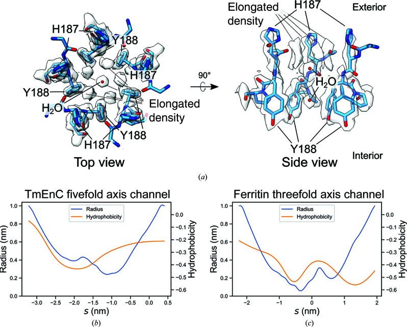
The fivefold axis channel is formed by five histidine residues that point towards the exterior (His187) and five tyrosine residues that point towards the interior (Tyr188) [Fig. 2 ▸(a)]. The five histidine residues on the exterior of the channel are intriguing because multiple histidines can coordinate metals such as iron and nickel, as employed in immobilized metal-affinity chromatography purification of recombinant histidine-tagged proteins (Block et al., 2009 ▸). The density for His187 is not well resolved in the structure, suggesting that it may be conformationally dynamic. Despite this, His187 is not conserved in other encapsulins, leaving the function of this residue unclear. The radius of the fivefold axis channel is about 2.4 Å at the smallest point, which could accommodate Fe2+ (ionic radius of 75 pm) [Fig. 2 ▸(b)] (Shannon, 1976 ▸). Compared with the threefold axis channel of ferritin, which is known to allow iron transport, the fivefold axis channel in TmEnc is larger in radius but is more hydrophobic [Figs. 2 ▸(b) and 2 ▸(c)] (Takahashi & Kuyucak, 2003 ▸; Chandramouli et al., 2016 ▸). Density for two solvent molecules or ions in the fivefold axis channel suggests that iron transport through this channel could be slowed by the hydrophobic sides of the tyrosine side chain, in contrast to the ferritin threefold axis channel, which contains polar residues [Fig. 2 ▸(a)] (Pozzi et al., 2015 ▸). Because it lacks polar residues to reduce the energetic barrier of desolvation, the fivefold axis channel may allow iron transport, but the rate is probably slower than free diffusion. Additionally, above the two molecules in the fivefold axis channel is elongated density with fivefold symmetry due to the symmetric cryo-EM 3D reconstruction. The identity of this elongated density could not be assigned confidently.
Figure 2.
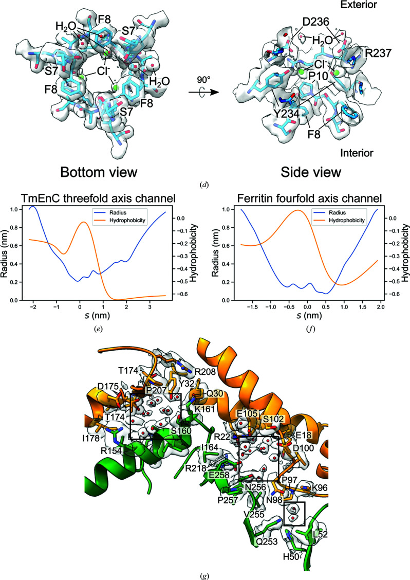
Putative iron-transport channels. (a) The fivefold axis channel consists of His187 and Tyr188. The left image shows the top view looking at the channel from the exterior into the interior of TmEnc and the right image is a cut-through side view. Density corresponding to two solvent molecules or ions is present in the channel. Above the two molecules are fivefold-symmetric elongated densities that could not be identified confidently. (b, c) Radius and hydrophobicity plots using data from CHAP of (b) the TmEnc fivefold axis channel and (c) the ferritin threefold axis channel as a function of probe position s. More negative s values correspond to positions closer to the exterior and vice versa. Hydrophobicity is measured with the Wimley–White scale (Wimley & White, 1996 ▸). Scales for each graph are identical for comparison. (d) The left image shows the threefold axis channel as viewed from the interior of TmEnc looking outwards. The right image is a cut-through side view. The threefold axis channel is formed by residues Asp236, Arg237, Pro10, Tyr234 and Phe8. Density for solvent (red spheres) and chloride ions (green spheres) is present in the channel (Skitchenko et al., 2020 ▸). (e, f) Radius and hydrophobicity plots of (e) the TmEnc threefold axis channel and (f) the ferritin fourfold axis channel as in (b) and (c). (g) Channels in the dimer interface are indicated in boxes, with water molecules shown as red spheres. Residues within 5 Å of the channel and their side-chain densities from the cryo-EM map are shown. Red spheres represent water molecules.
The threefold axis channel is large enough to accommodate an iron ion, with a radius of 2.9 Å at the smallest point, but the channel has three phenylalanine residues (Phe8) on the interior side that seem to preclude rapid iron transport into the encapsulin [Fig. 2 ▸(d)]. The threefold channel of TmEnc has similar hydrophobicity to the fourfold axis channel of ferritin, which is lined with hydrophobic residues and does not transport iron [Figs. 2 ▸(e) and 2 ▸(f)] (Takahashi & Kuyucak, 2003 ▸). Density for solvent and ion molecules is present in the threefold axis channel of TmEnc, which may be trapped and prevented from entering the interior of the compartment by the hydrophobic Phe8 residues. Therefore, the structure of the threefold axis channel suggests that it either blocks or only allows a slow rate of iron transport. Unlike the fivefold and threefold channels, the channels between a dimer consist only of hydrophilic residues and are not occluded by hydrophobic areas [Fig. 2 ▸(g)]. Two of the channels have a radius of 2.0 Å and the last has a radius of 1.2 Å, which are plausible sizes for iron-ion transport when compared with the 0.6 Å radius of the ferritin threefold axis channel. Taken together, we hypothesize that the fivefold axis channel and the dimer channels allow the transport of iron, but the threefold axis channel is unlikely to be permeable to iron.
3.3. Flavin ligand
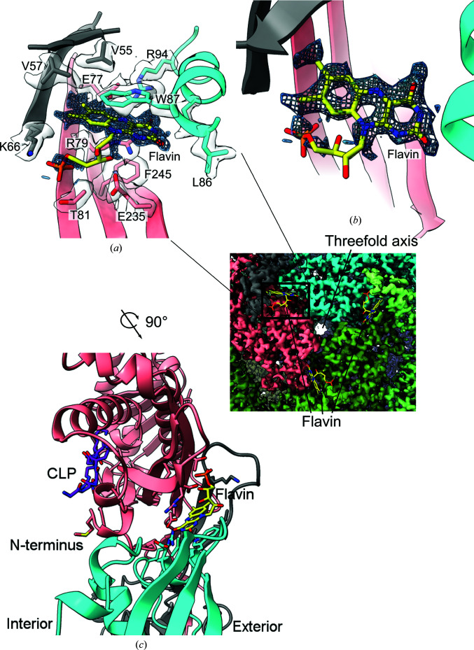
Inspection of density on the exterior of TmEnc surrounding the threefold axis revealed a tricyclic ring consistent with a flavin ligand [Figs. 3 ▸(a) and 3 ▸(b)]. This flavin is the first small-molecule ligand to be seen in an encapsulin structure and explains the yellow color of purified TmEnc. The UV–Vis spectrum of TmEnc has peaks at 360 and 450 nm, consistent with the spectra of flavins [Supplementary Fig. S1(c)] (Schwinn et al., 2020 ▸). Although the exact identity of the flavin ligand is ambiguous, flavin mononucleotide (FMN) seems to be consistent with the density and has been modeled into the structure. The flavin-binding site consists of residues of three different subunits, but these three subunits are distinct from the trimer that forms the threefold axis [Fig. 3 ▸(a), inset]. The structure was entered into the CSM-lig web server, which predicted a −log10(K d|K i) affinity of 13.1, which is higher than those of FMN-binding fluorescent protein and FMN-binding protein from Desulfovibrio vulgaris (predicted affinities of 10.1 and 9.6, respectively; Pires & Ascher, 2016 ▸; Möglich & Moffat, 2007 ▸; Suto et al., 2000 ▸). This high affinity is also shown by the retention of the flavin ligand during the purification of TmEnc from the E. coli lysate through the 80°C incubation step and into the final cryo-EM sample (see Section 2). Trp87 forms an aromatic stacking interaction with the flavin and is likely to be important for flavin binding [Fig. 3 ▸(a)]. Using an HMMER search for 150 homologous proteins, the ConSurf web server showed that Trp87 was 72% conserved (Ashkenazy et al., 2016 ▸). When the TmEnc sequence was aligned with its broader protein family Linocin_M18 (Pfam PF04454_rp55), Trp is only found at this position in 10% of sequences. Instead, the most common amino acids at this position are Arg, Glu and Ala, which appear in 26%, 17% and 13% of sequences, respectively. Using the mCSM-lig web server to predict the change in binding affinity with mutation, W87R, W87E and W87A are all predicted to be destabilizing mutations for flavin binding relative to Trp (Pires et al., 2016 ▸). The lack of conservation of Trp87 in the broader encapsulin alignment suggests that flavin binding is conserved in closely related encapsulins but may not be conserved in more distantly related encapsulins, perhaps those with different types of cargo proteins and functions. For example, the cargo protein of the MhEnc encapsulin is a DyP protein and does not have Trp87, and appears not to have a flavin-binding site (Lončar et al., 2020 ▸). Another interesting aspect of the flavin-binding site is that it is directly opposite the cargo-loading peptide-binding site on the interior of TmEnc, although the significance of this placement is not clear [Fig. 3 ▸(b)]. Although the cryo-EM structure of TmEnc revealed this high-affinity flavin-binding site, the role of the flavin in the context of the function of encapsulin needs further investigation.
Figure 3.
(a) Density for a tricyclic ligand was found in the cryo-EM map, consistent with flavin mononucleotide (FMN). Amino acids within 5 Å that form the binding pocket are labeled. Only density for FMN and the amino-acid side chains is shown for clarity. Different colors correspond to different subunits. The inset shows three flavin-binding sites on the exterior of TmEnc around the threefold axis. (b) An overhead view of the flavin density and the modeled FMN with the surrounding amino acids hidden. (c) The view from (a) is rotated 90° clockwise along the z axis pointing out of the page and shows that the flavin-binding pocket on the exterior is on the opposite side to the cargo-loading peptide (CLP) and N-terminus in the interior of TmEnc. The cargo protein is not present in the cryo-EM structure, so the CLP from the crystal structure was aligned and placed in the cryo-EM structure. The distance between the centroids of the FMN and CLP is 21.4 Å, which is slightly less than the thickness of the TmEnc compartment.
4. Discussion
The high-resolution structure of TmEnc presented here allows detailed inspection of the structural origins of thermostability, putative iron-transport channels and a newly discovered flavin-binding site. TmEnc contains many of the patterns associated with thermostable proteins versus their mesophilic counterparts, including more charged and hydrophobic residues, many ion pairs and shorter, more structured N- and C-termini. The increased thermostability of TmEnc relative to other encapsulins could be a result of additional hydrophobic and aromatic interactions in the E-loop interface. A cation–π intermolecular interaction seems to be a major contributor to the stability of this interface. This finding has implications for engineering protein–protein interfaces, as it suggests that complementary aromatic–aromatic stacking and cation–π interactions allow more stable interfaces beyond what is possible with just ionic, polar and hydrophobic interactions.
TmEnc natively encapsulates a ferritin-like-protein that stores iron, and a major question is how iron is transported from the exterior to the interior of the compartment. TmEnc has several channels that may allow iron transport: the fivefold axis channel, the threefold axis channel and several channels in the dimer interface. The fivefold axis channel is more likely to transport iron than the threefold axis channel because it is less hydrophobic. Nonetheless, the bottom of the fivefold axis channel consists of the hydrophobic sides of tyrosine side chains, suggesting that iron may not be able to freely diffuse through. This is consistent with a report that the encapsulated ferritin-like protein has an entry site that prevents rapid passage to the ferroxidase center (Piergentili et al., 2020 ▸). Iron transport may be regulated such that iron ions are conducted along a defined pathway in TmEnc towards the ferritin-like protein, as opposed to random diffusion.
This structure provides the first direct evidence for a small-molecule ligand-binding site in an encapsulin nanocompartment. This flavin ligand raises the possibility that the encapsulin shell itself could directly be involved in regulating iron storage and release, instead of just merely providing a compartmental function. For comparison, the addition of free flavins to ferritin is known to promote the release of iron (Satoh et al., 2019 ▸). This observation suggests that one possible role of the flavin ligand is to serve as an electron sink for iron oxidation or as a donor for iron reduction. This behavior may also explain how encapsulins with an Flp cargo help cells to resist oxidative stress, as flavins are known to be free-radical scavengers (McHugh et al., 2014 ▸; Sinha et al., 2020 ▸). Because T. maritima can use iron(III) as a terminal electron acceptor in metabolism, TmEnc and its associated Flp may be involved in recycling iron(II) back to iron(III); therefore, TmEnc could be intimately involved in the energy metabolism of T. maritima (Vargas et al., 1998 ▸). The identification of the flavin ligand in TmEnc lays the groundwork for a biochemical investigation of these newly hypothesized roles for Flp-containing encapsulin proteins.
This data set was collected in super-resolution mode with a Gatan K3 detector, which enabled the reconstruction to surpass the physical Nyquist limit (2.0 Å resolution, compared with the 2.158 Å Nyquist limit from a 1.079 Å physical pixel size; Feathers et al., 2019 ▸). Because the resolution is beyond the physical Nyquist limit, the resolution of this data set is most probably limited by the pixel size of the micrographs and not by the sample, as is the case for most macromolecules in cryo-EM. If another data set was collected at higher magnification, the resolution of TmEnc could surpass 2 Å (Cheng et al., 2015 ▸). To test the robustness of TmEnc in cryo-EM, random particle subsets ranging from 91 to 185 459 particles were refined in RELION. Only 363 particles were needed to reach <3 Å, 5796 for <2.5 Å and 46 365 particles for 2.11 Å resolution, indicating a high degree of homogeneity (Supplementary Fig. S2). Also, the resulting Henderson–Rosenthal plot has a relatively low B factor of 51 Å2 (Rosenthal & Henderson, 2003 ▸). Due to the ease of purification, in vitro stability, favorable cryo-EM behavior and high symmetry, we propose that TmEnc could be used as a model protein to test the resolution limits of cryo-EM structure determination.
5. Data availability
The cryo-EM structure of TmEnc has been deposited in the Protein Data Bank as entry 7kq5. The cryo-EM density maps can be accessed from the Electron Microscopy Data Bank as entry EMD-22992. Raw movie files are available from the Electron Microscopy Public Image Archive as entry EMPIAR-10674.
Supplementary Material
PDB reference: T. maritima encapsulin, 7kq5
EMDB reference: T. maritima encapsulin, EMD-22992
Supplementary Figures and Table. DOI: 10.1107/S2052252521001949/pw5017sup1.pdf
Acknowledgments
Cryo-EM data collection was supported by NIH grant U24GM129547 and was performed at the PNCC at OHSU with the assistance of Theo Humphreys and accessed through EMSL (grid.436923.9), a DOE Office of Science User Facility sponsored by the Office of Biological and Environmental Research. The authors acknowledge the use of facilities and instrumentation at the UC Irvine Materials Research Institute (IMRI), which is supported in part by the National Science Foundation through the UC Irvine Materials Research Science and Engineering Center (DMR-2011967).
Funding Statement
This work was funded by National Institutes of Health, National Institute of General Medical Sciences grants 1R01GM123275 and 2T32GM007240.
References
- Afonine, P. V., Poon, B. K., Read, R. J., Sobolev, O. V., Terwilliger, T. C., Urzhumtsev, A. & Adams, P. D. (2018). Acta Cryst. D74, 531–544. [DOI] [PMC free article] [PubMed]
- Ashkenazy, H., Abadi, S., Martz, E., Chay, O., Mayrose, I., Pupko, T. & Ben-Tal, N. (2016). Nucleic Acids Res. 44, W344–W350. [DOI] [PMC free article] [PubMed]
- Block, H., Maertens, B., Spriestersbach, A., Brinker, N., Kubicek, J., Fabis, R., Labahn, J. & Schäfer, F. (2009). Methods Enzymol. 463, 439–473. [DOI] [PubMed]
- Cassidy-Amstutz, C., Oltrogge, L., Going, C. C., Lee, A., Teng, P., Quintanilla, D., East-Seletsky, A., Williams, E. R. & Savage, D. F. (2016). Biochemistry, 55, 3461–3468. [DOI] [PubMed]
- Chandramouli, B., Bernacchioni, C., Di Maio, D., Turano, P. & Brancato, G. (2016). J. Biol. Chem. 291, 25617–25628. [DOI] [PMC free article] [PubMed]
- Cheng, Y., Grigorieff, N., Penczek, P. A. & Walz, T. (2015). Cell, 161, 438–449. [DOI] [PMC free article] [PubMed]
- Emsley, P., Lohkamp, B., Scott, W. G. & Cowtan, K. (2010). Acta Cryst. D66, 486–501. [DOI] [PMC free article] [PubMed]
- Feathers, J. R., Spoth, K. A. & Fromme, J. C. (2019). bioRxiv, 675397.
- Giessen, T. W., Orlando, B. J., Verdegaal, A. A., Chambers, M. G., Gardener, J., Bell, D. C., Birrane, G., Liao, M. & Silver, P. A. (2019). eLife, 8, e46070. [DOI] [PMC free article] [PubMed]
- Giessen, T. W. & Silver, P. A. (2017). Nat. Microbiol. 2, 17029. [DOI] [PubMed]
- Jones, J. A. & Giessen, T. W. (2021). Biotechnol. Bioeng. 118, 491–505. [DOI] [PMC free article] [PubMed]
- Jubb, H. C., Higueruelo, A. P., Ochoa-Montaño, B., Pitt, W. R., Ascher, D. B. & Blundell, T. L. (2017). J. Mol. Biol. 429, 365–371. [DOI] [PMC free article] [PubMed]
- Klesse, G., Rao, S., Sansom, M. S. P. & Tucker, S. J. (2019). J. Mol. Biol. 431, 3353–3365. [DOI] [PMC free article] [PubMed]
- Liebschner, D., Afonine, P. V., Baker, M. L., Bunkóczi, G., Chen, V. B., Croll, T. I., Hintze, B., Hung, L.-W., Jain, S., McCoy, A. J., Moriarty, N. W., Oeffner, R. D., Poon, B. K., Prisant, M. G., Read, R. J., Richardson, J. S., Richardson, D. C., Sammito, M. D., Sobolev, O. V., Stockwell, D. H., Terwilliger, T. C., Urzhumtsev, A. G., Videau, L. L., Williams, C. J. & Adams, P. D. (2019). Acta Cryst. D75, 861–877.
- Lončar, N., Rozeboom, H. J., Franken, L. E., Stuart, M. C. A. & Fraaije, M. W. (2020). Biochem. Biophys. Res. Commun. 529, 548–553. [DOI] [PubMed]
- McHugh, C. A., Fontana, J., Nemecek, D., Cheng, N., Aksyuk, A. A., Heymann, J. B., Winkler, D. C., Lam, A. S., Wall, J. S., Steven, A. C. & Hoiczyk, E. (2014). EMBO J. 33, 1896–1911. [DOI] [PMC free article] [PubMed]
- Möglich, A. & Moffat, K. (2007). J. Mol. Biol. 373, 112–126. [DOI] [PMC free article] [PubMed]
- Morin, A., Eisenbraun, B., Key, J., Sanschagrin, P. C., Timony, M. A., Ottaviano, M. & Sliz, P. (2013). eLife, 2, e01456. [DOI] [PMC free article] [PubMed]
- Nichols, R., LaFrance, B., Phillips, N., Oltrogge, L., Valentin-Alvarado, L., Bischoff, A., Nogales, E. & Savage, D. (2020). bioRxiv, 2020.05.24.113720.
- Nichols, R. J., Cassidy-Amstutz, C., Chaijarasphong, T. & Savage, D. F. (2017). Crit. Rev. Biochem. Mol. Biol. 52, 583–594. [DOI] [PubMed]
- Pettersen, E. F., Goddard, T. D., Huang, C. C., Couch, G. S., Greenblatt, D. M., Meng, E. C. & Ferrin, T. E. (2004). J. Comput. Chem. 25, 1605–1612. [DOI] [PubMed]
- Piergentili, C., Ross, J., He, D., Gallagher, K. J., Stanley, W. A., Adam, L., Mackay, C. L., Baslé, A., Waldron, K. J., Clarke, D. J. & Marles-Wright, J. (2020). J. Biol. Chem. 295, 15511–15526. [DOI] [PMC free article] [PubMed]
- Pires, D. E. V. & Ascher, D. B. (2016). Nucleic Acids Res. 44, W557–W561. [DOI] [PMC free article] [PubMed]
- Pires, D. E. V., Blundell, T. L. & Ascher, D. B. (2016). Sci. Rep. 6, 29575. [DOI] [PMC free article] [PubMed]
- Pozzi, C., Di Pisa, F., Bernacchioni, C., Ciambellotti, S., Turano, P. & Mangani, S. (2015). Acta Cryst. D71, 1909–1920. [DOI] [PubMed]
- Rohou, A. & Grigorieff, N. (2015). J. Struct. Biol. 192, 216–221. [DOI] [PMC free article] [PubMed]
- Rosenthal, P. B. & Henderson, R. (2003). J. Mol. Biol. 333, 721–745. [DOI] [PubMed]
- Satoh, J., Kimata, S., Nakamoto, S., Ishii, T., Tanaka, E., Yumoto, S., Takeda, K., Yoshimura, E., Kanesaki, Y., Ishige, T., Tanaka, K., Abe, A., Kawasaki, S. & Niimura, Y. (2019). J. Gen. Appl. Microbiol. 65, 308–315. [DOI] [PubMed]
- Scheres, S. H. W. (2012). J. Struct. Biol. 180, 519–530. [DOI] [PMC free article] [PubMed]
- Schwinn, K., Ferré, N. & Huix-Rotllant, M. (2020). Phys. Chem. Chem. Phys. 22, 12447–12455. [DOI] [PubMed]
- Shannon, R. D. (1976). Acta Cryst. A32, 751–767.
- Sinha, T., Naash, M. I. & Al-Ubaidi, M. R. (2020). Front. Cell. Dev. Biol. 8, 861. [DOI] [PMC free article] [PubMed]
- Skitchenko, R. K., Usoltsev, D., Uspenskaya, M., Kajava, A. V. & Guskov, A. (2020). Bioinformatics, 36, 3064–3071. [DOI] [PMC free article] [PubMed]
- Suto, K., Kawagoe, K., Shibata, N., Morimoto, Y., Higuchi, Y., Kitamura, M., Nakaya, T. & Yasuoka, N. (2000). Acta Cryst. D56, 368–371. [DOI] [PubMed]
- Sutter, M., Boehringer, D., Gutmann, S., Günther, S., Prangishvili, D., Loessner, M. J., Stetter, K. O., Weber-Ban, E. & Ban, N. (2008). Nat. Struct. Mol. Biol. 15, 939–947. [DOI] [PubMed]
- Takahashi, T. & Kuyucak, S. (2003). Biophys. J. 84, 2256–2263. [DOI] [PMC free article] [PubMed]
- Terwilliger, T. C., Ludtke, S. J., Read, R. J., Adams, P. D. & Afonine, P. V. (2020). Nat. Methods, 17, 923–927. [DOI] [PMC free article] [PubMed]
- Tracey, J. C., Coronado, M., Giessen, T. W., Lau, M. C. Y., Silver, P. A. & Ward, B. B. (2019). Sci. Rep. 9, 20122. [DOI] [PMC free article] [PubMed]
- Vargas, M., Kashefi, K., Blunt-Harris, E. L. & Lovley, D. R. (1998). Nature, 395, 65–67. [DOI] [PubMed]
- Vieille, C. & Zeikus, G. J. (2001). Microbiol. Mol. Biol. Rev. 65, 1–43. [DOI] [PMC free article] [PubMed]
- Wagner, T., Merino, F., Stabrin, M., Moriya, T., Antoni, C., Apelbaum, A., Hagel, P., Sitsel, O., Raisch, T., Prumbaum, D., Quentin, D., Roderer, D., Tacke, S., Siebolds, B., Schubert, E., Shaikh, T. R., Lill, P., Gatsogiannis, C. & Raunser, S. (2019). Commun. Biol. 2, 218. [DOI] [PMC free article] [PubMed]
- Weng, G., Wang, E., Wang, Z., Liu, H., Zhu, F., Li, D. & Hou, T. (2019). Nucleic Acids Res. 47, W322–W330. [DOI] [PMC free article] [PubMed]
- Williams, E. M., Jung, S. M., Coffman, J. L. & Lutz, S. (2018). ACS Synth. Biol. 7, 2514–2517. [DOI] [PubMed]
- Wimley, W. C. & White, S. H. (1996). Nat. Struct. Mol. Biol. 3, 842–848. [DOI] [PubMed]
- Zivanov, J., Nakane, T. & Scheres, S. H. W. (2020). IUCrJ, 7, 253–267. [DOI] [PMC free article] [PubMed]
Associated Data
This section collects any data citations, data availability statements, or supplementary materials included in this article.
Supplementary Materials
PDB reference: T. maritima encapsulin, 7kq5
EMDB reference: T. maritima encapsulin, EMD-22992
Supplementary Figures and Table. DOI: 10.1107/S2052252521001949/pw5017sup1.pdf
Data Availability Statement
The cryo-EM structure of TmEnc has been deposited in the Protein Data Bank as entry 7kq5. The cryo-EM density maps can be accessed from the Electron Microscopy Data Bank as entry EMD-22992. Raw movie files are available from the Electron Microscopy Public Image Archive as entry EMPIAR-10674.


