Abstract
Objectives
To investigate the efficiency and movement pattern of upper arch expansion using Invisalign aligners. The correlation between the amount of designed expansion and the efficiency of bodily expansion was evaluated, as were the initial molar torque and efficiency of bodily expansion.
Materials and Methods
Twenty Chinese adult patients who underwent arch expansion with Invisalign aligners were included in this study. Records of pretreatment (T0 stage) and immediately after completing the expansion phase (T1 stage) were collected, including digital models and cone-beam computed tomography. Dolphin 3D, Geomagic Studio 12.0, and Meazure software were employed to measure data and calculate differences between the expected and actual outcomes.
Results
There were significant differences between the expected and actual expansion amounts (P< .05). The average expansion efficiencies of the upper canine crown, first premolar crown, second premolar crown, and first molar crown were 79.75 ± 15.23%, 76.1 ± 18.32%, 73.27 ± 19.91%, and 68.31 ± 24.41%, respectively. The average efficiency of bodily expansion movement for the maxillary first molar was 36.35 ± 29.32%. Negative correlations were found between preset expansion amounts and the efficiency of bodily expansion movement (P < .05), and between initial maxillary first molar torque and efficiency of bodily expansion movement (P < .05).
Conclusions
Aligners could increase the arch width, but expansion was achieved by tipping movement. The evaluation of initial position and preset of sufficient root-buccal torque of posterior teeth were necessary due to the lower efficiency of bodily buccal expansion by the Invisalign system.
Keywords: Clear treatment, Expansion, Efficiency, Three-dimensional
INTRODUCTION
In 1946, Kesling proposed to manufacture a series of removable appliances called “aligners.” The underlying concept was to move teeth in a series of planned individual stages using positioners fabricated by thermoplastic material molding technology.1 In 1997, the company Align Technology converted Kesling's idea into a feasible treatment approach: a series of clear aligners, combining appliance production with computer-aided design/manufacturing (CAD/CAM) stereolithographic technology. Since its introduction as an esthetic alternative to fixed labial braces, the Invisalign appliance has evolved. Its unique advantages over traditional appliances include esthetics, comfort, removal for better hygiene, shorter appointment times, and 3D control of tooth movement.2,3 However, a portion of Invisalign patients require mid-course correction, case refinement, or conversion to fixed appliances before the end of treatment,4 so some doubts remain among clinicians about the efficiency and accuracy of teeth movement with the appliance.
Dental crowding is a leading reason that people seek orthodontic treatment. Expansion of a compressed arch as a method of resolving crowding can increase arch length, thus providing more space for tooth alignment. It can also improve the transverse dimension of the smile or correct dentoalveolar posterior crossbites.5,6 Some literature7,8 has stated that buccal expansion can be achieved by Invisalign to relieve dental crowding, as an alternative to interproximal reduction or to modify the arch form. Malik et al.9 reported in 2013 that expansion using Invisalign is indicated when having to resolve 1–5 mm of crowding and is also recommended for blocked out teeth.
However, there are only a limited number of studies10–14 on the efficiency of tooth movement with Invisalign, especially in transverse expansion. This lack of research has made it difficult for clinicians to characterize transverse expansion efficiency with Invisalign objectively. In addition, in existing published papers, the method used to quantify predictability of expansion movement is only by model measurements for the crown, without assessment of root movement.
Therefore, the primary objectives of this study were to quantify the efficiency of arch expansion using the Invisalign system in patients, to investigate the movement patterns by comparing actual expansion outcomes of crown and root with virtual planned expansion in ClinCheck software (Align Technology, Inc., Santa Clara, CA , USA), and to ascertain whether the preset expansion amount and initial molar torque correlated with the efficiency of bodily expansion movement.
MATERIALS AND METHODS
Subjects and Study Design
All patients signed informed consent to participate in this study, which was approved by the ethics committee on human research at the Stomatological Hospital of Shandong University. The mixed-gender study sample of adult patients treated with arch expansion using Invisalign aligners was recruited from the clientele of a single board-certificated orthodontist at the Stomatological Hospital of Shandong University.
All subjects met the following inclusion criteria: (1) age between 20 and 45 years old; (2) permanent dentition with second molars fully erupted; (3) good tooth contour, with sufficient height of clinical crowns; (4) a case in which arch expansion with Invisalign system had been planned; and (5) good compliance during treatment as assessed by the practitioner. The exclusion criteria were as follows: (1) systematic disease or drug-taking history affecting tooth movement; (2) orofacial malformation syndromes; (3) periodontal disease; (4) signs and/or symptoms of temporomandibular disorders (TMDs); (5) missing teeth (except for third molars); (6) extraction cases; (7) auxiliary treatment during arch expansion stage such as crossbite elastics; and (8) posterior interproximal reduction.
The final sample consisted of 20 Chinese adult patients including five males and 15 females (28.5 ± 6.3 years old). The ClinCheck for each case was planned consistently with the same expansion treatment procedure: arch expansion of 0.15 ± 0.5 mm per stage was performed for the upper arch with the first N aligners. N + 1 and N + 2 aligners were planned as passive aligners.
All maxillary first molars of participants were distributed into three groups according to the preset unilateral expansion amount for each first molar: (1) Group A (n = 14): expansion amount ≤1 mm; (2) Group B (n = 20): expansion amount between 1 and 2 mm; (3) Group C (n = 6): expansion amount was 2 mm. They were further divided into groups according to baseline torque as measured by a root vector analysis program using Dolphin software (Dolphin Imaging, Chatsworth, CA):15 (1) Group D (n = 11): faciolingual inclination ≤-2°; (2) Group E (n = 16): faciolingual inclination between -2° and 2°; (3) Group F (n = 13): faciolingual inclination ≥2° and <6°.
Data Collection
Pre-expansion (T0) and post-expansion (immediately after finishing N+2 aligner) (T1) records for each participant were collected, consisting of the following:
(1) Digital models from stereolithography (.stl) files created from plaster models converted from polyvinyl siloxane (PVS) impressions by Optimics scanner (Activity 880, Smart Optics, Germany). (2) T0 digital dentition models in ClinCheck provided as .stl files from Align Technology (Santa Clara, CA , USA). (3) Cone-beam computed tomography (CBCT) records as digital imaging and communications in medicine (DICOM) files, taken by GALILEOS Viewer (Sirona, Germany), in the participant's natural head position (NHP).
Measurements
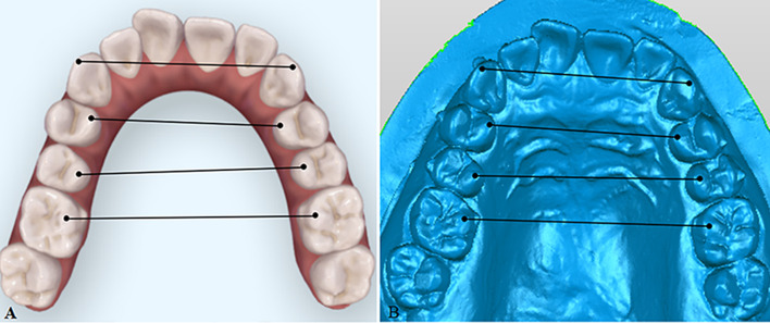
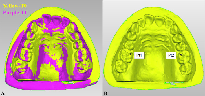
The .stl files of digital models were uploaded into Geomagic Studio 12.0 software (Research Triangle Park, NC) for measurement. The Meazure application was performed, similarly to that proposed by Solano-Mendoza,14 to analyze the virtual treatment goal in ClinCheck. Interdental width linear measurements at T0 and T1 stages were recorded, including intercanine width from the cusp tips, interpremolar widths from the palatal cusp tips of upper first premolars and second premolars, and intermolar width from the mesiolingual cusp tips of the upper first molars (Figure 1). In addition, T0 and T1 digital models were oriented using a consistent reference coordinate system (x, y, z axes) that described tooth position in three dimensions, and superimposed over the palatal rugae region as reference points in Geomagic studio 12 (Figure 2A). The point coordinates (x, y, z) for the mesiolingual cusp tip of the upper first molars were then determined exactly (Figure 2B). In doing so, the Δx was calculated to describe clinically-achieved crown expansion movements of each upper first molar unilaterally. In cases of wear and restored facets, the estimated cusp tip was used.
Figure 1.
Interdental width linear measurements. (A) Measurements in ClinCheck; (B) Measurements in Geomagic Studio Software.
Figure 2.
Analysis of expansion movement for individual maxillary first molars. (A) Superimposition of digital models at T0 and T1; (B) Determination of coordinate point for the mesiolingual cusp tip on the maxillary first molar.
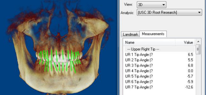
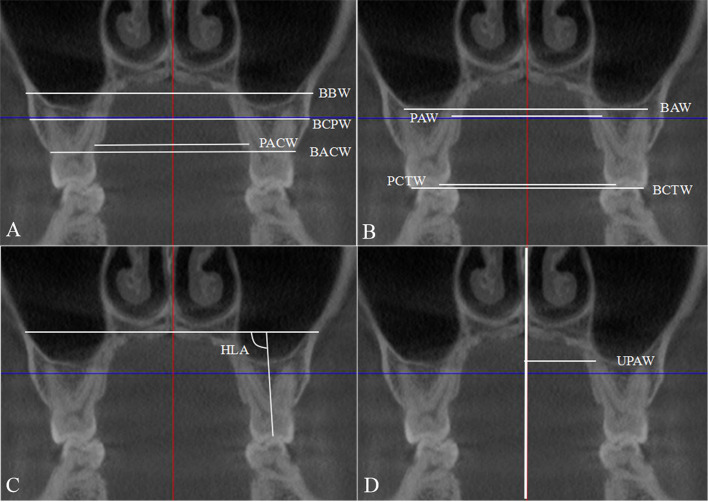
The DICOM data of the CBCTs were imported, and volumetric images were obtained using Dolphin 3D software. Standard orientation of the craniofacial structure was established using the Frankfort horizontal line as the x-axis, transporionic line as the y-axis, and a midsagittal line passing through the nasion point as the z-axis. With the standard orientation, pretreatment faciolingual inclination of each upper first molar (the angle formed by the projection of its long axis on the faciolingual plane and the line of intersection between the faciolingual and mesiodistal planes) was measured by a root vector analysis software program15 (Figure 3). To assess the achieved buccal movement of the upper first molars in three dimensions, the transverse liner and angular measurements for T0 and T1 were recorded in the coronal view after reorientation (Figure 4). The expansion efficiency for crown was calculated as a percentage:
 |
Figure 3.
Measurements of initial torque for the maxillary first molar using root vector analysis.
Figure 4.
CBCT measurements. (A) Maxillary basal bone width and alveolar bone width. (B) Maxillary dental arch width; (C) Maxillary first molar tipping; (D) Maxillary unilateral dental arch width. CBCT indicates cone-beam computed tomography.
In addition, efficiency of bodily expansion movement was calculated as:
 |
Statistical Analysis
All measurements were performed by one investigator. A randomly selected 20% of the sample were remeasured after a minimum interval of 15 days and analyzed by intraclass correlation coefficient (ICC) to assess reliability. A value of 0.9 or higher was considered to signify reliability. Sample normality was tested by the Shapiro-Wilk test. The paired-sample t test was used to examine the accuracy of transverse dimension variable measurements between the T0 ClinCheck and T0 digital models, as well as to compare designed and achieved expansion movements. The degree of correlation between the preset expansion amount, as well as the initial maxillary first molar torque, and bodily expansion movement efficiency was detected by Spearman correlation analysis. The differences in efficiency of bodily expansion movement within the A, B, and C groups and the D, E, and F groups were determined using a nonparametric test. SPSS (Statistical Product and Service Solutions), version 19.0 (IBM Corp, Armonk, NY) was used to analyze the data. A P value <.05 was considered statistically significant.
RESULTS
The ICCs ranged between 0.90 and 0.95, showing a high level of intraoperator reliability. There was no significant difference (P > .05) in the transverse dimension variables between the T0 ClinCheck and digital models, indicating good accuracy of measurement for ClinCheck models using the Meazure application.
Table 1 shows that there was a significant difference (P < .05) between the amount of designed expansion and the amount of achieved expansion for the canine, first premolar, second premolar, and first molar. The efficiencies of crown expansion movement for the canine, first premolar, second premolar, and first molar were 79.75%, 76.10%, 73.27%, and 68.31%, respectively.
Table 1.
Crown Movement Efficiency and Comparison Between Designed and Achieved Expansion
| Designed |
Achieved |
Difference |
Efficiency |
|||||||
| Tooth Type |
N |
Mean |
SD |
Mean |
SD |
Mean |
SD |
P Value |
Mean |
SD |
| Canine | 20 | 1.78 | 0.63 | 1.44 | 0.60 | 0.33 | 0.26 | .009** | 79.75 | 15.23 |
| First premolar | 20 | 2.27 | 0.86 | 1.74 | 0.84 | 0.53 | 0.45 | .005** | 76.10 | 18.32 |
| Second premolar | 20 | 2.12 | 1.03 | 1.57 | 0.96 | 0.65 | 0.76 | .017* | 73.27 | 19.91 |
| First molar | 20 | 2.32 | 1.07 | 1.58 | 0.97 | 0.74 | 0.73 | .007** | 68.31 | 24.41 |
P = .05; ** P = .01.
When comparing the effect of skeletal arch expansion in CBCT (Table 2), no significant change (P > .05) was observed in maxillary basal bone width. Regarding the maxillary alveolar bone width, the buccal alveolar crest width (BACW) and lingual alveolar crest width (LACW) increased significantly by 0.87 and 0.75 mm, respectively (P < .05), while the maxillary alveolar bone width at the level of the most convex point of the buccal alveolar ridge crest (BCPW) showed no significant difference between T0 and T1 (P > .05). The buccolingual inclination of the maxillary molar (HLA) significantly increased by 2.07° after arch expansion.
Table 2.
Comparison of Maxillary Linear Measurements Between T0 and T1 in CBCTa,b
| T0 |
T1 |
Difference |
||||||
| Variable |
N |
Mean |
SD |
Mean |
SD |
Mean |
SD |
P Value |
| Maxillary basal bone width | ||||||||
| BBW | 20 | 65.84 | 3.72 | 65.88 | 3.74 | 0.04 | 0.18 | .519 |
| Maxillary alveolar bone width | ||||||||
| BCPW | 20 | 65.00 | 3.91 | 65.09 | 3.99 | 0.09 | 0.28 | .314 |
| BACW | 20 | 58.40 | 3.20 | 59.27 | 0.94 | 0.87 | 0.63 | .001*** |
| PACW | 20 | 37.45 | 3.41 | 38.20 | 3.00 | 0.75 | 0.80 | .011* |
| Dental arch width | ||||||||
| BAW | 20 | 58.45 | 3.44 | 59.04 | 3.43 | 0.59 | 0.61 | .009** |
| BCTW | 20 | 56.31 | 4.03 | 57.78 | 3.54 | 1.47 | 0.98 | .001*** |
| PAW | 20 | 38.55 | 3.99 | 39.08 | 3.78 | 0.54 | 0.51 | .006** |
| PCTW | 20 | 42.15 | 3.82 | 43.64 | 3.60 | 1.48 | 1.02 | .001*** |
| UPAW | 40 | 19.01 | 1.97 | 19.30 | 2.01 | 0.29 | 0.36 | .010** |
| Maxillary first molar tipping | ||||||||
| HLA | 40 | 90.41 | 6.81 | 92.49 | 5.72 | 2.07 | 3.27 | .034* |
P = .05; ** P = .01; *** P = .001.
BBW, maxillary width between the most concave points of bilateral buccal basal bone close to the apex for the maxillary first molar; BCPW, maxillary alveolar bone width at the level of the most convex point of buccal aspect for alveolar bone; BACW, maxillary alveolar bone width at the level of buccal alveolar ridge crest; PACW, maxillary alveolar bone width at the level of palatal alveolar ridge crest. BAW, dental arch width measured at the buccal apex; BCTW, dental arch width measured at the buccal cusp tip; PAW, dental arch width measured at palatal apex; PCTW, dental arch width measured at the palatal cusp tip; UPAW, unilateral dental width measured relative to the midsagittal plane at the palatal apex; HLA, the angle between the horizontal reference line parallel to patient's palatal plane and the long axis passing the root furcation and central fossa of maxillary first molar.
CBCT indicates cone-beam computed tomography.
For the maxillary first molar, the amounts of expansion achieved for the crown and root were 1.06 ± 0.51 mm and 0.29 ± 0.36 mm, respectively, and the efficiency of bodily expansion movement was 36.35 ± 29.32% (Table 3).
Table 3.
Amount of Achieved Expansion Movement and Efficiency of Bodily Expansion Movement for Unilateral Maxillary First Molar
| Variable |
Mean |
SD |
Maximum |
Minimum |
| Achieved expansion movement | ||||
| Crown | 1.06 | 0.51 | 2.07 | 0.51 |
| Root | 0.29 | 0.36 | 1 | -0.7 |
| Efficiency of bodily expansion movement | 36.35 | 29.32 | 106.54 | -35 |
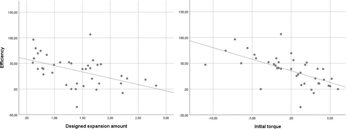
The preset expansion amount and initial maxillary first molar torque were significantly negatively correlated with efficiency of bodily expansion movement (P < .05). The correlation coefficients were −0.543 and −0.690, respectively (Table 4 and Figure 5).
Table 4.
Spearman Correlation Analysis Between Bodily Expansion Movement Efficiency and Designed Expansion Amount as well as Initial Torque for the Maxillary First Molar
| Variable |
N |
Spearman Correlation |
P Value |
| Designed expansion amount vs bodily expansion efficiency | 40 | −0.543 | .000* |
| Initial torque vs bodily expansion efficiency | 40 | −0.690 | .000* |
P = .0001.
Figure 5.
Results of Spearman correlation analysis. (A) Designed expansion amount and efficiency of bodily expansion movement; (B) Initial maxillary first molar torque and efficiency of bodily expansion movement.
As seen in Table 5, the efficiency of group A was 56.03 ± 19.79%, of group B was 29.76 ± 30.77% and of group C was 12.40 ± 11.95%. There was a significant difference in average bodily expansion efficiency between groups A and B (P < .05) and between groups A and C (P < .05), while no significant difference was found between groups B and C (P > .05). The efficiency in group D was 56.37 ± 24.68%, in group E was 37.03 ± 30.87%, and in group F was 14.42 ± 18.67%. There were significant differences in average bodily expansion efficiency between each pair (D and E, D and F, E and F) (P < .05).
Table 5.
Efficiency of Bodily Expansion Movement for Each Group
| Group |
N |
Efficiency |
Kruskal-Wallis Test P Value |
Mann-Whitney U-Test P Value |
|
| Mean |
SD |
||||
| A | 14 | 56.03 | 19.79 | .002** | A vs B .005** |
| B | 20 | 29.76 | 30.77 | A vs C .001*** | |
| C | 6 | 12.40 | 11.95 | B vs C .324 | |
| D | 11 | 56.37 | 24.68 | .001*** | D vs E .044* |
| E | 16 | 37.03 | 30.87 | D vs F .000**** | |
| F | 13 | 14.42 | 18.67 | E vs F .012* | |
P = .05; ** P = .01; *** P = .001; **** P = .0001.
DISCUSSION
Understanding the efficiency and effects of arch expansion treatment using Invisalign aligners on skeletal and dental components is paramount for clinicians. However, there are only a few published articles13,14 on expansion efficiency or predictability of clear aligners. Previously, interdental width was only evaluated by 3D digital model measurements for the crown, while the buccal movement for root and skeletal effects were not evaluated. Therefore, 3D digital model measurements and CBCT measurements were integrated in this study to assess the efficiency and movement pattern of upper arch expansion achieved with Invisalign aligners comprehensively and objectively. The correlations with preset expansion amount as well as the initial molar torque and efficiency of bodily expansion movement also were evaluated.
The subjects selected for this study were adults ranging between 25 and 45 years old, thereby ruling out an increase in dental arch width caused by growth and development and eliminating the influence of bone metabolism on the movement of teeth during puberty and perimenopause. Graber et al.16 pointed out that for patients with mild maxillary transverse deficiency or mild crowding, orthodontic archwires and other special devices can be used for dentoalveolar expansion. In the current study, all subjects had dental constriction or skeletal constriction of less than 3 mm. Each subject's periodontal condition was assessed by clinical examination and CBCT analysis at baseline.
It has been reported that the expansion extent with Invisalign should be 2–4 mm.17 Ali et al.7 indicated that the range of dental expansion should be limited to 2–3 mm in each quadrant to reduce the risk of gingival recession. In the current study, the prescribed amount of expansion for each participant was customized based on measurements of the dentition and CBCT to be within a safe range. Each Invisalign aligner can achieve 0.25–0.33 mm tooth movement in 14 days.18 However, Clements et al.19 showed that the Invisalign aligners have the least control in posterior teeth when correcting for the lateral dimension. Therefore, the expansion amount for each aligner was set at 0.15 ± 0.05 mm according to the total expansion amount needed and the baseline periodontal condition.
The results of this study showed that Invisalign aligners can achieve arch expansion, as the expansion efficiencies were 79.75 ± 15.23% at the canine cusp tip, 76.1 ± 18.32% at the first premolar cusp tip, 73.27 ± 19.91% at the second premolar cusp tip, and 68.31 ± 24.41% at the first molar cusp tip. The efficiencies were slightly lower than those found by Houle et al.,13 in which the efficiencies of expansion for the canine, first premolar crown, second premolar, and the first molar were 88.7%, 84.7%, 81%, and 76.6%, respectively. The reason for these differences might be that the evaluation periods in the two studies were different. Due to the hysteresis of Invisalign appliances, the efficiency of expansion at the end of an entire Invisalign treatment sequence may be slightly higher than at the end of expansion treatment. Additionally, making absolute comparisons between the present research and previous studies is difficult due to differences in clinical protocols, treatment plans, and sample sizes, etc.
The efficiency of expansion decreased from the canine to the first molar. This finding was consistent with the results of Houle et al.13 This may be due to differences in root anatomy and cortical bone thickness, the higher occlusal load, and the greater soft tissue resistance from the cheeks in the posterior region. Another possibility is that the mechanical efficiency for delivering effective buccally-directed force by the aligner decreases from anterior to posterior.
Regardless of what type of expansion is desired, buccal tipping of the posterior teeth should be minimized in most cases. Therefore, it is vitally important to investigate efficiency of bodily expansion movement. Thus, in this research, the maxillary first molars were selected for assessing bodily expansion efficiency. The results of this study showed that the bodily expansion efficiency of the maxillary first molar was 36.35 ± 29.32%, and the ratio of the expansion movement between the root and crown was approximately 2:5. The maxillary first molar buccally tipped 2.07 ± 3.27° after expansion. Arch expansion with Invisalign aligners was mainly due to tipping movement, which has important significance for guiding the application of Invisalign aligners. Thus, according to the initial torque of the posterior teeth, an appropriate amount of negative torque in the crown could be preset in ClinCheck to improve bodily expansion efficiency. There was no statistically significant increase in either the width of maxillary basal bone or the width between the most convex points on the buccal aspect of the maxillary alveolar bone (P > .05). Interdental width and width at the buccal and lingual alveolar crests significantly increased (P < .05), indicating that the expansion effect from Invisalign aligners was mainly buccal movement of teeth.
It was previously stated that the difference in predictability among groups was based on the magnitude of planned expansion.14 This study showed, by means of Spearman correlation analysis, that there was a negative correlation between the preset expansion amount and bodily expansion efficiency (P < .05). Therefore, for patients who need a large amount of expansion, clinicians should consider reducing the amount of expansion for each aligner to ensure periodontal health and preset more negative torque to achieve better crown and root control to achieve bodily expansion. The Spearman correlation analysis also showed a negative correlation between the initial torque and bodily expansion efficiency (P < .05). These findings have paramount value for clinical design, suggesting that clinicians should pay close attention to evaluating the initial position of molars. For posterior teeth with a larger buccal inclination, it is necessary to reduce magnitudes of expansion appropriately and preset more negative crown torque, to prevent adverse effects on the occlusion caused by excessive buccal inclination of posterior teeth. Additionally, it indicates that dental compensation for maxillary transverse deficiency has reached a limit for those posterior teeth with large buccal inclinations, and traditional dental expansion treatment should be avoided.
CONCLUSIONS
The Invisalign system can increase arch width by tipping movement of posterior teeth. The efficiency of bodily buccal expansion for maxillary first molars averaged 36.35%.
The preset amount of expansion movement and initial torque are negative correlated with bodily expansion efficiency. Thus, it is necessary to preset sufficient buccal root torque of posterior teeth according to the preset amount of expansion and initial torque.
ACKNOWLEDGMENT
The authors note that they did not receive any specific grants or funding from a commercial firm or other outside entities.
REFERENCES
- 1.Kesling HD. Coordinating the predetermined pattern and tooth positioner with conventional treatment. Am J Orthod Oral Surg. 1946;32(5):285–293. doi: 10.1016/0096-6347(46)90053-1. [DOI] [PubMed] [Google Scholar]
- 2.Azaripour A, Weusmann J, Mahmoodi B, et al. Braces versus Invisalign®: gingival parameters and patients' satisfaction during treatment: a cross-sectional study. BMC Oral Health. 2015;15:69. doi: 10.1186/s12903-015-0060-4. [DOI] [PMC free article] [PubMed] [Google Scholar]
- 3.Shalish M, Cooperkazaz R, Ivgi I, et al. Adult patients adjustability to orthodontic appliances. Part I: a comparison between Labial, Lingual, and Invisalign. Eur J Orthod. 2012;34(6):724–730. doi: 10.1093/ejo/cjr086. [DOI] [PubMed] [Google Scholar]
- 4.Rossini G, Parrini S, Castroflorio T, Deregibus A, Debernardi CL. Efficacy of clear aligners in controlling orthodontic tooth movement: a systematic review. Angle Orthod. 2015;85(5):881–889. doi: 10.2319/061614-436.1. [DOI] [PMC free article] [PubMed] [Google Scholar]
- 5.Krishnan V, Daniel ST, Lazar D, Asok A. Characterization of posed smile by using visual analog scale, smile arc, buccal corridor measures, and modified smile index. Am J Orthod Dentofacial Orthop. 2008;133(4):515–523. doi: 10.1016/j.ajodo.2006.04.046. [DOI] [PubMed] [Google Scholar]
- 6.Giancotti A, Mampieri G. Unilateral canine crossbite correction in adults using the Invisalign method: a case report. Orthodontics (Chic) 2012;13(1):122–127. [PubMed] [Google Scholar]
- 7.Ali S A, Miethke H R. Invisalign, an innovative invisible orthodontic appliance to correct malocclusions: advantages and limitations. Dent Update. 2012;39(4):254–256. doi: 10.12968/denu.2012.39.4.254. 258–260. [DOI] [PubMed] [Google Scholar]
- 8.Vlaskalic V, Boyd R. Orthodontic treatment of a mildly crowded malocclusion using the Invisalign System. Aust Orthod J. 2001;17(1):41–46. [PubMed] [Google Scholar]
- 9.Malik OH, Mcmullin A, Waring DT. Invisible orthodontics part 1: Invisalign. Dent Update. 2013;40(3):203. doi: 10.12968/denu.2013.40.3.203. [DOI] [PubMed] [Google Scholar]
- 10.Kravitz ND, Kusnoto B, Begole E, Obrez A, Agran B. How well does Invisalign work? A prospective clinical study evaluating the efficacy of tooth movement with Invisalign. Am J Orthod Dentofacial Orthop. 2009;135(1):27–35. doi: 10.1016/j.ajodo.2007.05.018. [DOI] [PubMed] [Google Scholar]
- 11.Simon M, Keilig L, Schwarze J, Jung BA, Bourauel C. Treatment outcome and efficacy of an aligner technique–regarding incisor torque, premolar derotation and molar distalization. BMC Oral Health. 2014;14:68. doi: 10.1186/1472-6831-14-68. [DOI] [PMC free article] [PubMed] [Google Scholar]
- 12.Papadimitriou A, Mousoulea S, Gkantidis N, Kloukos D. Clinical effectiveness of Invisalign® orthodontic treatment: a systematic review. Prog Orthod. 2018;19(1):37. doi: 10.1186/s40510-018-0235-z. [DOI] [PMC free article] [PubMed] [Google Scholar]
- 13.Houle JP, Piedade L, Todescan R, Jr, Pinheiro FH. The predictability of transverse changes with Invisalign. Angle Orthod. 2017;87(1):19–24. doi: 10.2319/122115-875.1. [DOI] [PMC free article] [PubMed] [Google Scholar]
- 14.Solano-Mendoza B, Sonnemberg B, Solano-Reina E, Iglesias-Linares A. How effective is the Invisalign system in expansion movement with Ex30′ aligners? Clin Oral Investig. 2017;21(5):1475–1484. doi: 10.1007/s00784-016-1908-y. [DOI] [PubMed] [Google Scholar]
- 15.Tong H, Enciso R, Van Elslande D, Major PW, Sameshima GT. A new method to measure mesiodistal angulation and faciolingual inclination of each whole tooth with volumetric cone-beam computed tomography images. Am J Orthod Dentofacial Orthop. 2012;142(1):133–143. doi: 10.1016/j.ajodo.2011.12.027. [DOI] [PubMed] [Google Scholar]
- 16.Graber TM, Vanarsdall RL, Jr, Vig KWL. Orthodontics Current Principles and Techniques 4th ed. St. Louis, MO: Elsevier; 2005. [Google Scholar]
- 17.Boyd RL. Esthetic orthodontic treatment using the Invisalign appliance for moderate to complex malocclusions. J Dent Educ. 2008;72(8):948–967. [PubMed] [Google Scholar]
- 18.Muggiano F, Quaranta A. The aesthetic alternative in orthodontics with sequential aligners: the Invisalign system. WebmedCentral ORTHODONTICS. 2013;4(10):WMC004434 [Google Scholar]
- 19.Clements KM, Bollen AM, Huang G, King G, Hujoel P, Ma T. Activation time and material stiffness of sequential removable orthodontic appliances. Part 2: dental improvements. Am J Orthod Dentofacial Orthop. 2003;124(5):502–508. doi: 10.1016/s0889-5406(03)00577-8. [DOI] [PubMed] [Google Scholar]


