Abstract
Objective
To evaluate the perception of esthetic orthodontic appliances by means of eye-tracking measurements and survey investigation.
Materials and Methods
En face and close-up images with different orthodontic appliances (aligner appliance [a], aligner appliance and attachments [b], lingual appliance [c], ceramic brackets [d], no appliance [e; control]) were shown to 140 participants. Eye movement and gaze direction was recorded by eye-tracking system. For different anatomical areas and areas of the appliances, time to first fixation and total fixation time were recorded. The questions included in a visual analog scale regarding individual sentiency were answered by the participants.
Results
For all groups, the anatomical landmarks were inspected in the following order: (1) eyes, (2) mouth, (3) nose, (4) hair, and (5) ears. Only in group d, first fixation was on the mouth region (1.10 ± 1.05 seconds). All appliances except the lingual appliance (1.87 ± 1.31 seconds) resulted in a longer fixation on the mouth area (a, 2.97 ± 1.32 seconds; b, 3.35 ± 1.38 seconds; d, 3.29 ± 1.36 seconds). For close-up pictures, the fastest (0.58 seconds) and longest (3.14 seconds) fixation was found for group d, followed by group b (1.02 seconds/2.3 seconds), group a (2.57 seconds/0.83 seconds), and group c (3.28 seconds/0.05 seconds). Visual analog scale scoring of questions on visibility were consistent with eye-tracking measurements. With increasing visibility, the feeling of esthetic impairment was considered higher.
Conclusions
Lingual orthodontic appliances do not change how the face is perceived. Other esthetic orthodontic appliances may change the pattern of facial inspection and are different in subjective perception.
Keywords: Aligner, Lingual braces, Esthetic, Orthodontic, Eye tracking, Survey
INTRODUCTION
During the past decades, there has been an increased demand in orthodontic and dentofacial orthopedic treatment in adults.1 Young adults and female patients especially appear to increasingly seek therapy for various types of malocclusion.2 Work-related and professional factors contribute to the interest in less-visible treatment options such as ceramic brackets and lingual or aligner appliances.3
Although multiple studies on effectiveness,4–6 patient comfort,7–9 and laboratory/material components10–12 have been conducted for the various appliance systems, there are few data on the visual perception of esthetic orthodontic appliances. Most of these studies were cross-sectional and included questionnaires on the visual characteristics of various orthodontic appliances.13–15
Modern digital oculometric measuring procedures (eye-tracking methods) are widely used in medical and psychological research as well as in marketing and computer/robotic science. The current study applied eye-tracking measurements to investigate the objective perception of different esthetic orthodontic appliances. In addition, a questionnaire regarding the visibility and personal impact was performed. The hypothesis of the study was that there would be different patterns of view of varying esthetic orthodontic appliances and they would differ in visibility.
MATERIALS AND METHODS
Image Preparation
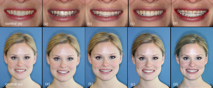
Prior to the eye-tracking process, images of a 24-year-old eugnathic female subject were prepared using a neutral blue background (Canon EOS 600D digital single lens reflex camera (Canon, Tokyo, Japan) and a SIGMA EM-140 DG optical flash (SIGMA, Kawasaki, Japan). The subject had no orthodontic treatment and no facial asymmetries or other saliences. Pictures were taken with (a) aligners in situ (Scheu Dental, Iserlohn, Germany), (b) aligners in situ and attachments (Tetric EvoFlow, A2, Ivoclar Vivadent, Ellwangen, Germany) bonded, (c) lingual brackets (WIN, DW Lingual Systems, Bad Essen, Germany) bonded, and (d) ceramic brackets bonded and wire (.016 × .022 stainless steel wire without coating) in place (Clarity Advanced, 3M Unitek, Landsberg, Germany). For all appliances, both complete en face photos of the smiling face as well as close-up images of the smiling oral region were produced under uniform conditions. Images with no appliance in situ were taken as a control (e; Figure 1).
Figure 1.
Close-up and facial images of the different appliances in situ: (a) aligner, (b) aligner + attachments, (c) lingual appliance, (d) ceramic brackets, and (e) control.
Subjects and Application
The photos of the subject were shown in randomized order to a group of 140 participants for a period of 7 seconds on a computer screen connected to the eye-tracking system. During inspection, the eye movement was recorded with a contact-free monocular eye-tracking system (Monocular System Eyegaze Edge Systems, LC Technologies, Inc., Fairfax, VA, USA; 60 Hz, 0.3–0.5° angular error). After the initial scanning process, the close-up images were shown for a period of 30 seconds and the question form had to be completed for each appliance photo. The process was carried out for each participant alone. The answering process of the questionnaire was performed afterward.
Fields of Interest and Landmarks
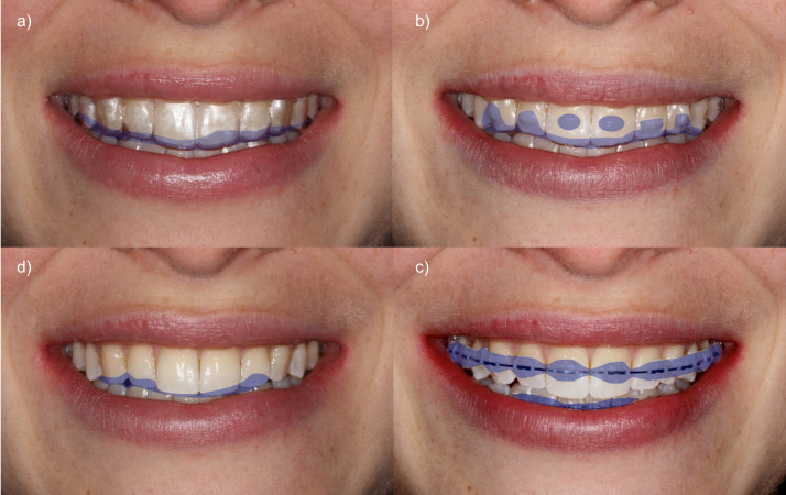
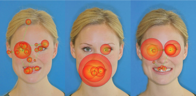
For the full facial images, different anatomical landmarks were defined: oral and lip region, nose, eyes, hair, ears. For the smiling close-up, the areas of the appliance were defined (Figure 2). For all areas, time to first fixation (seconds) and total fixation time (duration; seconds) were recorded (Figure 3). The landmarks were prepared only for analysis and not visible to the participants.
Figure 2.
Areas of interest of the different appliances: (a) aligner, (b) aligner + attachments, (c) ceramic brackets, and (d) lingual appliance.
Figure 3.
Facial images showing the eye-tracking patterns of control (left), aligner (middle), and lingual (right).
Survey
The following questions regarding the close-up images were answered by 140 participants using a 10-cm visual analog scale (VAS):
How would you rate the visibility of the orthodontic appliance?
How much does the orthodontic appliance affect the visual appearance of the patient shown in the picture?
Please rate the personal impairment as if you had to wear this type of braces yourself?
Statistical Analysis
Time to first fixation and total fixation time were described by median, mean, standard deviation, and range. Kaplan-Meier curves were employed to describe time to first fixation data. The questionnaires were analyzed descriptively. The primary endpoint was time to first fixation on the area of interest based on the close-up images. This was analyzed using multivariable cox regression analyses with frailty terms to account for the paired and time-to-event data structure. To account for multiplicity, a Bonferroni correction was applied for the six primary pairwise confirmatory analyses. Accordingly, the adjusted significance level was α = .008.
Secondary endpoints comprised the time to first fixation on the mouth in the en face images that were also analyzed with multivariable cox regressions with frailty terms. Further secondary endpoints were the total fixation time on the area of interest in the close-ups and the total fixation time on the mouth in the en face images with pairwise comparisons of appliances based on paired Wilcoxon tests. All secondary endpoint analyses were regarded as exploratory with P values < .05 considered as indications of difference.
Statistical analyses were performed using SPSS for Windows version 23.0 (SPSS Inc., Chicago, Ill) and R version 3.2.5 (R Foundation for Statistical Computing, Vienna, Austria). Data from the survey were analyzed descriptively.
Informed Consent, Ethical Approval, and Subject Releases
The subject (L.K.) signed the inform consent for showing and publishing her images. All applicable subject releases were obtained and are on file with the corresponding author. All participants gave their agreement to take part in the study. This study was performed according to the guidelines of the Ethical Review Committee of Rhineland-Palatinate (Germany), and that committee gave permission to conduct the study.
RESULTS
A total of 139 participants underwent the eye-tracking procedure, and 140 completed the questionnaire (1 person could not be adjusted for the eye-tracking system, thus there was 1 drop out). The mean age of the test persons was 25.4 ± 5.3 years; 44% were men and 56% women; 70 (50%) had a professional background in orthodontics.
Eye Tracking
Description of fixation in the close-up and the en face images.
Table 1 shows the description of time to first fixation and total fixation time for the areas of interest in the close-up smiling images. The area of interest of the lingual brackets was fixed only by 10% of the test persons, followed by aligner appliances with 84% fixation. Areas of interest with the aligner + attachments and with the ceramic brackets were fixed by all participants within the 7 seconds.
Table 1.
Description of Fixation Within 7 Seconds on the Areas of Interest in the Close-Up Images
| Group |
Number of Fixations |
Time to First Fixation |
Total Fixation Timea |
| a: aligner | |||
| n (%) or mean ± SD | 117 (84.2) | 2.57 ± 1.64 | 0.99 ± 0.74 |
| Median (range) | 2.26 (0.07–6.52) | 0.85 (0.12–2.90) | |
| b: aligner + attachments | |||
| n (%) or mean ± SD | 139 (100.0) | 1.02 ± 0.88 | 2.36 ± 1.29 |
| Median (range) | 0.75 (0.00–5.02) | 2.29 (0.33–5.70) | |
| c: lingual brackets | |||
| n (%) or mean ± SD | 14 (10.1) | 3.28 ± 2.03 | 0.53 ± 0.67 |
| Median (range) | 3.03 (0.70–6.09) | 0.32 (0.13–2.69) | |
| d: ceramic | |||
| N (%) or mean ± SD | 139 (100.0) | 0.58 ± 0.53 | 3.14 ± 1.14 |
| Median (range) | 0.42 (0.03–4.07) | 3.04 (0.50–5.84) | |
Only patients who had a fixation of the respective area, mean ± standard deviation, and median and range. SD indicates standard deviation.
Table 2 shows the results for time to first fixation and the total fixation time for the anatomic landmarks in the en face images. In both aligner appliances (a), aligner with attachments (b), and the lingual appliance (c), the face was inspected in the sequence of eyes first and then mouth/lips, nose, hair, and ears. The image with the ceramic brackets (d) was looked at in the order of mouth/lip first and then eyes, nose, hair, and ears.
Table 2.
Description of Fixation, Time to First Fixation, and Total Fixation Time on the Different Anatomic Landmarks in the En Face Images: Eyes, Mouth, Nose, Ears, and Hair
| Group |
Eyes |
Mouth |
||||
| Fixation |
Time to First Fixation |
Total Fixation Timea |
Fixation |
Time to First Fixation |
Total Fixation Timea |
|
| a: aligner | ||||||
| n | 131 | 138 | ||||
| Mean ± SD | 0.94 ± 1.16 | 1.66 ± 1.10 | 1.17 ± 0.96 | 2.97 ± 1.32 | ||
| Median (range) | 0.42 (0.00–6.75) | 1.41 (0.12–5.77) | 0.74 (0.25–5.39) | 2.98 (0.33–5.81) | ||
| b: aligner + attachments | ||||||
| n | 133 | 138 | ||||
| Mean ± SD | 0.75 ± 1.01 | 1.42 ± 1.02 | 1.03 ± 0.85 | 3.35 ± 1.38 | ||
| Median (range) | 0.37 (0.00–5.30) | 1.18 (0.10–5.29) | 0.70 (0.20–4.43) | 3.39 (0.20–6.07) | ||
| c: lingual brackets | ||||||
| n | 132 | 120 | ||||
| Mean ± SD | 0.82 ± 1.00 | 2.40 ± 1.17 | 1.64 ± 1.63 | 1.87 ± 1.31 | ||
| Median (range) | 0.46 (0.03–5.54) | 2.28 (0.22–5.27) | 0.99 (0.13–6.20) | 1.63 (0.13–5.50) | ||
| d: ceramic | ||||||
| n | 128 | 135 | ||||
| Mean ± SD | 1.32 ± 1.66 | 1.50 ± 0.97 | 1.10 ± 1.05 | 3.29 ± 1.36 | ||
| Median (range) | 0.42 (0.05–6.75) | 1.29 (0.10–4.39) | 0.65 (0.12–5.56) | 3.33 (0.38–5.78) | ||
| e: control | ||||||
| n | 133 | 130 | ||||
| Mean ± SD | 0.84 ± 1.10 | 2.42 ± 1.23 | 1.48 ± 1.39 | 1.72 ± 1.30 | ||
| Median (range) | 0.42 (0.02–5.51) | 2.34 (0.28–5.89) | 1.03 (0.02–5.99) | 1.38 (0.17–6.45) | ||
Only patients who had a fixation of the respective area, mean ± standard deviation, and median and range. SD indicates standard deviation.
Table 2.
Extended
| Nose |
Ears |
Hair |
||||||
| Fixation |
Time to First Fixation |
Total Fixation Timea |
Fixation |
Time to First Fixation |
Total Fixation Timea |
Fixation |
Time to First Fixation |
Total Fixation Timea |
| 90 | 15 | 19 | ||||||
| 2.10 ± 1.94 | 0.50 ± 0.37 | 4.74 ± 1.33 | 0.28 ± 0.14 | 4.00 ± 1.78 | 0.52 ± 0.33 | |||
| 1.53 (0.02–6.67) | 0.43 (0.08–1.59) | 5.01 (1.93–6.52) | 0.25 (0.04–0.53) | 4.43 (0.05–6.40) | 0.43 (0.13–1.33) | |||
| 86 | 17 | 24 | ||||||
| 1.91 ± 1.77 | 0.45 ± 0.39 | 4.91 ± 1.65 | 0.36 ± 0.27 | 4.27 ± 1.78 | 0.46 ± 0.33 | |||
| 1.26 (0.10–6.76) | 0.32 (0.10–2.09) | 5.89 (1.95–6.74) | 0.29 (0.15–1.34) | 5.89 (1.95–6.74) | 0.41 (0.10–1.61) | |||
| 95 | 28 | 38 | ||||||
| 2.14 ± 1.94 | 0.55 ± 0.42 | 4.75 ± 1.33 | 0.44 ± 0.27 | 4.21 ± 2.04 | 0.56 ± 0.34 | |||
| 1.4 (0.18–6.71) | 0.47 (0.08–2.30) | 4.95 (2.06–6.74) | 0.37 (0.10–1.16) | 4.70 (0.02–6.66) | 0.46 (0.13–1.38) | |||
| 95 | 10 | 14 | ||||||
| 2.17 ± 2.12 | 0.53 ± 0.48 | 4.35 ± 1.07 | 0.33 ± 0.16 | 4.07 ± 1.41 | 0.49 ± 0.55 | |||
| 1.1 (0.01–6.69) | 0.37 (0.08–2.82) | 4.40 (2.37–5.78) | 0.31 (0.13–0.63) | 4.14 (1.94–6.62) | 0.37 (0.02–2.29) | |||
| 111 | 27 | 68 | ||||||
| 1.76 ± 1.71 | 0.64 ± 0.61 | 4.11 ± 1.31 | 0.36 ± 0.29 | 3.69 ± 1.81 | 0.59 ± 0.45 | |||
| 1.18 (0.00–6.75) | 0.51 (0.08–5.13) | 4.07 (1.19–6.74) | 0.25 (0.02–1.31) | 3.30 (0.48–6.63) | 0.49 (0.15–3.35) | |||
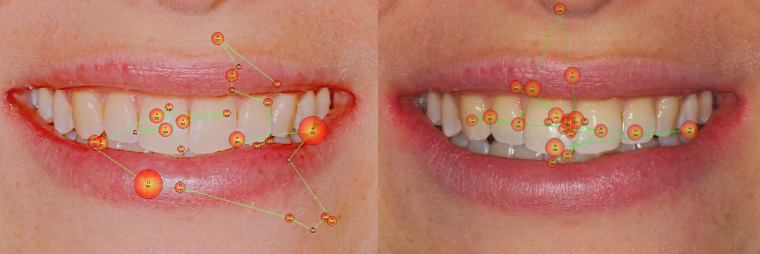
For the aligner appliances (a) and aligners with attachments (b) as well as for ceramic brackets (d), the maximum total fixation time (ie, looking at the landmark for the longest time) was the smiling mouth/lip area. For the lingual appliance (b), the participants inspected the eye region for the longest time. The lingual appliance (c) appeared to be the most similar with the untreated control images, both in time to first fixation and total fixation time testing (Figure 4).
Figure 4.
Example of eye-tracking patterns of close-up pictures with control (left) vs lingual (right).
Comparative analyses of appliances.
As shown in Figure 5, the risk for fixation on the area of interest in the close-up images was lowest with the lingual appliance (c) followed by the aligner (a) and the aligner with attachments (b) and highest with the ceramic brackets (d). The regression analyses showed these differences to be significant with 94% lower risk of fixation on the area of interest in the 7-second timeframe with the lingual appliance when compared with aligner, 88% lower when compared with aligner + attachments, and 82% lower when compared with the ceramic appliance (hazard ratio [HR] = 0.06, HR = 0.12, HR = 0.18, respectively, all P < .001; Table 3).
Figure 5.
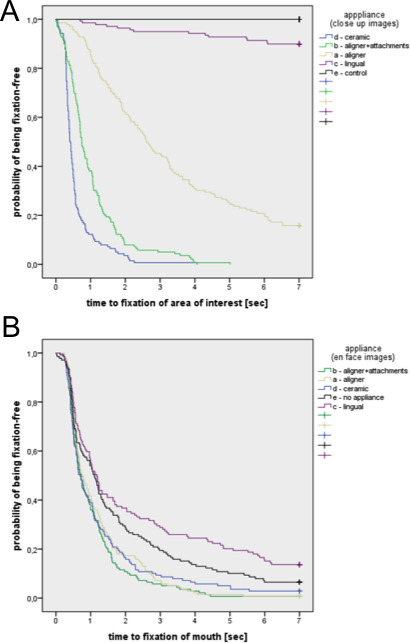
Kaplan–Meier curves of the time to first fixation of (A) the area of interest in the close-up images and (B) the mouth in the en face images for the different appliances.
Table 3.
Results of Multivariable Cox Regression Models for Time to First Fixation on Area of Interest in the Close-Up Images (Primary Endpoint) Comparing Appliances Pairwisea
| Group |
Hazard Ratio |
Confidence Interval |
P Value |
| b (aligner + attachments) vs d (ceramic) | 0.52 | 0.33–0.54 | <.001 |
| a (aligner) vs d (ceramic) | 0.32 | 0.27–0.38 | <.001 |
| c (lingual app.) vs d (ceramic) | 0.18 | 0.14–0.24 | <.001 |
| a (aligner) vs b (aligner + attachments) | 0.20 | 0.15–0.26 | <.001 |
| c (lingual app.) vs b (aligner + attachments) | 0.12 | 0.08–0.17 | <.001 |
| c (lingual app.) vs a (aligner) | 0.06 | 0.03–0.10 | <.001 |
All models adjusted for professional background of test persons; test vs control not applicable (no area of interest in control image). app. indicates appliance.
In the en face images, the risk for fixation on the mouth was also lowest with the lingual appliance (c) and the control (e) and higher with ceramic brackets (d), aligner (a), and aligner + attachments (b). The pairwise comparisons are shown in Table 4. Accordingly, the risk for fixation on the mouth was 18% lower with the lingual appliance when compared with aligner + attachments (HR = 0.82, P < .001), 23% lower when compared with aligner (HR = 0.77 and HR = 0.80, P < .001), and 30% when compared with ceramic (HR = 0.70, P < .001). These differences were comparable with those of the control images.
Table 4.
Results of Multivariable Cox Regression Models for Time to First Fixation on the Mouth in the En Face Images Comparing Appliances Pairwisea
| Group |
Hazard Ratio |
Conficence Interval |
P Value |
| a (aligner) vs b (aligner + attachments) | 0.80 | 0.61–1.05 | .110 |
| d (ceramic) vs b (aligner + attachments) | 0.96 | 0.83–1.09 | .510 |
| e (control) vs b (aligner + attachments) | 0.82 | 0.74–0.89 | <.001 |
| c (lingual app.) vs b (aligner + attachments) | 0.82 | 0.77–0.88 | <.001 |
| d (ceramic) vs a (aligner) | 1.11 | 0.84–1.45 | .470 |
| e (control) vs a (aligner) | 0.80 | 0.70–0.91 | <.001 |
| c (lingual app.) vs a (aligner) | 0.77 | 0.70–0.85 | <.001 |
| e (control) vs d (ceramic) | 0.61 | 0.47–0.80 | <.001 |
| c (lingual app.) vs d (ceramic) | 0.70 | 0.61–0.80 | <.001 |
| c (lingual app.) vs e (control) | 0.73 | 0.55–0.96 | .023 |
app. indicates appliance.
There was also evidence for differences in the total fixation time on the area of interest in the close-up images as well as the total fixation time on the mouth in the en face images in all pairwise comparisons (P < .001) except ceramic (d) vs aligner + attachments (b) and lingual (c) vs control (e) in the en face images.
Survey
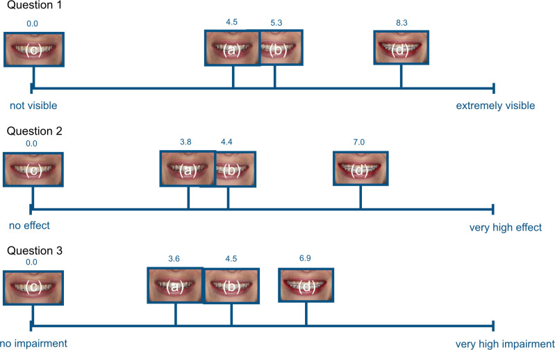
Figure 6 shows the average values of the VAS markings for questions 1 to 3. For the lingual appliance, the questions regarding visibility, discomfort, and willingness for personal use were all answered with VAS scores of 0.0 (no visibility, no negative effect on smile esthetics, no problem with wearing this type of appliance). Both aligner appliances (with and without attachments) showed mean scores of 4.5 to 5.34 (question 1), 3.8 to 4.4 (question 2), and 3.64 to 4.57 (question 3). The values for aligners with attachments appeared to be slightly higher. The highest scores for visibility were found with the ceramic brackets (8.3), which also had the highest scores for negative effect on the smile (7.01) and the average score for feeling toward using the appliance themselves (6.9).
Figure 6.
Diagrammatic representation showing average scoring of the visual analog scale for questions 1 to 3.
DISCUSSION
The specific aim of this study was to evaluate differences in the perception of the face and smiling mouth region when influenced by orthodontic appliances. Eye-tracking systems are capable of providing quantitative measurements of visual attention.16 The visual process when inspecting an image can broadly be divided into two aspects: 90% of the viewing time is spent with fixation, with the remaining 10% showing the aspect of repositioning eye movements such as saccades and/or relocation movements. For eye-tracking data analysis, typically the fixation time is used to assess visual attention while minimizing data complexity.17
Rosvall et al.13 and Ziuchkovski et al.14 investigated the attractiveness of orthodontic appliances by means of digital images and cross-sectional data using VAS. They showed that attractiveness ratings can be grouped in the hierarchy of lingual appliances and aligners followed by ceramic appliances, ceramic self-ligation appliances, and stainless-steel appliances.13,14 This was in agreement with the current eye-tracking findings.
In 2011, Jeremiah et al.15 showed that intellectual ability was associated with the appearance of different orthodontic appliances. They also used a cross-sectional analytical questionnaire study with color photographs of different appliances. They found no differences in social competence and psychological adjustment influenced by orthodontic appliances.
In 2010, Hickman et al.18 investigated the visual attention of the face in orthodontic patients with the help of eye tracking for the first time. They concluded that the mouth region only played a minor role in visual fixation of orthodontically treated patients. Other recent studies evaluated smile esthetics and treatment need with the use of eye tracking.19,20 It was shown that higher treatment need scores were associated with more attention to the oral mouth region and, in addition, orthodontic treatment was able to influence scan paths during facial detection. It has to be stated that female subjects are generally judged more critically with regard to facial attractiveness and affecting factors.21,22
For the en face images, it was shown that, for both the aligner and lingual appliances, the first and fastest fixation was on the area of the eyes followed by the mouth as well as in the control group. This was in agreement with other eye-tracking studies in the field of orthodontics22 and neurobiological investigations.23,24 Ceramic brackets in combination with a steel arch wire changed this chronological perception to first fixation on the mouth. The duration of fixation on the mouth region varied significantly between ceramic brackets/aligners and the lingual appliance, with values close to the control image. This might be relevant for a patient and affect the decision regarding the choice of orthodontic appliance.
For the close-up images, it was shown that ceramic brackets had the most duration of fixation followed by aligner with attachments. Significantly less time was spent inspecting the aligner without attachments and the lingual appliance. This has to be put in context with the size of the area of interest and one might argue that there is a lack of display of the lingual appliance at all. The control picture was compared with the image of the lingual appliance, and it was determined that the darker incisor edge and darker areas in the occlusion were the areas of interest within the lingual appliance, which was considered rather critical. This approach was comparable to the area of interest assigned to the aligner (aligner thickness and reflections).
VASs have been used in medical science to investigate a variety of subjective questioning.25 This study assessed the subjective visibility of esthetic appliances. It may be assumed that lingual appliances in this setup were subjectively not seen by the tested individuals, therefore they were not perceived as esthetically disturbing and the willingness to undergo orthodontic procedures with this type of appliance was the highest. Of course, other factors may contribute to the overall experience and discomfort while undergoing lingual treatment.26,27
This was the first investigation to study this matter both objectively through eye tracking and on a personal level with the questionnaire. It can be assumed that ceramic brackets have a high average VAS score (>8) when assessed in a close-up image. Further studies might put this into context with regard to metal bracket appliances, but the focus of this investigation was on esthetic orthodontic appliances. Further studies might also be useful to analyze test–retest and intrarater reliability. Additional studies could be able to provide insight on specific visual phenomena connected to orthodontic treatment and be of help for patient information and consent in the area of growing interest in discreet orthodontic tooth movement.
CONCLUSIONS
Lingual orthodontic appliances do not change how the face is perceived. Other esthetic orthodontic appliances may change the pattern of facial inspection and are different in subjective perception.
ACKNOWLEDGMENT
This study was funded by the German Society of Lingual Orthodontics.
REFERENCES
- 1.Nattrass C, Sandy JR. Adult orthodontics—a review. Br J Orthod. 1995;22(4):331–337. doi: 10.1179/bjo.22.4.331. [DOI] [PubMed] [Google Scholar]
- 2.Whitesides J, Pajewski NM, Bradley TG, Iacopino AM, Okunseri C. Socio-demographics of adult orthodontic visits in the United States. Am J Orthod Dentofacial Orthop. 2008;133:489.e9–489.e14. doi: 10.1016/j.ajodo.2007.08.016. [DOI] [PubMed] [Google Scholar]
- 3.Hohoff A, Wiechmann D, Fillion D, Stamm T, Lippold C, Ehmer U. Evaluation of the parameters underlying the decision by adult patients to opt for lingual therapy: an international comparison. J Orofac Orthop. 2003;64:135–144. doi: 10.1007/s00056-003-0217-7. [DOI] [PubMed] [Google Scholar]
- 4.Fuck LM, Wiechmann D, Drescher D. Comparison of the initial orthodontic force systems produced by a new lingual bracket system and a straight-wire appliance. J Orofac Orthop. 2005;66:363–376. doi: 10.1007/s00056-005-0442-3. [DOI] [PubMed] [Google Scholar]
- 5.Gorman JC, Smith RJ. Comparison of treatment effects with labial and lingual fixed appliances. Am J Orthod Dentofacial Orthop. 1991;99:202–209. doi: 10.1016/0889-5406(91)70002-E. [DOI] [PubMed] [Google Scholar]
- 6.Lagravère MO, Flores-Mir C. The treatment effects of Invisalign orthodontic aligners: a systematic review. J Am Dent Assoc. 2005;136:1724–1729. doi: 10.14219/jada.archive.2005.0117. [DOI] [PubMed] [Google Scholar]
- 7.Caniklioglu C, Öztürk Y. Patient discomfort: a comparison between lingual and labial fixed appliances. Angle Orthod. 2005;75:86–91. doi: 10.1043/0003-3219(2005)075<0086:PDACBL>2.0.CO;2. [DOI] [PubMed] [Google Scholar]
- 8.Hohoff A, Fillion D, Stamm T, Goder G, Sauerland C, Ehmer U. Oral comfort, function and hygiene in patients with lingual brackets. A prospective longitudinal study. J Orofac Orthop. 2003;64:359–371. doi: 10.1007/s00056-003-0307-6. [DOI] [PubMed] [Google Scholar]
- 9.Miller KB, McGorray SP, Womack R, et al. A comparison of treatment impacts between Invisalign aligner and fixed appliance therapy during the first week of treatment. Am J Orthod Dentofacial Orthop. 2007;131:1–9. doi: 10.1016/j.ajodo.2006.05.031. [DOI] [PubMed] [Google Scholar]
- 10.Eliades T, Pratsinis H, Athanasiou AE, Eliades G, Kletsas D. Cytotoxicity and estrogenicity of Invisalign appliances. Am J Orthod Dentofacial Orthop. 2009;136:100–103. doi: 10.1016/j.ajodo.2009.03.006. [DOI] [PubMed] [Google Scholar]
- 11.Martorelli M, Gerbino S, Giudice M, Ausiello P. A comparison between customized clear and removable orthodontic appliances manufactured using RP and CNC techniques. Dent Mater. 2013;29:1–10. doi: 10.1016/j.dental.2012.10.011. [DOI] [PubMed] [Google Scholar]
- 12.Clements KM, Bollen AM, Huang G, King G, Hujoel P, Ma T. Activation time and material stiffness of sequential removable orthodontic appliances. Part 2: dental improvements. Am J Orthod Dentofacial Orthop. 2003;124:502–508. doi: 10.1016/s0889-5406(03)00577-8. [DOI] [PubMed] [Google Scholar]
- 13.Rosvall M, Fields H, Ziuchkovski J, Rosenstiel S, Johnston W. Attractiveness, acceptibility, and value of orthodontic appliances. Am J Orthod Dentofacial Orthop. 2009;135:276–286. doi: 10.1016/j.ajodo.2008.09.020. [DOI] [PubMed] [Google Scholar]
- 14.Ziuchkovski J, Fields H, Johnston W, Lindsey D. Assessment of perceived orthodontic appliance attractiveness. Am J Orthod Dentofacial Orthop. 2008;133:68–78. doi: 10.1016/j.ajodo.2006.07.025. [DOI] [PubMed] [Google Scholar]
- 15.Jeremiah H, Bister D, Newton J. Social perceptions of adults wearing orthodontic appliances: a cross-sectional study. Eur J Orthod. 2011;33:476–482. doi: 10.1093/ejo/cjq069. [DOI] [PubMed] [Google Scholar]
- 16.Duchowski A. Eye tracking methodology Theory and Practice 2nd ed. London: Springer; 2007. [Google Scholar]
- 17.Salvucci D, Goldberg J. Palm Beach Gardens, Florida, USA: Nov 06–08, 2000. Identifying fixations and saccades in eye-tracking protocols. Paper presented at: Eye Tracking Research and Applications Symposium. [Google Scholar]
- 18.Hickman L, Firestone AR, Beck FM, Speer S. Eye fixations when viewing faces. J Am Dent Assoc. 2010;141:40–46. doi: 10.14219/jada.archive.2010.0019. [DOI] [PubMed] [Google Scholar]
- 19.Wang X, Cai B, Cao Y, et al. Objective method for evaluating orthodontic treatment from the lay perspective: an eye-tracking study. Am J Orthod Dentofacial Orthop. 2016;150:601–610. doi: 10.1016/j.ajodo.2016.03.028. [DOI] [PubMed] [Google Scholar]
- 20.Johnson EK, Fields HW, Jr, Beck FM, Firestone AR, Rosenstiel SF. Role of facial attractiveness in patients with slight-to-borderline treatment need according to the Aesthetic Component of the Index of Orthodontic Treatment Need as judged by eye tracking. Am J Orthod Dentofacial Orthop. 2017;151:297–310. doi: 10.1016/j.ajodo.2016.06.037. [DOI] [PubMed] [Google Scholar]
- 21.Geron S, Wasserstein A. Influence of sex on the perception of oral and smile esthetics with different gingival display and incisal plane inclination. Angle Orthod. 2015;75:778–784. doi: 10.1043/0003-3219(2005)75[778:IOSOTP]2.0.CO;2. [DOI] [PubMed] [Google Scholar]
- 22.Richards MR, Fields HW, Beck FM, et al. Contribution of malocclusion and female facial attractiveness to smile esthetics evaluated by eye tracking. Am J Orthod Dentofacial Orthop. 2015;147:472–482. doi: 10.1016/j.ajodo.2014.12.016. [DOI] [PubMed] [Google Scholar]
- 23.Levy J, Foulsham T, Kingstone A. Monsters are people too. Biol Lett. 2013;9(1) doi: 10.1098/rsbl.2012.0850. [DOI] [PMC free article] [PubMed] [Google Scholar]
- 24.Peterson MF, Eckstein MP. Fixating the eyes is an optimal strategy across important face (related) tasks. J Vis. 2011;11:662. [Google Scholar]
- 25.Wewers ME, Lowe NK. A critical review of visual analogue scales in the measurement of clinical phenomena. Res Nurs Health. 1990;13:227–236. doi: 10.1002/nur.4770130405. [DOI] [PubMed] [Google Scholar]
- 26.Miyawaki S, Yasuhara M, Koh Y. Discomfort caused by bonded lingual orthodontic appliances in adult patients as examined by retrospective questionnaire. Am J Orthod Dentofacial Orthop. 1999;115:83–88. doi: 10.1016/s0889-5406(99)70320-3. [DOI] [PubMed] [Google Scholar]
- 27.Wiechmann D, Gerß J, Stamm T, Hohoff A. Prediction of oral discomfort and dysfunction in lingual orthodontics: a preliminary report. Am J Orthod Dentofacial Orthop. 2008;133:359–364. doi: 10.1016/j.ajodo.2006.03.045. [DOI] [PubMed] [Google Scholar]


