Abstract
African swine fever virus (ASFV) is the etiological agent of African swine fever (ASF), an often lethal disease in domestic and wild pigs. ASF represents a major threat to the swine industry worldwide. Currently, no commercial vaccine is available because of the complexity of ASFV or biosecurity concerns. Live attenuated viruses that are naturally isolated or genetically manipulated have demonstrated reliable protection against homologous ASFV strain challenge. In the present study, a mutant ASFV strain with the deletion of ASFV MGF-110-9L (ASFV-Δ9L) was generated from a highly virulent ASFV CN/GS/2018 parental strain, a genotype II ASFV. Relative to the parental ASFV isolate, deletion of the MGF-110-9L gene significantly decreased the ability of ASFV-Δ9L to replicate in vitro in primary swine macrophage cell cultures. The majority of animals inoculated intramuscularly with a low dose of ASFV-Δ9L (10 HAD50) remained clinically normal during the 21-day observational period. Three of five ASFV-Δ9L-infected animals displayed low viremia titers and low virus shedding and developed a strong virus-specific antibody response, indicating partial attenuation of the ASFV-Δ9L strain in pigs. The findings imply the potential usefulness of the ASFV-Δ9L strain for further development of ASF control measures.
Keywords: African swine fever virus (ASFV), MGF-110-9L, Mutant, Attenuated virulence, Pig
Introduction
African swine fever (ASF) is a significant disease with mortality rates approaching 100% in domestic pigs and wild boars (Gallardo et al. 2015). In contrast, African wild pig species, including warthogs and bush pigs, are resistant to infection with African swine fever virus (AFSV) (Keßler and Forth 2018). ASF is endemic in more than 20 sub-Saharan African, European, and Asian countries (Correa-Fiz et al. 2019). The extensive spread of the disease in Asian countries has had an unprecedented impact on the pig industry.
ASFV is a large enveloped virus containing an approximately 190 kb double-stranded (ds) DNA genome encoding over 150 proteins (Borca et al. 2020). The virus replicates predominantly in the cytoplasm of infected macrophages, which are the major natural host cells of ASFV (Alcamí et al. 1990; Dixon et al. 2013). ASFV replicates in several soft tick species that play an important role in virus transmission between wild pigs in Africa (Kleiboeker et al. 1999). In addition, the movement of infected pigs and pig products is a main cause of spread (Costard et al. 2013; Jori et al. 2013).
There is no vaccine available for ASF. Disease outbreaks have been quelled by animal quarantine and slaughter. Attempts to vaccinate animals using DNA vaccines, vector-based vaccines, or detergent-treated infected alveolar macrophages have failed to induce protective immunity (Lacasta et al. 2014; Lokhandwala et al. 2017). Moderately virulent or attenuated variants of ASFV obtained from the surviving pigs infected with the viral can confer long-term resistance to homologous virulent viruses, but rarely to heterologous ASFV challenge (Hamdy and Dardiri 1984). Protection of pigs has resulted from the use of live attenuated ASF viruses containing genetically engineered deletions of specific ASFV virulence-associated genes before challenge with homologous parental virus (Zsak et al. 1996; Moore et al. 1998; Lewis et al. 2000; O'Donnell et al. 2015a, b; O'Donnell et al. 2017). In addition, the European Commission confirmed the use of attenuated strains as the most plausible approach to developing an effective ASF vaccine in the short/medium term (https://ec.europa.eu/food/animals/animal-diseases/control-measures/asf_en).
ASFV multigene family 110 (MGF-110) located at the left end of the ASFV genome and contained a hydrophobic NH2-terminal sequence and a conserved cysteine-rich domain (Almendral et al. 1990). Expression of MGF-110 family members could involve in host range or the viral virulence and MGF-110 gene product had role in preparing the endoplasmic reticulum for its role in viral morphogenesis (Netherton et al. 2004). However, the functions of MGF-110 family members remain to be unclear.
ASFV MGF-110-9L belongs to the ASFV MGF 110 family. ASFV MGF-110-9L is a genotype II ASFV that contains approximately 873 nucleotides. The amino acid sequence of ASFV MGF-110-9L is highly conserved among genotype II ASFV. In this study, we demonstrate that deletion of the ASFV gene MGF-110-9L (ASFV-Δ9L and hereafter designated as 9L) from the highly virulent ASFV CN/GS/2018 isolate resulted in partial attenuation in swine. Swine inoculated with the ASFV-Δ9L virus lacking the 9L gene developed a strong virus-specific antibody response. Importantly, most of the swine survived after inoculation with ASFV-Δ9L, while all pigs inoculated with parental ASFV died.
Materials and Methods
Cell Culture and Viruses
Porcine alveolar macrophages (PAM) prepared by bronchoalveolar lavage as previously described (Carrascosa et al. 1982) were grown in Dulbecco’s modified Eagle’s medium (DMEM) supplemented with 2 mmol/L L-glutamine, 100 U/mL gentamicin, non-essential amino acids, and 10% porcine serum. Cells were grown at 37 °C in a 7% CO2 atmosphere saturated with water vapor. ASFV CN/GS/2018 propagated on PAM. Briefly, subconfluent PAM cells were cultivated in p150 plates and infected with ASFV at a multiplicity of infection (MOI) of 0.01 in DMEM supplemented with 10% porcine serum. At 96 h post-infection (hpi), the cells were recovered and centrifuged at 775 rcf for 15 min. The cell pellet was discarded. The supernatant containing the viruses was clarified at 16,873 rcf for 6 h at 4 °C, resuspended in medium, and stored at −80 °C. Infection was performed after ASFV viral adsorption at 37 °C for 90 min, when the inoculum was removed and fresh medium was added. Cells were incubated at 37 °C for defined times.
Growth curves between ASFV CN/GS/2018 and ASFV-Δ9L viruses were obtained in primary swine macrophage cell cultures. Preformed monolayers were prepared in 24-well plates and infected at an MOI of 0.01 (based on the 50% hemadsorption unit [HAD50] that was previously determined in primary swine macrophage cell cultures). After 1 h of adsorption at 37 °C in an atmosphere of 5% CO2, the inoculum was removed and the cells were rinsed twice with phosphate buffered saline. The monolayers were rinsed with macrophage medium and incubated for 0, 12, 24, 36, and 48 h at 37 °C in an atmosphere of 5% CO2. At appropriate hpi, the cells were frozen at −80 °C and then thawed. The thawed lysates were used to determine virus load by quantitative polymerase chain reaction (qPCR) in PAM cell cultures. All samples were run simultaneously to avoid inter-assay variability.
qPCR
ASFV genomic DNA was extracted from cell supernatants, tissue homogenates, or EDTA-treated whole peripheral blood using GenElute™ Mammalian Genomic DNA Miniprep Kits (Sigma Aldrich, St. Louis, MO, USA). qPCR was carried out on a QuantStudio 5 system (Applied Biosystems, Franklin Lakes, NJ, USA) according to the procedure recommended by the World Organisation for Animal Health.
Pig Studies
Landrace-crossed pigs approximately 50-days-of-age weighing 80 to 90 pounds were obtained from the Laboratory Animal Center of the Lanzhou Veterinary Research Institute (LVRI, Lanzhou, China). The pigs were tested to ensure they were negative for porcine respiratory and reproductive syndrome, classical swine fever (CSF), ASFV, and pseudorabies virus (PRV).
To evaluate the virulence of the gene-deleted ASFV-Δ9L in pigs, pigs were inoculated intramuscularly with 10 HAD50 of each test virus. The pigs were monitored daily for 19 days for temperature and mortality.
Virus Titration
The wild-type CN/GS/2018 virus was quantified using the HAD assay as described previously (Malmquist and Hay 1960) with minor modifications. PAMs were seeded in 96-well plates. The samples were added to the wells and titrated in triplicate using tenfold serial dilutions. The red blood cells were added into the plates before ASFV produced cytopathic effect in PAMs. HAD was determined on day 7 post-inoculation (p.i.), and 50% HAD doses (HAD50) were calculated as previously described (Muench 1938).
Samples from the gene-deleted ASFV-infected cell supernatants were quantified by testing their 50% tissue culture infectious dose (TCID50). PAMs were seeded into 96-well plates, and three days later tenfold serially diluted samples were added into each well in triplicate. After seven days of culture, the fluorescent protein expression was assessed by using fluorescence microscopy. TCID50 was calculated by using the method of Reed and Muench.
Biosafety Statement and Facility
All experiments with live ASF viruses were conducted within the enhanced biosafety level 3 (P3) facilities at LVRI and were approved by the Ministry of Agriculture and Rural Affairs and the China National Accreditation Service for Conformity Assessment.
Plasmid Design for Traditional Recombination
Plasmid pUC19 lacking multiple cloning was used as a backbone. The recombination cassette was inserted at the SalI and NdeI restriction sites after the T7 promoter. The recombination cassette contains a left recombination arm that is 1000 bp upstream of the 9L open reading frame identical to ASFV CN/GS/2018 nucleotide positions nt10561 to nt11560, followed by the p72 promoter identical to ASFV CN/GS/2018 nucleotide positions on the negative strand nt105677 to nt105465, enhanced green fluorescent protein (eGFP), and an SV40 termination sequence, and a right recombination arm 1077 bp downstream of 9L identical to ASFV CN/GS/2018 nucleotide positions nt12400 to nt13476.
CRISPR/Cas9 Transfection
CRISPR/Cas9 experiments were conducted after a 1 h virus adsorption at 37 °C in an atmosphere of 5% CO2. The inoculum was discarded and the indicated plasmids were transfected with Fugene HD following the manufacturer’s protocol (http://www.promega.com/techserv/tools/FugeneHdTool/). A 3:1 Fugene:DNA ratio was used with 3.3 μg of DNA and 9.9 μL of Fugene HD. The complex was mixed carefully by pipetting and incubated for 10 min. Then, 150 μL of the complex was added dropwise to the cells. The cells were incubated at 37 °C in an atmosphere of 5% CO2, observed for the presence of GFP fluorescence, frozen 24 h following transfection, thawed, and titrated. The frequency of transfection was calculated as the ratio between the titer of the recombinant and parental viruses.
Complete Next-Generation Sequencing (NGS) of ASFV Genomes
PAM cells were seeded as described above and infected with ASFV. When the cytopathic effect was evident throughout the monolayer, DNA was isolated as described above from cells infected with ASFV. The extracted DNA was used to completely sequence the virus DNA, as previously described (Krug et al. 2015). In brief, the viral DNA was sheared using enzymatic reactions that produced variously sized fragments. Identifying barcodes were ligated using an adapter sequence and added to the DNA fragments. The required size range of the library was collected using a Pippin Prep™ (Sage Science, Beverly, MA, USA) and normalized. The DNA library for NGS uses the NextSeq system (Illumina, San Diego, CA, USA) following the manufacture’s protocol. Sequence analysis was performed using CLC Genomics Workbench software (CLCBio, Waltham, MA, USA).
Blocking ELISA
ELISA microtitre plates were coated using an optimal concentration (8 μg/mL) of p30 protein (100 μL/well) in 0.05 mol/L carbonate buffer solution (pH 9.6) and incubated overnight at 4 °C. Antigen-coated plates were washed three times with PBST (PBS containing 0.5% (v/v) Tween-20) and nonspecific binding sites were blocked with 200 μL of blocking buffer (2.5% (w/v) non-fat dry milk in PBST) incubated overnight at 4 °C. After three washes with PBST, 100 μL of test samples, positive serum samples, negative serum samples diluted (1:2) in blocking buffer, were added separately to each well in duplicate. Next, the plates were incubated for 30 min at 37 °C followed by three washes and addition of 100 μL/well of p30-mAb-HRP at the dilution (1:25,000), with incubation at 37 °C for an additional 30 min. Following a final three washes, 100 μL/well of 3,3',5,5'-Tetramethylbenzidine (TMB) substrate, made from mixing of two solutions, A and B (A: 205 mmol/L potassium citrate (pH 4.0); B: 41 mmol/L TMB) in a ratio of A: B (v/v) of 39:1 was added to each well and the plates were incubated in the dark for 15 min at 37 °C. As a final step, 2 mol/L H2SO4 (50 μL/well) was used to stop the colorimetric reaction and the OD450nm values were read using an automated ELISA plate reader.
ELISA
The secreted lgG and lgM in sera from ASFV-Δ9L-infected pigs were analyzed with ELISA kits (Porcine IgG ELISA Kit, SEKP-0012, Solarbio life sciences, Beijing, China; Porcine IgM ELISA KIT, SEKP-0014, Solarbio Life Sciences, Beijing, China) according to the manufacturer’s instruction.
Results
Sequence Conservation of the 9L Gene in Different ASFV Isolates
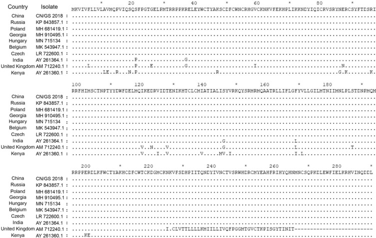
ASFV CN/GS/2018 open reading frame (ORF) MGF-110-9L (abbreviated to 9L) encodes for a 290 amino acids protein and is positioned on the reverse strand between nucleotide position nt11627 and nt12499 of the ASFV CN/GS/2018 genome. To assess the degree of conservation of 9L, amino acid sequences from 10 African, European, and Caribbean pathogenic virus isolates were analyzed. The 290 amino acid 9L protein was detected in most of the isolates. A few isolates featured a truncated C-terminus of the sequence (Fig. 1). The results indicated that 9L is highly conserved among the isolates.
Fig. 1.
Multiple sequence alignment of the indicated ASFV isolates of viral protein 9L. Gaps in the sequence are represented by ‘.’
9L Is Transcribed Early in the Virus Replication Cycle
To determine the time course of the transcription of the 9L gene, the expression of 9L at the mRNA level was determined at 3, 6, 9, 12, 15, 18, 21, and 24 h post-infection (h.p.i.). The expression of ASFV protein p30 and p72, which occurs early and late in the virus replication cycle respectively, was determined as a control. The p30 and p72 proteins were expressed at approximately 3 or 10 h.p.i. respectively. The expression pattern of 9L was similar to p30 protein (Fig. 2). These results suggested that the 9L gene is transcribed at an early stage in the virus replication cycle.
Fig. 2.

Time course of 9L gene transcriptional activity. ASFV p30, p72, and 9L open reading frame RNA were prepared from ex vivo pig macrophages infected with ASFV at 3, 6, 9, 12, 15, 18, 21, and 24 h post infection.
Construction of the ASFV 9L Deletion Mutant
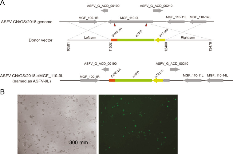
To further determine the role of 9L, an ASFV-Δ9L mutant was generated by genetic modification of the highly virulent ASFV CN/GS/2018 isolate. The 9L gene was deleted from the ASFV CN/GS/2018 virus using the CRISPR-Cas9 protocol. The 9L gene was replaced with a cassette containing the eGFP fluorescent gene under the control of the ASFV p72 promoter (Fig. 3A). The mutant virus was obtained after 11 rounds of purification. The mutant viruses obtained from the last round of purification were replicated in primary swine macrophage cells to obtain a virus stock. The absence of the 9L gene in the ASFV-Δ9L mutant was confirmed by sequence analyses of both the parental and mutant viruses. Green fluorescence was observed in ASFV-Δ9L-infected PAM cells at 12 h.p.i. (Fig. 3B).
Fig. 3.
Construction of ASFV-Δ9L virus. A Diagram indicating the position of the 9L open reading frame in the ASFV CN/GS/2018 genome. The donor plasmid with the homologous arms to ASFV CN/GS/2018 and GFP under control of the p72 promoter in the orientation as indicated. The final genomic changes introduced to develop ASFV–Δ9L where the sequence of the donor plasmid GFP reporter was introduced to replace the ORF of 9L as indicated. B Primary PAMs infected with ASFV–Δ9L that expressed green fluorescence are shown.
Replication of ASFV-Δ9L in Primary Swine Macrophages
To evaluate the growth characteristics of ASFV-Δ9L in vitro, replication of both parental ASFV CN/GS/2018 and ASFV-Δ9L in primary swine macrophages was examined at 0, 12, 24, 36, and 48 h.p.i. (Fig. 4). ASFV-Δ9L virus displayed significantly slower growth kinetics than that of the parental ASFV CN/GS/2018 virus. ASFV-Δ9L yields were fivefold to tenfold lower than those of the parental virus. These results suggested that deletion of the 9L gene significantly hindered the ability of virus replication in vitro in primary swine macrophage cell cultures.
Fig. 4.

In vitro growth characteristics of ASFV-Δ9L and parental ASFV CN/GS/2018. Primary swine macrophage cell cultures were infected (MOI = 0.01) with each of the viruses and virus titers at the indicated times post-infection. Data represent means from three independent experiments.
Assessment of ASFV-Δ9L Virulence in Pigs
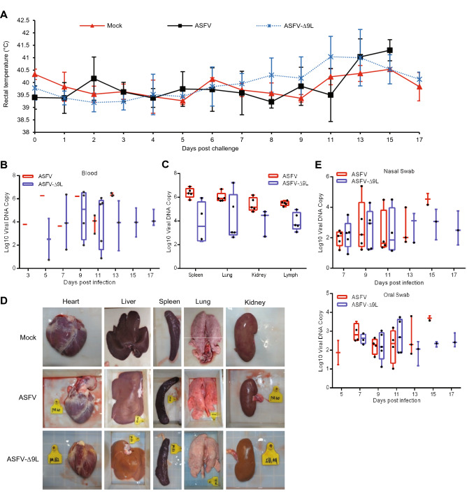
To examine the pathogenesis and virulence of ASFV-Δ9L, pigs were inoculated intramuscularly with 10 HAD50 of the parental virus ASFV CN/GS/2018 or ASFV-Δ9L. All five animals inoculated with ASFV CN/GS/2018 displayed increased body temperature at the time of death and displayed clinical signs associated with the disease, including anorexia, depression, purple skin discoloration, staggering gait, and diarrhea (Table 1). Signs of the disease became progressively aggravated over time and the animals died by 7 or 15 days p.i. Two of five animals inoculated intramuscularly with ASFV-Δ9L displayed these same symptoms. However, the other three animals developed fever for only a short time and then the temperature returned to normal (Fig. 5A). In addition, all the sentinel animals remained clinically normal (Fig. 5A).
Table 1.
Swine survival and fever response following infection with 10 HAD50 doses of ASFV-Δ9L or parental ASFV CN/GS/2018
| Virus and dose (HAD50) | No. of survivors/total | Mean time to death (days ± SD) | Fever | ||
|---|---|---|---|---|---|
| No. of days to onset (days ± SD) | Duration No. of days to onset (days ± SD) | Maximum daily temp (°C ± SD) | |||
| ASFV CN/GS/2018 | 0/5 | 12 (3.34) | 9 (3) | 3 (0.84) | 40.71 (0.66) |
| ASFV-Δ9L | 3/5 | 11 (0) | 9 (0) | 2 (0) | 40.65 (0.81) |
Fig. 5.
Kinetics of body temperature values and virus loads in pigs intramuscularly inoculated with 10 HAD50 of ASFV-Δ9L or 10 HAD50 of ASFV CN/GS/2018. A Kinetics of body temperature values in pigs inoculated intramuscularly with 10 HAD50 of ASFV-Δ9L, mock inoculated (sentinels, shown in red) or 10 HAD50 of parental ASFV. B–C Viral DNA detection in blood (B) and different tissues (C) of pigs. D Histomorphologic change of lung, spleen, kidney, and liver in control pigs, ASFV-infected pigs, or ASFV-Δ9L-infected pigs. E Viral DNA detection of oral and nasal swab. Each curve represents values from individual animals in each group.
Animals infected with ASFV CN/GS/2018 presented with high viral load in blood at the time of death (Fig. 5B). Two of five animals inoculated with ASFV-Δ9L developed pronounced viremia. Their viral load had decreased at the time of death (Fig. 5B). Three of the five animals inoculated with ASFV-Δ9L showed remarkably lower virus loads in blood than that of the parental virus (Fig. 5B). The results indicated that the deletion of the 9L gene partly attenuated the virulence of the ASFV CN/GS/2018 strain.
Animals inoculated with ASFV CN/GS/2018 displayed high viral loads in tissue samples for the spleen, lung, kidney, and lymph at the time of death (Fig. 5C). Two of five animals infected with ASFV-Δ9L developed remarkably high viremia in spleen and lung samples, but not in kidney and mesenteric lymph samples at the time of death (Fig. 5C). Three of five animals inoculated with ASFV-Δ9L displayed remarkably low virus loads in different tissues (spleen, lung, kidney, and lymph) (Fig. 5C). In addition, spleens of pigs that survived ASFV-Δ9L infection were normal compared with those of the control, whereas the spleen of dead pigs after ASFV infection was longer than control spleens (Fig. 5D). Consistently, the lungs of pigs that survived ASFV-Δ9L infection were normal compared with those of the control, whereas the infected pigs that died showed pulmonary congestion compared with control pigs (Fig. 5D). The liver, kidney, and heart were normal in ASFV or ASFV-Δ9L-infected pigs compared with control pigs (Fig. 5D). To investigate viral shedding in the ASFV-Δ9L group, viral loads in nasal and oral swabs were determined. The results showed consistent patterns to those observed in the blood experiment (Fig. 5E).
Antibody Response in Pigs Infected with ASFV-Δ9L
Previous studies have shown that there is no definite association between host immune mechanisms mediating protection and virulent strains of ASFV in animals infected with attenuated strains of virus (Ruiz Gonzalvo et al. 1986; Onisk et al. 1994; Oura et al. 2005). In order to detect if ASFV-Δ9L-infected animals induced ASFV-specific antibody response, we found that the surviving pigs infected with ASFV-Δ9L displayed a gradual increase in p30 antibody at the late stage of infection and ASFV-infected pigs did not induce p30 antibody response (Fig. 6A). In addition, to explore ASFV-Δ9L-induced immune response in vivo, we detected the lgG and lgM expression in pig serum. The results showed that lgG and lgM antibodies can be detected in the surviving pigs infected with ASFV-Δ9L by 5 to 11 days post-inoculation (Fig. 6B and 6C). The results indicated that the surviving pig infected with ASFV-Δ9L is likely to be associated with increasing host antibody response and immune response.
Fig. 6.
ASFV p30, lgG, and lgM antibodies were detected by ELISA. A Anti-p30 antibody detected by ELISA in pigs intramuscularly inoculated with 10 HAD50 of ASFV-Δ9L or 10 HAD50 of ASFV CN/GS/2018. B–C lgG and lgM antibodies were detected by ELISA in pigs intramuscularly inoculated with 10 HAD50 of ASFV-Δ9L. Each curve represents values from individual animals in each group.
Discussion
The lack of available vaccines against ASFV limits the control of ASFV. The use of attenuated strains is currently the most plausible approach to developing an effective ASF vaccine in protecting pigs against challenge with homologous virulent isolates. Several attenuated viruses have been produced by sequential passages in cell cultures or by genetic manipulation. Attenuated viruses have been obtained by genetic manipulation consisting of deletions of a single gene or group of genes. Simultaneous deletion of ASFV MGF360 and MGF505 genes, or 9GL and UK genes attenuated the virus and confer protection against parental virus challenge (O'Donnell et al. 2015a, b; O'Donnell et al. 2017). Independent deletions of the NL (DP71L), the UK (DP69R), TK (A240L), and 9GL (B119L) gene, or the I177L gene from ASFV has produced recombinant deletion mutant viruses with significantly reduced virulence in swine that conferred protection against homologous parental virus (Zsak et al. 1996, 1998; Moore et al. 1998; Lewis et al. 2000; Neilan et al. 2004). These observations indicate that the rational development of effective live attenuated ASFV recombinant viruses by genetic manipulation of target genes is an effective approach for vaccines against ASFV.
Presently, several lines of evidence suggest that deletion of the 9L gene from the highly virulent ASFV CN/GS/2018 virus partially attenuates the virus in swine. First, three of five animals survived when infected with ASFV-Δ9L. Second, most animals inoculated with ASFV-Δ9L displayed a lower viremia in blood and tissue samples than the viremia values of the animals in the control group. Third, most animals inoculated with ASFV-Δ9L presented a lower viral load in oral and nasal swabs than the control group. The experiments were performed using pigs approximately 50-days-of-age and 80 to 90 lb in weight. Piglets do not have a fully functional immune system and ASFV preferentially infects pig monocytes and macrophages. Therefore, whether the ASFV-Δ9L virus similarly infects growing fattening pigs and piglets needs to be further studied.
Interestingly, animals die when infected with either parental virus or its deletion virus if they had experienced a constantly high temperature (> 40 °C). The body temperature of most of the ASFV-Δ9L-infected animals increased to a peak and then subsided to normal in the survivors. The body temperature of animals does not change when infected with ASFV-G-ΔI177L and these animals can survive (Borca et al. 2020). These observations indicate that high temperature is one of the most important factors in the death of animals infected with ASFV.
Virus load was detected at the time of death when animals were infected with either the parental or the deletion virus. ASFV-Δ9L was not detected from all tissues after infecting at the time of sacrifice. In addition, the mesenteric lymph is more sensitive to ASFV than spleen, lung, and kidney tissues. However, the lymph tissue does not harbor the highest virus load. The relationship between ASFV and infected tissue needs further study.
The results implicate ASFV-Δ9L as a vaccine candidate to protect animals against the ASFV Georgia isolate and its derivatives. These viruses are the cause of current outbreaks in a wide geographical area from central Europe to China and Southeast Asia. We are planning to study if ASFV-Δ9L induces protection against challenge with the parental virus.
Acknowledgements
This work was supported by grants from the National Key Research and Development Program (2018YFC0840402), National Natural Science Foundation of China (31941002), Special Fund for Basic Scientific Research of Chinese Academy of Agricultural Sciences (Y2019YJ07-01), Science and technology innovation engineering major scientific research program of Chinese Academy of Agricultural Sciences (CAAS-ZDRW202006-03), and State Key Laboratory of Veterinary Etiological Biology Major achievements cultivation project of Chinese Academy of Agricultural Sciences (SKLVEB2020CGPY02).
Author contributions
DL, HXZ and ZJL designed the experiments. YGL, XLQ, YW, PL, ZM, and YJL carried out the experiments. DL, HXZ, ZJL, YGL, and XLQ analyzed the data. DL, HXZ, ZJL, YGL, and XLQ wrote the paper. DL, HXZ, and ZJL checked and finalized the manuscript. All authors read and approved the final manuscript.
Compliance with Ethical Standards
Conflict of Interest
The authors declare no conflict of interest.
Animal and Human Rights Statement
All animal experimental procedures have been reviewed and approved by the Animal Care and Use Committee of Lanzhou Veterinary Research Institute of the Chinese Academy of Agricultural Sciences (approval ID: SYXK(Gan) 2015–0003). All pigs were supplied with water, food, and healthcare throughout the study period.
Footnotes
Dan Li, Yinguang Liu and Xiaolan Qi contributed equally to this work.
Contributor Information
Haixue Zheng, Email: haixuezheng@163.com.
Zhijie Liu, Email: liuzhijie@caas.cn.
References
- Alcamí A, Carrascosa AL, Viñuela E. Interaction of African swine fever virus with macrophages. Virus Res. 1990;17:93–104. doi: 10.1016/0168-1702(90)90071-I. [DOI] [PubMed] [Google Scholar]
- Almendral JM, Almazán F, Blasco R, Viñuela E. Multigene families in African swine fever virus: family 110. J Virol. 1990;64:2064–2072. doi: 10.1128/jvi.64.5.2064-2072.1990. [DOI] [PMC free article] [PubMed] [Google Scholar]
- Borca MV, Ramirez-Medina E, Silva E, Vuono E, Rai A, Pruitt S, Holinka LG, Velazquez-Salinas L, Zhu J, Gladue DP. Development of a highly effective african swine fever virus vaccine by deletion of the i177l gene results in sterile immunity against the current epidemic eurasia strain. J Virol. 2020;94:e02017–e2019. doi: 10.1128/JVI.02017-19. [DOI] [PMC free article] [PubMed] [Google Scholar]
- Carrascosa AL, Santarén JF, ViUela E. Production and titration of African swine fever virus in porcine alveolar macrophages. J Virol Methods. 1982;3:303–310. doi: 10.1016/0166-0934(82)90034-9. [DOI] [PubMed] [Google Scholar]
- Correa-Fiz F, Blanco-Fuertes M, Navas MJ, Lacasta A, Bishop RP, Githaka N, Onzere C, Le Potier MF. Comparative analysis of the fecal microbiota from different species of domesticated and wild suids. Sci Rep. 2019;9:13616. doi: 10.1038/s41598-019-49897-1. [DOI] [PMC free article] [PubMed] [Google Scholar]
- Costard S, Mur L, Lubroth J, Sanchez-Vizcaino JM, Pfeiffer DU. Epidemiology of African swine fever virus. Virus Res. 2013;173:191–197. doi: 10.1016/j.virusres.2012.10.030. [DOI] [PubMed] [Google Scholar]
- Dixon LK, Chapman DAG, Netherton CL, Upton C. African swine fever virus replication and genomics. Virus Res. 2013;173:3–14. doi: 10.1016/j.virusres.2012.10.020. [DOI] [PubMed] [Google Scholar]
- Gallardo MC, Reoyo AT, Fernández-Pinero J, Iglesias I, Muñoz MJ, Arias ML. African swine fever: a global view of the current challenge. Porcine Health Manag. 2015;1:21. doi: 10.1186/s40813-015-0013-y. [DOI] [PMC free article] [PubMed] [Google Scholar]
- Hamdy FM, Dardiri AH. Clinical and immunologic responses of pigs to African swine fever virus isolated from the Western Hemisphere. Am J Vet Res. 1984;45:711–714. [PubMed] [Google Scholar]
- Jori F, Vial L, Penrith ML, Pérez-Sánchez R, Etter E, Albina E, Michaud V, Roger F. Review of the sylvatic cycle of African swine fever in sub-Saharan Africa and the Indian ocean. Virus Res. 2013;173:212–227. doi: 10.1016/j.virusres.2012.10.005. [DOI] [PubMed] [Google Scholar]
- Keßler C, Forth JH. The intracellular proteome of African swine fever virus. Sci Rep. 2018;8:14714. doi: 10.1038/s41598-018-32985-z. [DOI] [PMC free article] [PubMed] [Google Scholar]
- Kleiboeker SB, Scoles GA, Burrage TG, Sur J. African swine fever virus replication in the midgut epithelium is required for infection of Ornithodoros ticks. J Virol. 1999;73:8587–8598. doi: 10.1128/JVI.73.10.8587-8598.1999. [DOI] [PMC free article] [PubMed] [Google Scholar]
- Krug PW, Holinka LG, O'Donnell V, Reese B, Sanford B, Fernandez-Sainz I, Gladue DP, Arzt J, Rodriguez L, Risatti GR, Borca MV. The progressive adaptation of a georgian isolate of African swine fever virus to vero cells leads to a gradual attenuation of virulence in swine corresponding to major modifications of the viral genome. J Virol. 2015;89:2324–2332. doi: 10.1128/JVI.03250-14. [DOI] [PMC free article] [PubMed] [Google Scholar]
- Lacasta A, Ballester M, Monteagudo PL, Rodríguez JM, Salas ML, Accensi F, Pina-Pedrero S, Bensaid A, Argilaguet J, López-Soria S, Hutet E, Le Potier MF, Rodríguez F. Expression library immunization can confer protection against lethal challenge with African swine fever virus. J Virol. 2014;88:13322–13332. doi: 10.1128/JVI.01893-14. [DOI] [PMC free article] [PubMed] [Google Scholar]
- Lewis T, Zsak L, Burrage TG, Lu Z, Kutish GF, Neilan JG, Rock DL. An African swine fever virus ERV1-ALR homologue, 9GL, affects virion maturation and viral growth in macrophages and viral virulence in swine. J Virol. 2000;74:1275–1285. doi: 10.1128/JVI.74.3.1275-1285.2000. [DOI] [PMC free article] [PubMed] [Google Scholar]
- Lokhandwala S, Waghela SD, Bray J, Sangewar N, Charendoff C, Martin CL, Hassan WS, Koynarski T, Gabbert L, Burrage TG, Brake D, Neilan J, Mwangi W. Adenovirus-vectored novel african swine fever virus antigens elicit robust immune responses in swine. PLoS ONE. 2017;12:e0177007. doi: 10.1371/journal.pone.0177007. [DOI] [PMC free article] [PubMed] [Google Scholar]
- Malmquist WA, Hay D. Hemadsorption and cytopathic effect produced by African swine fever virus in swine bone marrow and buffy coat cultures. Am J Vet Res. 1960;21:104–108. [PubMed] [Google Scholar]
- Moore DM, Zsak L, Neilan JG, Lu Z, Rock DL. The African swine fever virus thymidine kinase gene is required for efficient replication in swine macrophages and for virulence in swine. J Virol. 1998;72:10310–10315. doi: 10.1128/JVI.72.12.10310-10315.1998. [DOI] [PMC free article] [PubMed] [Google Scholar]
- Muench H. A simple method of estimating fifty percent endpoints. Am J Hyg. 1938;27:493–497. [Google Scholar]
- Neilan JG, Zsak L, Lu Z, Burrage TG, Kutish GF, Rock DL. Neutralizing antibodies to African swine fever virus proteins p30, p54, and p72 are not sufficient for antibody-mediated protection. Virol. 2004;319:337–342. doi: 10.1016/j.virol.2003.11.011. [DOI] [PubMed] [Google Scholar]
- Netherton C, Rouiller I, Wileman T. The subcellular distribution of multigene family 110 proteins of African swine fever virus is determined by differences in C-terminal KDEL endoplasmic reticulum retention motifs. J Virol. 2004;78:3710–3721. doi: 10.1128/JVI.78.7.3710-3721.2004. [DOI] [PMC free article] [PubMed] [Google Scholar]
- O'Donnell V, Holinka LG, Gladue DP, Sanford B, Krug PW, Lu X, Arzt J, Reese B, Carrillo C, Risatti GR, Borca MV. African swine fever virus georgia isolate harboring deletions of mgf360 and mgf505 genes is attenuated in swine and confers protection against challenge with virulent parental virus. J Virol. 2015;89:6048–6056. doi: 10.1128/JVI.00554-15. [DOI] [PMC free article] [PubMed] [Google Scholar]
- O'Donnell V, Holinka LG, Krug PW, Gladue DP, Borca MV. African swine fever virus georgia 2007 with a deletion of virulence-associated gene 9GL (B119L), when Administered at low doses, leads to virus attenuation in swine and induces an effective protection against homologous challenge. J Virol. 2015;89:8556–8566. doi: 10.1128/JVI.00969-15. [DOI] [PMC free article] [PubMed] [Google Scholar]
- O'Donnell V, Risatti GR, Holinka LG, Krug PW, Carlson J, Velazquez-Salinas L, Azzinaro PA, Gladue DP, Borca MV. Simultaneous deletion of the 9GL and UK genes from the african swine fever virus Georgia 2007 isolate offers increased safety and protection against homologous challenge. J Virol. 2017;91:e01760–e1816. doi: 10.1128/JVI.01760-16. [DOI] [PMC free article] [PubMed] [Google Scholar]
- Onisk DV, Borca MV, Kutish G, Kramer E, Irusta P, Rock DL. Passively transferred African swine fever virus antibodies protect swine against lethal infection. Virol. 1994;198:350–354. doi: 10.1006/viro.1994.1040. [DOI] [PubMed] [Google Scholar]
- Oura CAL, Denyer MS, Takamatsu H, Parkhouse RME. In vivo depletion of CD8+ T lymphocytes abrogates protective immunity to African swine fever virus. J Gen Virol. 2005;86:2445–2450. doi: 10.1099/vir.0.81038-0. [DOI] [PubMed] [Google Scholar]
- Ruiz Gonzalvo F, Carnero ME, Caballero C, Martínez J. Inhibition of African swine fever infection in the presence of immune sera in vivo and in vitro. Am J Vet Res. 1986;47:1249–1252. [PubMed] [Google Scholar]
- Zsak L, Lu Z, Kutish GF, Neilan JG, Rock DL. An African swine fever virus virulence-associated gene NL-S with similarity to the herpes simplex virus ICP34.5 gene. J Virol. 1996;70:8865–8871. doi: 10.1128/jvi.70.12.8865-8871.1996. [DOI] [PMC free article] [PubMed] [Google Scholar]
- Zsak L, Caler E, Lu Z, Kutish GF, Rock DL. A nonessential African swine fever virus gene UK Is a significant virulence determinant in domestic swine. J Virol. 1998;72:1028–1035. doi: 10.1128/JVI.72.2.1028-1035.1998. [DOI] [PMC free article] [PubMed] [Google Scholar]


