



Abstract
Background:
Clinicodemographic and laboratory parameters predicting the outcome of Stevens–Johnson syndrome (SJS)/toxic epidermal necrolysis (TEN) may vary among populations owing to genotypic and environmental variations. There is a scarcity of studies evaluating these parameters in Indian population.
Aims:
To analyze clinicodemographic and laboratory parameters predicting disease outcome in patients of SJS/TEN.
Materials and Methods:
Clinical records of patients admitted with a diagnosis of SJS/TEN from January 2014 to December 2018 were reviewed retrospectively with respect to data pertaining to clinicodemographic details, laboratory parameters, and disease outcome.
Results:
Of 51 patients included in the study, 24 (47.06%) were females. Anticonvulsants [phenytoin (19.6%), carbamazepine (13.7%), others (5.88%)] were the most commonly implicated drugs followed by NSAIDs (19.6%). The overall mortality was 21.6% [SJS (0%), SJS-TEN overlap (18.8%), and TEN (28.6%)]. The mean detached body surface area (BSA) (35.4% ± 10.4% vs. 25.7% ± 11.8%; P = 0.02) was significantly higher among patients with mortality. Blood urea nitrogen, serum HCO3− levels, and random blood sugar were significantly associated with mortality. Presence of sepsis during the disease course was associated with higher mortality (9/12 vs. 2/39; P = 0.001). Other components of SCORTEN like age and heart rate were not significantly associated with poor outcome in our study. None of our patients had associated malignancy.
Conclusion:
A higher detached BSA, presence of sepsis, higher blood urea nitrogen and random blood sugar, and lower serum HCO3− levels were associated with mortality. Refinement of scoring systems predicting the outcome of SJS-TEN is needed for better disease prognostication.
Keywords: SCORTEN, Steven–Johnson syndrome, toxic epidermal necrolysis
Introduction
Adverse cutaneous drug reactions are relatively uncommon and sometimes, serious complications encountered in clinical practice. Cutaneous manifestations are immunologically mediated reactions seen in 2%–3% of patients suffering from adverse drug reactions.[1] The clinical presentation ranges from benign maculopapular rash to severe cutaneous adverse reactions (SCARs) including Stevens–Johnson syndrome (SJS), toxic epidermal necrolysis (TEN), and drug rash with eosinophilia and systemic symptoms (DRESS).
Incidence rates of SJS/TEN range from 1.4–12.7/million person-years in different studies[2,3,4] with a mortality rate of 10% in SJS spectrum to more than 40% in TEN.[5] Several clinical, demographic, and laboratory parameters have been found to predict disease outcome in several previous studies.[2,4] The epidemiological characteristics of SJS-TEN and factors predicting the outcome vary among different populations and are influenced by the community genetic makeup, drug prescription policy, medical infrastructure, and other environmental factors. There is a scarcity of data pertaining to clinicodemographic and laboratory parameters predicting the outcome of SJS/TEN among Indian population. Hence, we performed this retrospective study to delineate these factors in a better way among our SJS/TEN patients.
Materials and Methods
This was a single-center retrospective study performed at a tertiary care institute in north India wherein the records of patients admitted in dermatology ward with a diagnosis of SJS, SJS-TEN overlap, and TEN from January 2014 to December 2018 were reviewed. The diagnosis and classification of patients were based on the criteria proposed by Bastuji-Garin et al.[6] Data pertaining to clinicodemographic details including patient's age, gender, duration of hospitalization, suspected drug, time interval between the drug intake and onset of symptoms, associated comorbidities, involved body surface area (BSA), SCORTEN (SCORe of toxic epidermal necrolysis; performed on the day of admission and day 2) and its components were recorded. Management, complications, and clinical outcome were also assessed. Drugs taken by patients within 4 weeks of the onset of symptoms were considered “causative” and in cases with history of polypharmacy, the causative drug was decided based on the score on ALDEN algorithm.[7] The disease was defined as inactive on the basis of clinical observations like the absence of new lesions and perilesional erythema, and lack of progression of existing lesions and onset of reepithelization from erosion margins.
Statistical analysis
Statistical analysis was carried out using Statistical Package for Social Sciences (SPSS version 22.0 for Windows, IBM Corporation, Armonk, NY, USA). The normalcy of continuous data was checked using the Kolmogorov–Smirnov Test. Mean and standard deviation were calculated for all normally distributed continuous variables, e.g., age, duration, and percentage of body surface area (BSA) affected. Comparison of two groups was done using student's t-test for normally distributed data and Mann–Whitney U test for skewed data. Qualitative or categorical data were compared using Chi-square or Fisher's exact test whichever was applicable. Multivariate logistic regression was applied to predict the factors affecting outcome in SJS-TEN. A two-tailed P value <0.05 was considered statistically significant with 95% confidence interval.
Results
During the study period, 51 patients with SJS/TEN were admitted at our center and, hence, were recruited in our study. The mean age of the study population was 38.2 ± 17.6 years (range: 13–77 years) with males accounting for 52.9% cases (27/51). Seven patients (13.7%) had SJS, 16 patients (31.4%) SJS-TEN overlap, and 28 patients (54.9%) TEN. Only 1 patient had family history of SJS/TEN. Clinicodemographic parameters of patients are summarized in Table 1.
Table 1.
Clinicodemographic characteristics of patients of SJS/TEN
| Characteristics | SJS/TEN patients (n=51) |
|---|---|
| Age, in years (Mean±SD) | 38.2±17.6 |
| Sex (male:female) | 27:24 |
| Diagnosis | |
| SJS | 7 (13.7%) |
| SJS-TEN overlap | 16 (31.4%) |
| TEN | 28 (54.9%) |
| Associated comorbidity | |
| Seizure disorder | 13 (25.5%) |
| Joint symptoms (arthralgia/arthritis) | 12 (23.5%) |
| Infectious illness | 9 (17.6%) |
| HIV | 7 (13.7%) |
| Head injury | 3 (0.6%) |
| Miscellaneous (mood disorder, depression, tinea corporis, dental caries, chest pain, subarachnoid hemorrhage) | 7 (13.7%) |
| Family history | 1/51 |
| Delay in presentation (days, mean±SD) | 5.04±3.35 |
| Detached BSA at presentation (%, mean±SD) | 27.7±12.1 |
| SCORTEN (mean±SD) | |
| At presentation | 2.16±1.06 |
| At 48 h after admission | 1.86±0.95 |
| Duration of drug interval; (days, mean±SD) | 16.31±10.76 |
| Treatment (no. of patients) | |
| Corticosteroids | 21 (41.2%) |
| Cyclosporine | 24 (47.1%) |
| IVIG | 3 (0.6%) |
| Supportive treatment only | 3 (0.6%) |
| Duration of treatment; (days, mean±SD) | 6.67±3.03 |
| Days for reepithelization; (days, mean±SD) | 6.53±2.59 |
| Duration of hospital stay; (days, mean±SD) | 8.49±4.51 |
| Complications | |
| Acute kidney injury | 3 (0.6%) |
| Transaminitis | 3 (0.6%) |
| Pneumonia | 1 (0.02%) |
| Leucopenia | 1 (0.02%) |
*BSA: Body surface area, IVIG: Intravenous immunoglobulin, SCORTEN: SCORe of toxic epidermal necrolysis, SD: Standard deviation, SJS: Stevens-Johnson Syndrome, TEN: Toxic epidermal necrolysis
Clinical characteristics
Among 51 patients, seizure disorder (13/51, 25.5%) was the commonest indication for which the offending drugs were taken, followed by joint symptoms (arthralgias, arthritis; 9/51, 17.6%), fever (7/51, 13.7%), HIV (antiretroviral therapy-7/51, 13.7%), and head injury (3/51, 5.9%). Anticonvulsants [phenytoin (19.6%), carbamazepine (13.7%), others (5.88%)] were the most commonly implicated drugs followed by NSAIDs (19.6%), nevirapine (13.7%), allopurinol (11.8%), and cephalosporins (7.8%). Other less common causative drugs included penicillins, febuxostat, and itraconazole [Table 2]. Associated comorbidities were present in 17/51 (66.7%) patients which included hypertension in 9 patients, diabetes in 4 patients, hypothyroidism in 3 patients, and chronic kidney disease, asthma, chronic obstructive pulmonary disease, dilated cardiomyopathy, and coronary artery disease in 1 patient each. The mean latency between drug intake and symptom onset was 16.3 ± 10.8 days (median-15 days). The delay between the onset of symptom and presentation to us was 5.04 ± 3.35 days. At presentation, the mean total affected BSA was 51.22 ± 22.02%, while mean detached/detachable BSA was 27.73 ± 12.08% (median-30%). Mucosal involvement was seen in all patients with number of mucosa involved being one/two in 23/51 (45.1%) patients, three in 21/51 (41.2%) patients, and four in 7/51 (13.7%) patients. Oral mucosal involvement was seen in 50/51 patients followed by ocular mucosa in 40/51 patients, genital mucosa in 35/51 patients, and nasal mucosa in 7/51 patients.
Table 2.
Drugs implicated in SJS/TEN
| Drug | SJS (n=7) | SJS-TEN overlap (n=16) | TEN (n=28) | Total (n=51) |
|---|---|---|---|---|
| Anticonvulsant | ||||
| Phenytoin | 2 | 3 | 5 | 10 |
| Carbamazepine | 0 | 3 | 4 | 7 |
| Lamotrigine | 1 | 0 | 1 | 2 |
| Valproate | 0 | 1 | 0 | 1 |
| Antimicrobial | ||||
| Nevirapine | 1 | 3 | 3 | 7 |
| Cephalosporin | 1 | 2 | 1 | 4 |
| Penicillin | 1 | 1 | 0 | 2 |
| Itraconazole | 0 | 0 | 1 | 1 |
| NSAIDs | ||||
| Paracetamol | 1 | 0 | 4 | 5 |
| Ibuprofen | 0 | 0 | 1 | 1 |
| Nimesulide | 0 | 1 | 0 | 1 |
| Diclofenac | 0 | 0 | 2 | 2 |
| Etoricoxib | 0 | 0 | 1 | 1 |
| Others | ||||
| Allopurinol | 0 | 1 | 5 | 5 |
| Febuxostat | 0 | 1 | 0 | 1 |
*SJS: Stevens-Johnson Syndrome, TEN: Toxic epidermal necrolysis
SCORTEN
SCORTEN was calculated at the time of presentation (SCORTEN-0) in all the 51 patients and after 48 h of admission in hospital (SCORTEN-2) in 50/51 (98.1%) patients as one patient had mortality within 2 days of admission. Mean SCORTEN-0 and SCORTEN-2 was 2.16 ± 1.06 and 1.86 ± 0.95. SCORTEN-0 in our study patients ranged from 0–5 (SCORTEN; score 0–1 patient, 1–12 patients, 2–20 patients, 3–13 patients, 4–4 patients, 5–1 patient).
Treatment
All patients were given adequate supportive care. Mean duration of treatment received was 6.67 ± 3.03 days (range: 2–15 days). Twenty-one (41.2%) patients received systemic corticosteroids (prednisolone 0.5–1 mg/kg/day) for a mean duration of 5.95 ± 2.63 days, while 24/51 (47.1%) patients were given oral cyclosporine 3–5 mg/kg with a mean duration of 7.17 ± 2.73 days. Only 3 (5.9%) patients received intravenous immunoglobulin (IVIG) 2 g/kg over a period of 5 days. Three (5.9%) patients were treated with supportive care only. Mean duration for reepithelization and hospital stay was 6.53 ± 2.59 days and 8.49 ± 4.51 days, respectively.
Outcome and complications
Among 51 patients, 11/51 (21.6%) patients had unfavorable outcome (death). All the 7 patients of SJS survived, while the mortality rate observed in SJS-TEN overlap and TEN was 3/16 (18.8%) and 8/28 (28.6%), respectively. Causative drugs leading to SJS/TEN in patients with mortality as the outcome were phenytoin (3 patients), NSAIDs (3 patients), allopurinol (2 patients), and penicillin, carbamazepine, and lamotrigine in one patient each. Mortality in patients with comorbidity was observed among 6/17 (35.3%) patients in comparison to 5/34 (14.7%) patients without any comorbidity (P-0.15). Death was reported in 4/21, 3/24, 2/3, and 2/3 patients treated with corticosteroids, cyclosporine, IVIG, and supportive treatment, respectively. Twelve (23.5%) patients developed sepsis before or after admission out of which, 9 (75%) patients died. Source of infection in most patients was hospital-acquired organisms isolated from both blood and swab culture. Commonly isolated organisms were Staphylococcus aureus (both methicillin sensitive and resistant), Pseudomonas aeruginosa, Acinetobacter baumanii, Staphylococcus epidermidis, and Klebsiella pneumoniae. Antibiotics given in culture-confirmed cases included clindamycin, piperacillin-tazobactam, vancomycin, teicoplanin, meropenem, and colistin, while 8 patients with negative cultures but suspected clinical infection received empirical antibiotics which included amoxicillin-clavulanate, cephalosporins, clindamycin, and piperacillin-tazobactam. Systemic complications reported in our patients were transaminitis (3/51, 5.9%), acute kidney injury (3/51, 5.9%), pneumonia (1/51; 1.9%), and leucopenia in 1 patient (1.9%).
Factors influencing outcome
Mean percentage of detached BSA was significantly higher in patients with mortality (35.37 ± 10.38%; median-40%) as compared to patients who survived (25.65 ± 11.79%, median-27.5%; P = 0.02). There was no statistical significant difference in mortality rate between patients with detached/detachable BSA >10% and patients with detached/detachable BSA <10% at presentation (11/44 vs. 0/7, P = 0.32) [Table 3]. However, this may be attributed to smaller number of patients with detached/detachable BSA <10%, i.e., 7 patients. On univariate analysis of components of SCORTEN, blood urea nitrogen >28 mg/dL (RR, 3.03; CI, 1.14–8.08), serum HCO3− levels <20 meq/mL (RR, 3.82; CI, 1.3–11.23), and random blood sugar (RBS) >252 mg/dL (RR, 5.26; CI, 2.34–11.82) were significantly associated with adverse outcome, while no significant association between age >40 years (RR, 1.58; CI, 0.55–4.52) and heart rate >120 bpm (RR, 2.71; CI, 1.0–7.32) and mortality was noted [Figure 1]. Further, multivariate logistic regression was applied to predict the independent factors for mortality in SJS-TEN patients. Due to multicollinearity between detached BSA and sepsis, we excluded sepsis in the final model as given in Table 4. After adjusting all significant predictors, only RBS (>252 mg/dL) came as an independent predictor for mortality in SJS TEN patients (adjusted OR = 28.555, 95% CI: 2.040 to 399.734; P value = 0.013). Moreover, malignancy was not present in any of the cases in our cohort. The mortality rate was significantly higher in patients with sepsis as compared to patients without sepsis [9/12 (75%) vs. 2/39 (5.1%), P = 0.001]. Of these 12 patients with sepsis, 9 patients had TEN and 3 SJS-TEN overlap. Detached BSA% was significantly higher in patients with sepsis (35.9 ± 9.7) as compared to patients without sepsis (25.2 ± 11.7) (P = 0.02). However, even after adjusting for detached BSA, sepsis was significantly associated with higher mortality (P = 0.001). However, there was no statistically significant difference in delay in seeking treatment, number of involved mucosa, and duration of hospital stay between patients who survived and patients with mortality.
Table 3.
Factors influencing outcome in patients of SJS/TEN
| Factors | Patients with mortality (n=11) | Patients who survived (n=40) | P |
|---|---|---|---|
| Delay in presentation (mean±SD, in days) | 5.18±2.27 | 5.00±3.62 | 0.87 |
| Diagnosis | |||
| SJS (n=7) | 0 (0%) | 7 (100%) | 0.324 |
| SJS-TEN overlap (n=16) | 3 (18.8%) | 13 (81.2%) | |
| TEN (n=28) | 8 (28.6%) | 20 (71.4%) | |
| Number of involved mucosa (mean±SD) | 2.55±1.04 | 2.53±0.85 | 0.95 |
| Detached BSA, %; mean±SD (median) | 35.37±10.38 (40) | 25.65±11.79 (27.5) | 0.02 |
| SCORTEN (mean±SD) | 3.45±0.82 | 1.85±0.77 | 0.001 |
| SCORTEN components | |||
| Age >40 years | 6/11 (54.5%) | 16/40 (40%) | 0.5 |
| Malignancy present | 0 | 0 | 0 |
| Detached BSA >10% | 11/11 (100%) | 33/40 (82.5%) | 0.32 |
| Blood urea nitrogen >28 mg/dL | 5/11 (45.4%) | 6/40 (15%) | 0.04 |
| Serum HCO3- <20 meq/mL | 7/11 (63.6%) | 9/40 (22.5%) | 0.02 |
| Heart rate >120 beats/min Random blood sugar >252 mg/dL | 5/11 (45.4%) | 7/40 (17.5%) | 0.1 |
| 4/11 (36.3%) | 1/40 (2.5%) | 0.006 | |
| Sepsis | 9/11 (81.8%) | 3/40 (7.5%) | 0.001 |
*BSA: Body surface area, IVIG: Intravenous immunoglobulin, SCORTEN: SCORe of toxic epidermal necrolysis, SD: Standard deviation, SJS: Stevens-Johnson Syndrome, TEN: Toxic epidermal necrolysis
Figure 1.
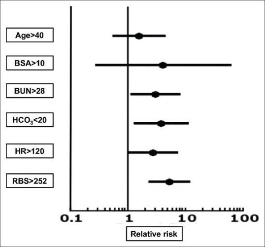
Forest plot showing relative risk ratio (after univariate logistic regression) of components of SCORTEN to mortality (*Relative risk of malignancy could not be calculated as none of the patients had malignancy in our study)
Table 4.
Multivariate logistic regression to predict mortality among SJS-TEN patients
| Independent predictors | Adjusted Odds ratio | 95% CI for adjusted Odds Ratio | P | |
|---|---|---|---|---|
| Lower | Upper | |||
| Blood urea nitrogen (> 28 mg/dL) | 2.785 | 0.379 | 20.452 | 0.314 |
| Serum HCO3- (<20 meq/mL) | 3.313 | 0.576 | 19.050 | 0.180 |
| Random blood sugar (>252 mg/dL) | 28.555 | 2.040 | 399.734 | 0.013 |
| Detached BSA (%) | 1.086 | 0.985 | 1.196 | 0.097 |
| Sepsis | Excluded from the final model due to multicollinearity between detached BSA (%) and sepsis | |||
| Constant | 0.0001 | 0.005 | ||
Comparison between corticosteroids and cyclosporine as treatment modality
Twenty-one (41.2%) patients were administered oral corticosteroids (prednisolone 0.5–1 mg/kg/day) and 24 (47.1%) patients were administered oral cyclosporine 3–5 mg/kg/day in two divided doses. Both steroids and cyclosporine were tapered/stopped abruptly over a period of 5–7 days once the disease activity had ceased. Baseline characteristics of patients receiving prednisolone and cyclosporine, i.e., age, sex, detached BSA, and SCORTEN at presentation were comparable. Patients in the cyclosporine group had delayed presentation (5.71 ± 4.12 days) as compared to patients in the prednisolone group (3.76 ± 1.79) but this was not statistically significant (P = 0.051). Mean duration of treatment was 5.95 ± 2.63 and 7.17 ± 2.73 days in patients who received prednisolone and cyclosporine, respectively. Time taken for reepithelization was slightly more in patients who received prednisolone than cyclosporine (6.71 ± 2.45 vs. 6.12 ± 2.27 days; P = 0.41), while the duration of hospital stay was more in patients who received cyclosporine. The incidence of sepsis was observed in 4 patients each in the prednisolone and cyclosporine group. Complications like acute kidney injury (AKI), transaminitis, and leucopenia were seen more in prednisolone group (6/21, 28.6%) than in cyclosporine group (1/24, 4.2%; P = 0.04). Mortality rate was higher in patients receiving prednisolone than cyclosporine, though not statistically significant (19% vs. 12.5%; P = 0.68) [Table 5].
Table 5.
Comparison of patients who received systemic corticosteroids vs. systemic cyclosporine
| Characteristics | Patients who received corticosteroids (n=21) | Patients who received cyclosporine (n=24) | P |
|---|---|---|---|
| Age (years, mean±SD) | 37±15.7 | 40.8±19.1 | 0.47 |
| Sex (male:female) | 12:9 | 13:11 | 1.0 |
| Diagnosis | |||
| SJS | 5 | 2 | 0.34 |
| SJS-TEN overlap | 6 | 6 | |
| TEN | 10 | 16 | |
| Duration of drug interval; (days, mean±SD) | 12.76±11.04 | 19.79±12.06 | 0.05 |
| Delay in presentation (days, mean±SD) | 3.76±1.79 | 5.71±4.12 | 0.05 |
| Detached BSA at presentation (%, mean±SD) | 26.61±14.19 | 28.25±10.75 | 0.66 |
| SCORTEN at presentation (mean±SD) | 2.24±1.3 | 2.04±0.69 | 0.51 |
| Duration of treatment; (days, mean±SD) | 5.95±2.63 | 7.17±2.73 | 0.13 |
| Days for reepithelization; (mean±SD) | 6.71±2.45 | 6.12±2.27 | 0.41 |
| Duration of hospital stay; (days, mean±SD) | 8±3.13 | 9.17±5.67 | 0.41 |
| Sepsis | 4/21 (19%) | 4/24 (16.7%) | 1.0 |
| Complications | 6/21 (28.6%) | 1/24 (4.2%) | 0.04 |
| Mortality | 4/21 (19%) | 3/24 (12.5%) | 0.69 |
*BSA: Body surface area, IVIG: Intravenous immunoglobulin, SCORTEN: SCORe of toxic epidermal necrolysis, SD: Standard deviation, SJS: Stevens-Johnson Syndrome, TEN: Toxic epidermal necrolysis
Discussion
Owing to the rarity of severe adverse drug reactions, controlled prospective studies are not very feasible. Hence, most of the knowledge and management guidelines about SJS/TEN are based on retrospective studies or case series. This study presents a retrospective analysis of 51 SJS/TEN patients seen over 5 years at a tertiary care hospital in north India. Female preponderance has been seen in previous studies,[8] while we observed a male preponderance (52.9%). Although SJS/TEN has been defined as severe cutaneous adverse reactions, a definitive drug history may not always be elicited.[9,10] In our study, all 51 patients had a temporal association with drug intake. Commonly implicated drugs in SJS/TEN reported in literature included anticonvulsants, antibiotics, and NSAIDs.[8,9,11] In our study as well, anticonvulsants (phenytoin, carbamazepine, lamotrigine, and valproate, 39.2%) were commonest cause for SJS/TEN followed by NSAIDs (paracetamol, ibuprofen, diclofenac, nimesulide, and etoricoxib, 19.6%). Antimicrobials have also been reported as a common cause of SJS/TEN in previous studies.[12] Antibiotics, i.e., cephalosporins and penicillins, were the cause in 6 patients and 7 patients on nevirapine antiretroviral therapy-developed SJS/TEN to nevirapine. Nevirapine has been found to be associated with the highest risk (approximately 0.3%–1%) of developing SJS/TEN among antiretroviral drugs.[13,14] In a study of 50 patients of SJS/TEN in HIV patients, nevirapine was found to be a causal drug in 42 (84%) of patients.[15]
SJS/TEN is usually associated with significant mortality and morbidity with estimated mortality ranging from 10% in SJS to more than 40% in TEN, with septicemia-related multi-organ failure being the commonest cause.[16,17] We observed a mortality rate of 28.6% in TEN and 18.8% in SJS-TEN overlap, while no mortality was seen in 7 patients with SJS. In a retrospective analysis of mortality rates in SJS/TEN, a significant decrease in overall mortality was seen over a period of the last 20 years.[18] In our study, sepsis was seen in 81.8% of patients with mortality, which is an important factor affecting the outcome in patients with SJS/TEN. Other factors significantly associated with poor outcome included more detached BSA and higher SCORTEN at presentation. However, delay in presentation and number of mucosa involved did not affect the outcome significantly. Among the seven components of SCORTEN, age and heart rate >120 bpm were not significantly associated with poor outcome in our study and none of our patients had associated malignancy. Thus, the findings of our study point toward the need for probable modification of the current scoring system in predicting mortality in patients of SJS/TEN and inclusion of sepsis as a significant parameter. A similar observation was made by Vaishampayan et al.[19] in which authors mentioned the need to include other comorbidities like diabetes and tuberculosis in the scoring system. Apart from these, AKI[20] and prior dialysis[21] have been proposed as significant factors to affect the mortality significantly in SJS/TEN. Moreover, SCORTEN has been reported to overestimate the mortality rates in many previous studies.[22,23] Another important reason for overestimating these rates is the scoring of detached/detachable BSA >10% as improvement in supportive measures, specialized centers for SJS/TEN, development of guidelines for the treatment of SJS/TEN, and standardized treatment protocols over past few years has led to decreased mortality in SJS/TEN patients with BSA 10%–30%.
Till now, there are no randomized controlled trials as far as management of SJS/TEN is concerned. Different studies have shown variable efficacy of systemic steroids, oral cyclosporine, IVIG, and supportive treatment only in the management of SJS/TEN. A recent meta-analysis of systemic therapies in SJS/TEN concluded that corticosteroids and cyclosporine are most promising therapeutic options.[24] In our study, supportive treatment was given to all patients and 88.2% patients received either prednisolone 0.5 mg/kg/day or cyclosporine 3–5 mg/kg/day along with supportive treatment. Corticosteroids and cyclosporine have to be started early in the course of the disease to arrest the progression of epidermal necrosis as a delay in seeking treatment can affect the outcome.[25] However, in our study, the outcome was not affected by the duration of delay in seeking treatment. Both corticosteroids and cyclosporine were comparable with respect to the duration of treatment required, mean time to reepithelization, and duration of hospital stay. However, mortality rate (19% vs. 12.5%; P = 0.69) and complication rate (28.6% vs. 4.2%; P = 0.04) were higher in patients who received corticosteroids than those treated with cyclosporine. The findings of our study were similar to the study by Singh et al. who observed slightly better efficacy of cyclosporine over corticosteroids in uncomplicated SJS/TEN.[26]
In our hospital, being a tertiary care and referral center, most patients present after receiving some treatment at local health care centers which leads to increase in the time interval before presentation, delayed withdrawal of culprit drug, and change in the actual initial presentation of the disease. Earliest withdrawal of culprit drug is of paramount importance in the management of SJS-TEN. Thus, these factors may affect the outcome in SJS-TEN patient and more clinicoepidemiological data is needed regarding time interval between onset of symptoms and presentation to the specialist, and time interval between onset of symptoms and withdrawal of offending drug for a better outcome in these patients.
The main limitations of our study were its retrospective design, small sample size, and single-center study. There may be inter-physician variations assessing the detached BSA leading to the heterogeneity of data. However, most of the previous studies had similar limitations.[8,23]
Conclusion
Anticonvulsants, NSAIDs, and antibiotics are the most commonly implicated drugs as seen in our study also. Immediate identification and removal of the possible causative agents is a key part of management. A higher detached BSA, presence of sepsis, higher blood urea nitrogen and RBS, and lower serum HCO3− levels were associated with increased mortality. Other components of SCORTEN like age and heart rate were not significantly associated with poor outcome in our study. None of our patients had associated malignancy. Although the calculation of a SCORTEN is helpful in predicting prognosis and mortality rate, slight modifications are needed for better prognostication.
Financial support and sponsorship
Nil.
Conflicts of interest
There are no conflicts of interest.
References
- 1.Bigby M, Jick S, Jick H, Arndt K. Drug-induced cutaneous reactions. A report from the Boston Collaborative Drug Surveillance Program on 15,438 consecutive inpatients, 1975 to 1982. JAMA. 1986;256:3358–63. doi: 10.1001/jama.256.24.3358. [DOI] [PubMed] [Google Scholar]
- 2.Diphoorn J, Cazzaniga S, Gamba C, Schroeder J, Citterio A, Rivolta AL, et al. Incidence, causative factors and mortality rates of Stevens-Johnson syndrome (SJS) and toxic epidermal necrolysis (TEN) in northern Italy: Data from the REACT registry. Pharmacoepidemiol Drug Saf. 2016;25:196–203. doi: 10.1002/pds.3937. [DOI] [PubMed] [Google Scholar]
- 3.Frey N, Jossi J, Bodmer M, Bircher A, Jick SS, Meier CR, et al. The Epidemiology of Stevens-Johnson syndrome and toxic epidermal necrolysis in the UK. J Invest Dermatol. 2017;137:1240–7. doi: 10.1016/j.jid.2017.01.031. [DOI] [PubMed] [Google Scholar]
- 4.Hsu DY, Brieva J, Silverberg NB, Silverberg JI. Morbidity and mortality of Stevens-Johnson syndrome and toxic epidermal necrolysis in United States Adults. J Invest Dermatol. 2016;136:1387–97. doi: 10.1016/j.jid.2016.03.023. [DOI] [PubMed] [Google Scholar]
- 5.Roujeau JC, Bastuji-Garin S. Systematic review of treatments for Stevens-Johnson syndrome and toxic epidermal necrolysis using the SCORTEN score as a tool for evaluating mortality. Ther Adv Drug Saf. 2011;2:87–94. doi: 10.1177/2042098611404094. [DOI] [PMC free article] [PubMed] [Google Scholar]
- 6.Bastuji-Garin S, Rzany B, Stern RS, Shear NH, Naldi L, Roujeau JC. Clinical classification of cases of toxic epidermal necrolysis, Stevens-Johnson syndrome, and erythema multiforme. Arch Dermatol. 1993;129:92–6. [PubMed] [Google Scholar]
- 7.Sassolas B, Haddad C, Mockenhaupt M, Dunant A, Liss Y, Bork K, et al. ALDEN, an algorithm for assessment of drug causality in Stevens-Johnson syndrome and toxic epidermal necrolysis: Comparison with case-control analysis. Clin Pharmacol Ther. 2010;88:60–8. doi: 10.1038/clpt.2009.252. [DOI] [PubMed] [Google Scholar]
- 8.Carrasquillo OY, Santiago-Vazquez M, Cardona R, Cruz-Manzano M, Figueroa LD. Stevens-Johnson syndrome and toxic epidermal necrolysis: A retrospective descriptive study. Int J Dermatol. 2019;58:1293–9. doi: 10.1111/ijd.14493. [DOI] [PubMed] [Google Scholar]
- 9.Schopf E, Stuhmer A, Rzany B, Victor N, Zentgraf R, Kapp JF. Toxic epidermal necrolysis and Stevens-Johnson syndrome. An epidemiologic study from West Germany. Arch Dermatol. 1991;127:839–42. doi: 10.1001/archderm.1991.01680050083008. [DOI] [PubMed] [Google Scholar]
- 10.Roujeau JC, Guillaume JC, Fabre JP, Penso D, Flechet ML, Girre JP. Toxic epidermal necrolysis (Lyell syndrome). Incidence and drug etiology in France, 1981-1985. Arch Dermatol. 1990;126:37–42. doi: 10.1001/archderm.126.1.37. [DOI] [PubMed] [Google Scholar]
- 11.Tan SK, Tay YK. Profile and pattern of Stevens-Johnson syndrome and toxic epidermal necrolysis in a general hospital in Singapore: Treatment outcomes. Acta Derm Venereol. 2012;92:62–6. doi: 10.2340/00015555-1169. [DOI] [PubMed] [Google Scholar]
- 12.Sanmarkan AD, Sori T, Thappa DM, Jaisankar TJ. Retrospective analysis of stevens-johnson syndrome and toxic epidermal necrolysis over a period of 10 years. Indian J Dermatol. 2011;56:25–9. doi: 10.4103/0019-5154.77546. [DOI] [PMC free article] [PubMed] [Google Scholar]
- 13.Warren KJ, Boxwell DE, Kim NY, Drolet BA. Nevirapine-associated Stevens-Johnson syndrome. Lancet. 1998;351:567. doi: 10.1016/S0140-6736(98)24008-6. [DOI] [PubMed] [Google Scholar]
- 14.Pollard RB, Robinson P, Dransfield K. Safety profile of nevirapine, a nonnucleoside reverse transcriptase inhibitor for the treatment of human immunodeficiency virus infection. Clin Ther. 1998;20:1071–92. doi: 10.1016/s0149-2918(98)80105-7. [DOI] [PubMed] [Google Scholar]
- 15.Rotunda A, Hirsch RJ, Scheinfeld N, Weinberg JM. Severe cutaneous reactions associated with the use of human immunodeficiency virus medications. Acta Derm Venereol. 2003;83:1–9. doi: 10.1080/00015550310002611. [DOI] [PubMed] [Google Scholar]
- 16.Sekula P, Dunant A, Mockenhaupt M, Naldi L, Bouwes Bavinck JN, Halevy S, et al. Comprehensive survival analysis of a cohort of patients with Stevens-Johnson syndrome and toxic epidermal necrolysis. J Invest Dermatol. 2013;133:1197–204. doi: 10.1038/jid.2012.510. [DOI] [PubMed] [Google Scholar]
- 17.Schwartz RA, McDonough PH, Lee BW. Toxic epidermal necrolysis: Part II.Prognosis, sequelae, diagnosis, differential diagnosis, prevention, and treatment. J Am Acad Dermatol. 2013;69:187. doi: 10.1016/j.jaad.2013.05.002. e1-16; quiz 203-4. [DOI] [PubMed] [Google Scholar]
- 18.Bettuzzi T, Penso L, de Prost N, Hemery F, Hua C, Colin A, et al. Trends in mortality rates for Stevens-Johnson syndrome and toxic epidermal necrolysis: Experience of a single centre in France between 1997 and 2017. Br J Dermatol. 2020;182:247–8. doi: 10.1111/bjd.18360. [DOI] [PubMed] [Google Scholar]
- 19.Vaishampayan SS, Das AL, Verma R. SCORTEN: Does it need modification? Indian J Dermatol Venereol Leprol. 2008;74:35–7. doi: 10.4103/0378-6323.38405. [DOI] [PubMed] [Google Scholar]
- 20.Lee TH, Lee CC, Ng CY, Chang MY, Chang SW, Fan PC, et al. The influence of acute kidney injury on the outcome of Stevens-Johnson syndrome and toxic epidermal necrolysis: The prognostic value of KDIGO staging. PLoS One. 2018;13:e0203642. doi: 10.1371/journal.pone.0203642. [DOI] [PMC free article] [PubMed] [Google Scholar]
- 21.Noe MH, Rosenbach M, Hubbard RA, Mostaghimi A, Cardones AR, Chen JK, et al. Development and validation of a risk prediction model for in-hospital mortality among patients with Stevens-Johnson syndrome/toxic epidermal necrolysis-ABCD-10. JAMA Dermatol. 2019;155:448–54. doi: 10.1001/jamadermatol.2018.5605. [DOI] [PMC free article] [PubMed] [Google Scholar]
- 22.Micheletti RG, Chiesa-Fuxench Z, Noe MH, Stephen S, Aleshin M, Agarwal A, et al. Stevens-Johnson syndrome/toxic epidermal necrolysis: A multicenter retrospective study of 377 adult patients from the United States. J Invest Dermatol. 2018;138:2315–21. doi: 10.1016/j.jid.2018.04.027. [DOI] [PubMed] [Google Scholar]
- 23.Imahara SD, Holmes JH, 4th, Heimbach DM, Engrav LE, Honari S, Klein MB, et al. SCORTEN overestimates mortality in the setting of a standardized treatment protocol. J Burn Care Res. 2006;27:270–5. doi: 10.1097/01.BCR.0000216532.71360.9B. [DOI] [PubMed] [Google Scholar]
- 24.Zimmermann S, Sekula P, Venhoff M, Motschall E, Knaus J, Schumacher M, et al. Systemic immunomodulating therapies for Stevens-Johnson syndrome and toxic epidermal necrolysis: A systematic review and meta-analysis. JAMA Dermatol. 2017;153:514–22. doi: 10.1001/jamadermatol.2016.5668. [DOI] [PMC free article] [PubMed] [Google Scholar]
- 25.Gupta LK, Martin AM, Agarwal N, D'Souza P, Das S, Kumar R, et al. Guidelines for the management of Stevens-Johnson syndrome/toxic epidermal necrolysis: An Indian perspective. Indian J Dermatol Venereol Leprol. 2016;82:603–25. doi: 10.4103/0378-6323.191134. [DOI] [PubMed] [Google Scholar]
- 26.Singh GK, Chatterjee M, Verma R. Cyclosporine in Stevens Johnson syndrome and toxic epidermal necrolysis and retrospective comparison with systemic corticosteroid. Indian J Dermatol Venereol Leprol. 2013;79:686–92. doi: 10.4103/0378-6323.116738. [DOI] [PubMed] [Google Scholar]