Abstract
Background:
Intradural extramedullary spinal cord tumors (IESCT) account for approximately two-thirds of largely benign intraspinal neoplasms. They occasionally present with acute neurological deterioration warranting emergent surgical intervention.
Methods:
Here, we reviewed a series of 31 patients with intradural extramedullary spinal tumors who underwent surgery from 2012 to 2019. Patients averaged 50.8 years of age, and there were 16 males and 15 females. Patients were followed for a minimum of 1 year. Multiple clinical outcome variables were studied (e.g., Karnofsky Performance Score [KPS], visual analog scale (VAS), and Frankel grade).
Results:
The majority of IESCT tumors were found in the thoracic spine 18 (58.06%) followed by the lumbar 8 (25.80%), cervical 1 (3.22%), and combined junctional tumors 4 (12.90%) (cervicothoracic-02 and thoracolumbar-02). Histopathological diagnoses included schwannomas-16 (51.61%), meningiomas-11 (35.48%), lipomas-2 (6.45%), hemangiomas-1 (3.22), and ependymomas-01 (03.22%). The VAS score was reduced in all cases, while KPS and Frankel grades were significantly improved. Complications included cerebrospinal fluid leakage, new/residual paresthesias, and tumor recurrence (12.50%).
Conclusion:
Most intradural extramedullary tumors are benign and are readily diagnosed utilizing MRI scans. Notably, good functional outcomes follow surgical intervention.
Keywords: Clinical outcome, Intradural extramedullary tumors, Spinal tumors, Surgical excision

INTRODUCTION
Primary spinal cord tumors constitute 2–4% of all central nervous system neoplasms; they are characterized as extradural, intradural extramedullary (IDEM: 65%), and intramedullary.[1] The most commonly seen IDEM tumors are schwannomas, neurofibromas, and meningiomas. The less frequently encountered lesions include ependymomas, lipomas, hemangiomas, metastatic deposits, paragangliomas, nerve sheath myxomas, and vascular tumors.[6] MRI is the investigation of choice, and the optimal management is gross total excision for symptomatic lesions.[9,10] Here, we examined the surgical outcomes of 31 patients with IDEM spinal cord tumors operated by a single surgeon and reviewed the clinical, radiological, surgical management, and histopathology of these lesions.
MATERIALS AND METHODS
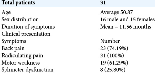
There were 31 intradural extramedullary tumors operated on between 2012 and 2019 by single surgeon. Karnofsky Performance Scores (KPSs), visual analog scales, and Frankel grades were studied pre-operatively, at the time of discharge, 3 and 12 months post-operatively. Patients averaged 50.87 years of age and included 16 males and 15 females. They were symptomatic on average for nearly a year (e.g., mean-11.56 months) before diagnosis and exhibited radiculopathy (RS: 100%), back pain (BP: 74.19%), motor deficits (MW: 61.29 %), and sphincter dysfunction (SD: 25.80 %) [Table 1].
Table 1:
Demographic data and clinical presentation on initial examination.
Radiological evaluation of location of tumors
MR studies determined the location and the size of these intradural lesions (TOIDS %) [Figures 1-5]. Most commonly, tumors were located in the thoracic (58.06%), followed by the lumbar (25.80%), junctional (CT-02, TL-02), and cervical (3.22%) regions [Table 2]. IDEM tumors were located anteriorly (4 cases), laterally (21 cases), and posteriorly (6 cases); anterior tumors were between “10 and 2 o’clock”, posterior tumors from “4 to 8 o’clock”, and lateral from “2 to 4 o’clock” or “8 to 10 o’clock.”[3] All four anterior tumors had produced significant neurological deficits [Table 3]. The tumor occupancy ratios were more than 80% in 45.16% and less than 80% in 54.84% patients.
Figure 1:
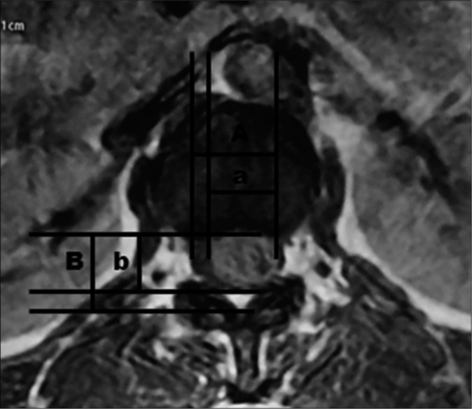
The percentage of tumor occupying the intradural space (TOIDS%) - Transverse diameter of the tumor mass + the longitudinal diameter of the tumor mass/the transverse diameter of the intradural space + the longitudinal diameter of the intradural space ×100.
Figure 5:
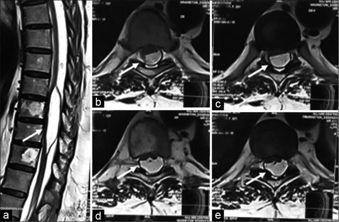
Lipoma (Arrow)– (a) (sagittal T2), (b-e) (axial T1 and axial T2 images) - T1/T2 hyperintense lesion occupying posterior part of the spinal canal compresses and displaces cord anteriorly. (Add fat sat and post-contrast images if possible).
Table 2:
Tumor incidence by anatomic location.

Table 3:
Association of axial location of the tumor within cord with neurological involvement.

Figure 2:
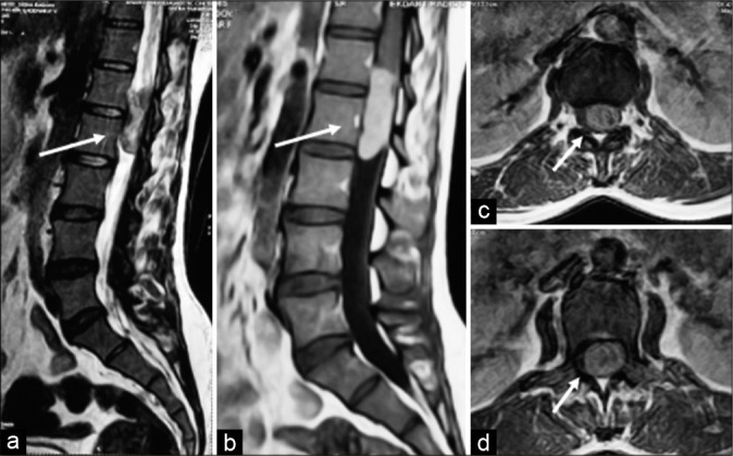
Schwannoma (Arrow) – (a) (sagittal T2), (b) (post-contrast T1) lobulated heterogenous signal intensity lesion shows intense enhancement. (c) (axial T2), and (d) (post-contrast T1) lobulated heterogenous T1/T2 hyperintense intradural extramedullary lesion occupying left half of the spinal canal displacing cauda equina nerve roots toward the right.
Figure 3:
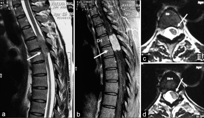
Meningioma (Arrow) – (a) (sagittal T2), (b) (post-contrast sagittal T1), (c) (axial T2), and (c) (post-contrast Axial 2D) - heterogenous mildly T2 hyperintense intradural extramedullary lesion with broad dural base and it shows intense contrast enhancement with small enhancing dural tail.
Figure 4:
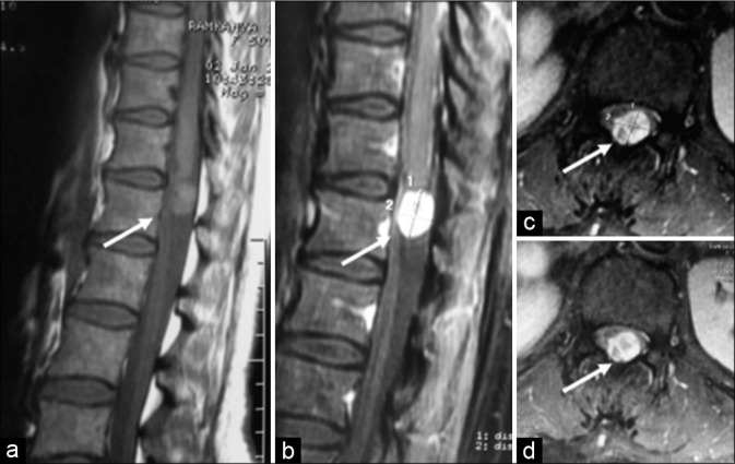
Hemangioma (Arrow) – (a) (sagittal T1), (b) (post-contrast sagittal T1), and (c and d) (post-contrast axial T1 images) - Fairly marginated extramedullary intradural lesion appearing isointense to the adjacent cord on sagittal T1 sequence (3a image) occupying postero-central part of spinal canal, compresses and displaces cord anteriorly, and shows intense contrast enhancement.
Surgery for 31 intradural extramedullary tumors
Under X-ray/fluoroscopic guidance and utilizing intraoperative neural monitoring, all patients underwent laminectomies.[2] For the anterior lesions, this additionally required unilateral medial facetectomies and cutting the paired denticulate ligament on one side to allow lateral retraction of the spinal cord. For lateral or posterior tumors, full facetectomies were not warranted.
Statistical analysis
The MedCalc version 19.4.0 was used. For continuous variables, means were compared using Mann–Whitney U-test, Wilcoxon signed-rank test, and Kruskal–Wallis test. Pearson’s correlation coefficient was used to calculate the correlation between tumor occupancy ratio and KPS. For categorized variables, Pearson’s chi-squared test or Fisher’s exact test were being used. P < 0.05 was considered to be statistically significant and < 0.005 as highly significant.
RESULTS
Pathology and location of intradural extramedullary tumors
The histopathological diagnoses included 16 schwannomas, 11 meningiomas, 2 lipomas, 1 hemangioma, and 1 ependymoma. Most meningiomas were located dorsally (81.81%) and schwannomas’ most common location is lower dorsal-lumbar group (93.75%).
Outcomes
All patients’ symptoms and neurological signs improved post-operatively. There were 1 patient with Frankel’s grade B, 10 patients with grade C, 8 patients with grade D, and 12 patients with grade E pre-operatively. Post-operatively, there were 6 patients with grade D and 25 with Grade E. The pre-operative neurological deficits improved within 8 postoperative weeks in most cases and within 1 year in all cases [Table 4]. Once the TOR increased, disability (KPS and visual analog scale [VAS]) also increased [Table 5]. VAS scores decreased, KPSs improved uniformly, while Frankel’s grade was better in all 19 neurologically involved cases out of 31 total cases [Table 6].
Table 4:
Functional outcome using the Frankel grade.

Table 5:
Association of tumor occupancy ratio with KPS and VAS functional score.
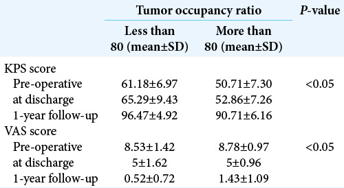
Table 6:
Functional scoring (n=31).

Complications
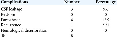
The post-operative complications included 3 CSF leaks, 4 with new or persistent paresthesias, and 1 with current tumor [Table 7].
Table 7:
Complication of surgery.
DISCUSSION
Intradural extramedullary tumors, that are typically benign, account for two-thirds of primary spinal tumors.[4] Their location determines the clinical presentation, ease of surgical excision, and outcome. Plain radiograph can help to identify pedicle erosion, vertebral body erosion, foraminal widening, and scoliosis. MRI imaging is the investigation of choice and readily shows their well-delineated intradural, extramedullary location; they appear isointense on T1W images and hyperintense on T2W images with homogeneously enhancement with gadolinium.[8] The optimal treatment is microscopic gross total surgical excision.
Here, we retrospectively evaluated 31 cases of IDEM tumors, 27 of which were totally removed, and 4 were partially excised. The mean age of presentation in our study was 50.87 years, and the mean duration of illness was 11.56 months, most patients were male (51.61%), and no significant difference was found between the clinical presentation and duration of illness between the genders. The literature shows the female preponderance in western countries while Asian studies[4,9] favors male preponderance although our study reflects more male ratio which may be because of social stigma or delayed follow-up of female patients in Indian population. The female preponderance of meningioma is a well-known entity and our study supports it. We found that meningioma favors the elderly female population while schwannoma is more in young male population similar to the previous literature.[9]
We have assessed the axial/sagittal location of tumors with outcome and complication. Ventrally located upper thoracic spine IDEM tumors were correlated with higher neurological involvement and poorer post-operative outcomes as in our four cases.[5] However, on sagittal location of IDEM tumors, upper thoracic spine group was likely to have more myelopathy which is explained by the higher cord-to-canal ratio, as well as the tenuous vascular supply to that region of the spinal cord. Schwannoma grows slowly compared to meningioma, so can be observed for longer period. However, meningiomas are constantly grows in size, and also, due to their common location in the thoracic spine and anterolateral axial location, surgery would be considered early.[7]
We have also analyzed the tumor occupancy ratio with the disability score and found that once the tumor occupancy increased, the disability also increased. We have divided TOR in two groups; if the TOR was above 80, KPS and VAS scores were poor while those in patients with ratios below 80 demonstrated better outcomes.[9]
We had done 27 gross total excisions and 4 partial excisions of tumors and found one recurrence case. We have done coagulation and cutting of involved rootlet in nerve sheath tumor and found no functional neurological deficit even in ventrally located tumors except sensory deficits in few cases in the involved region. Approximately 20% of patients in our study had residual focal deficits, none of which were disabling. Notably, two-thirds of patients had excellent postoperative results, a finding corroborated by the literature recovery rates (e.g., 62–88% cases).[1,4,9,10] The post-operative recurrence rate of intradural extramedullary spinal cord tumors varied between 5% and 16% in the literature and here was 5%.[1,4] Reflecting the following risk factors are ventral tumor location, high thoracic level, extradural invasion, nanogenic tumors, ependymomas, and incomplete removal of the dura mater in meningioma.
CONCLUSION
Intradural extramedullary tumors are mostly benign, are readily diagnosed on MRI scans, and are typically best managed with gross total surgical excision. Notably, anteriorly located lesions have the greatest pre-operative deficits and highest risk of post-operative residual neurological dysfunction.
Footnotes
How to cite this article: Patel P, Mehendiratta D, Bhambhu V, Dalvie S. Clinical outcome of intradural extramedullary spinal cord tumors: A single-center retrospective analytical study. Surg Neurol Int 2021;12:145.
Contributor Information
Pratik Patel, Email: patel.pratik323@gmail.com.
Dhanish Mehendiratta, Email: drdhanish@orthodocs.in.
Vivek Bhambhu, Email: vivekbhambhu88@gmail.com.
Samir Dalvie, Email: sdalvie@hotmail.com.
Declaration of patient consent
Patient’s consent not required as patients identity is not disclosed or compromised.
Financial support and sponsorship
Nil.
Conflicts of interest
There are no conflicts of interest.
REFERENCES
- 1.Ahsan MK, Awwal MA, Khan SI, Haque MH, Zaman N. Intradural extramedullary spinal cord tumours: A retrospective study of surgical outcomes. Bangabandhu Sheikh Mujib Med Univ J. 2016;9:11–9. [Google Scholar]
- 2.Chung JY, Lee JJ, Kim HJ, Seo HY. Characterization of magnetic resonance images for spinal cord tumors. Asian Spine J. 2008;2:15–21. doi: 10.4184/asj.2008.2.1.15. [DOI] [PMC free article] [PubMed] [Google Scholar]
- 3.Cohen-Gadol AA, Zikel OM, Koch CA, Scheithauer BW, Krauss WE. Spinal meningiomas in patients younger than 50 years of age: A 21-year experience. J Neurosurg. 2003;98(Suppl 3):258–63. doi: 10.3171/spi.2003.98.3.0258. [DOI] [PubMed] [Google Scholar]
- 4.Govind M, Mittal R, Sharma A, Gandhi A. Intradural extramedullary spinal cord tumors: A retrospective study at tertiary referral hospital. Rom Neurosurg. 2016;30:106–12. [Google Scholar]
- 5.Mehta AI, Adogwa O, Karikari IO, Thompson P, Verla T, Null UT, et al. Anatomical location dictating major surgical complications for intradural extramedullary spinal tumors: A 10-year single-institutional experience. J Neurosurg Spine. 2013;19:701–7. doi: 10.3171/2013.9.SPINE12913. [DOI] [PubMed] [Google Scholar]
- 6.Nizami FA, Mustafa SA, Nazir R, Salaria H, Singh GP, Gadgotra P. Intradural extramedullary spinal cord tumors: Surgical outcome in a newly developed tertiary care hospital. Int J Sci Study. 2017;5:48–53. [Google Scholar]
- 7.Ozawa H, Onoda Y, Aizawa T, Nakamura T, Koakutsu T, Itoi E. Natural history of intradural-extramedullary spinal cord tumors. Acta Neurol Belg. 2012;112:265–70. doi: 10.1007/s13760-012-0048-7. [DOI] [PubMed] [Google Scholar]
- 8.Schweitzer JS, Batzdorf U. Ependymoma of the cauda equina region: Diagnosis, treatment, and outcome in 15 patients. Neurosurgery. 1992;30:202–7. doi: 10.1227/00006123-199202000-00009. [DOI] [PubMed] [Google Scholar]
- 9.Singh PR, Pandey TK, Ahmad F, Chhabra DK. A prospective observational study of clinical outcome of operated patients of intradural extramedullary spinal cord tumor in our tertiary care center. Rom Neurosurg. 2018;32:632–40. [Google Scholar]
- 10.Song KW, Shin SI, Lee JY, Kim GL, Hyun YS, Park DY. Surgical results of intradural extramedullary tumors. Clin Orthop Surg. 2009;1:74–80. doi: 10.4055/cios.2009.1.2.74. [DOI] [PMC free article] [PubMed] [Google Scholar]


