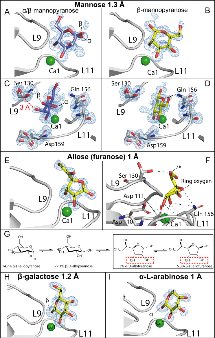
FIG 3.
Ligand-binding site of MpPA14 in complex with mannose, allose, galactose, and l-arabinose. Side (A) and top views (C) of α- and β-mannopyranose rings anchored to MpPA14 via their 3,4-diols. Electron density for only β-mannopyranose was seen for the binding mode via the 2,3-diol. (B) Side view; (D) top-down view. The distance between the β-carbon of serine on L9 and C-2 hydroxyl oxygen of α/β-mannopyranose is indicated by a red dashed line. (E) Side-view of β-allofuranose in the MpPA14 ligand-binding site. (F) Detailed polar interactions (black dashed lines) between β-allofuranose and MpPA14. Amino acid residues involved in allose-MpPA14 interaction are labeled. The color scheme is the same as that in Fig. 3. (G) Equilibria of allose anomers in aqueous solution at 30°C. The diol of allofuranose responsible for binding MpPA14 is indicated by a red dashed box. Side-view of β-galactose (H) and α-l-arabinose (I) in the MpPA14 ligand-binding site.

