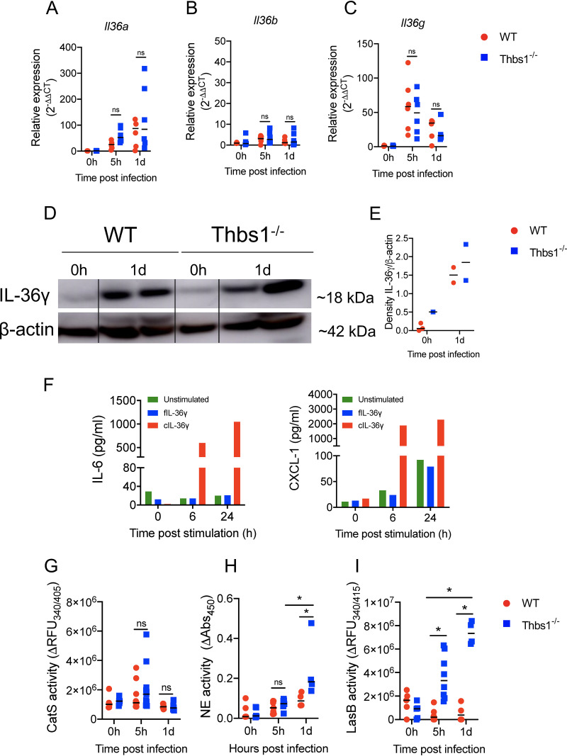
FIG 2.
TSP-1 does not alter IL-36 cytokines expression but downregulates the proteolytic environment required for activation. Thbs1−/− and WT mice were i.t. inoculated with P. aeruginosa at an inoculum of 106 CFU, and lung tissue (A) Il36a, (B) Il36b, and (C) Il36g transcripts were measured at 5 hpi and 1 dpi by quantitative reverse transcription-PCR (qRT-PCR) using gadph as the internal housekeeping gene. (D and E) IL-36γ expression in the lungs measured by Western blot at 1 dpi. Density expression of IL-36γ is normalized to β-actin. (F) IL-6 and CXCL-1 production by bone marrow-derived dendritic cells (BMDCs) after stimulation with full-length (fIL-36γ) or cleaved IL-36γ (cIL-36γ, S18 isoform). (G) Cathepsin S (CatS), (H) neutrophil elastase (NE), and (I) LasB activity were measured in the BALF of WT and Thbs1−/− mice at 5 hpi and 1 dpi using the specific substrates 2-aminobenzoyl-l-alanyl-glycyl-l-leucyl-l-alanyl-para-nitro-benzyl-amide, N-methoxysuccinyl-Ala-Ala-Pro-Val p-nitroanilide, and Mca-GRWPPMG∼LPWEK(Dnp)-D-R-NH2, respectively. *, P < 0.05, for single comparisons, the Shapiro-Wilk test was used to assess normal distribution, followed by a Mann-Whitney U test or a parametric t test. A two-way ANOVA test was followed by a post hoc test for multiple comparisons over time. Each data point represents an individual mouse, combined from two independent experiments, except for the Western blot and in vitro BMDC stimulation, which were performed once. Lines indicate the median.

