FIG 1.
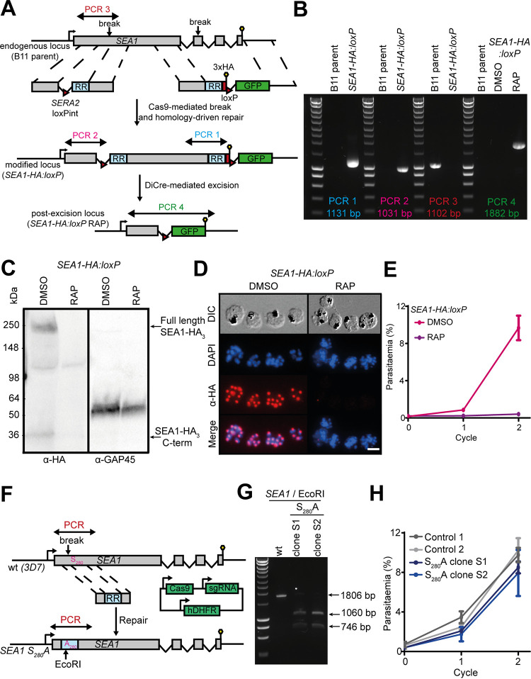
Epitope tagging and conditional disruption of the P. falciparum SEA1 gene confirms an essential role in asexual blood-stage parasite growth. (A) Schematic showing generation of the SEA1-HA:loxP line and RAP-induced gene disruption. Double-headed arrows indicate the regions targeted for amplification by diagnostic PCR. Red arrowheads, loxP sites. Lollipops, stop codons. RR denotes a recodonized region, and GFP denotes green fluorescent protein. Correct excision was expected to result in the expression of GFP fused to a severely truncated form of SEA1. However, GFP expression was not detectable by fluorescence microscopy. (B) PCR verifying the expected gene modifications and efficient excision of the floxed segment upon RAP treatment of an SEA1-HA:loxP clone. Amplified regions are illustrated in panel A. (C) Western blot detection of full-length SEA1-HA3 (∼250 kDa) along with a putative N-terminal processed fragment (∼30 kDa) in extracts of SEA1-HA:loxP schizonts and loss of the signals upon RAP treatment. The right-hand panel shows the same samples probed for GAP45 (PF3D7_1222700) as a loading control. (D) IFA showing epitope tagging and DiCre-mediated disruption of SEA1-HA3 in schizonts. Over 99% of all RAP-treated SEA1-HA:loxP trophozoites examined by IFA were HA negative. Scale bar, 10 μm. (E) Growth curves showing that RAP treatment of SEA1-HA:loxP parasites severely impaired their replication. (F) Schematic representation of Cas9-mediated generation of SEA1-HA:loxP S280A mutant parasites. Double-headed arrows indicate regions targeted for diagnostic PCR in panel G. RR, recodonized region; lollipops, stop codons. (G) PCR verifying correct integration of the recodonized region, including the S280A mutation and EcoRI restriction site. The region targeted for PCR amplification is indicated in panel F. (H) Growth curves comparing proliferation of SEA1-HA:loxP S280A parasites and control parental 3D7 parasites.

