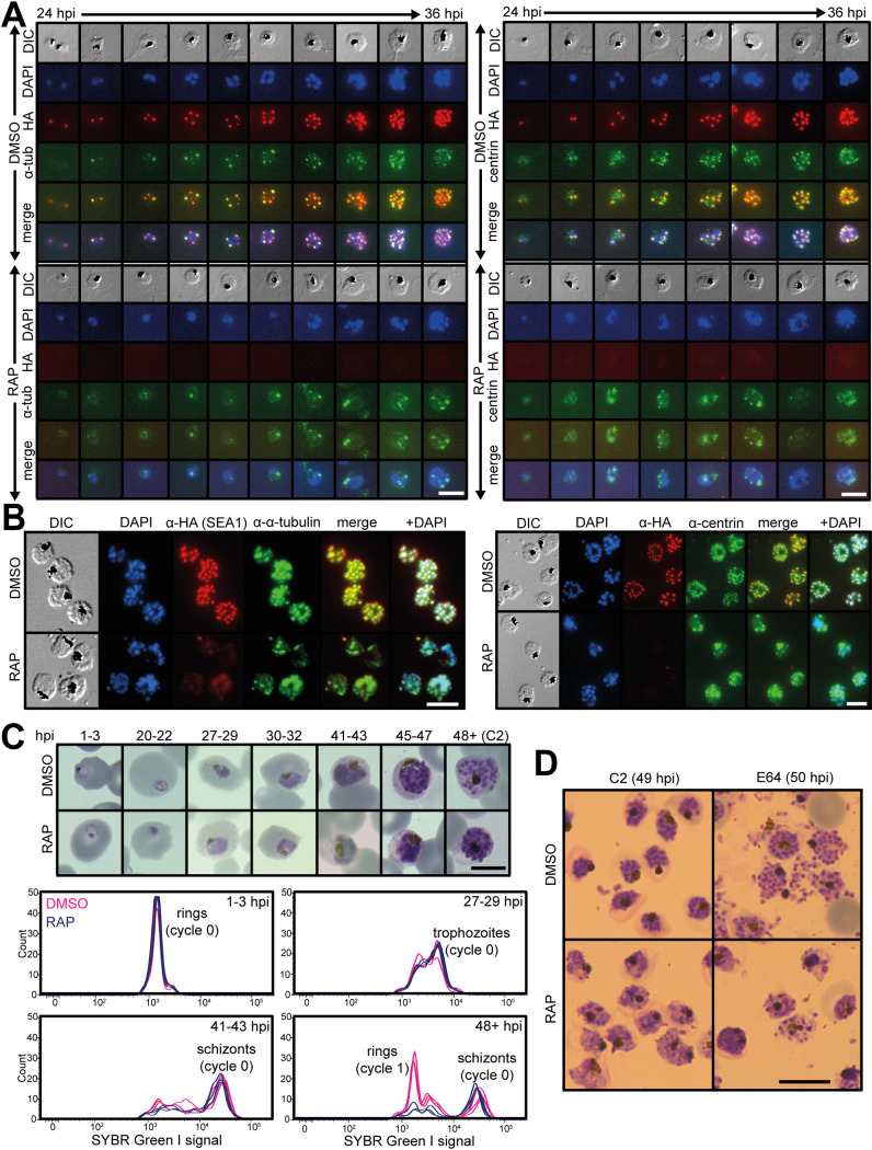
FIG 3.
SEA1-null parasites complete DNA replication but display unusual nuclear morphology. (A) IFA showing localization of SEA1-HA3 in proximity to α-tubulin and centrin throughout the development of SEA1-HA:loxP trophozoites/early schizonts. SEA1-HA3 is absent in RAP-treated parasites. Scale bar, 10 μm. (B) IFA showing localization of SEA1-HA3 in proximity to α-tubulin and centrin in SEA1-HA:loxP schizonts (∼45 h postinvasion, hpi). Scale bar, 10 μm. (C) Images from Giemsa-stained thin films showing the development of SEA1-HA:loxP parasites throughout the cycle of treatment with DMSO or RAP (cycle 0). Selected time points (hpi) are accompanied by plots displaying the DNA content of each infected RBC, as determined by SYBR green I staining and flow cytometry. Scale bar, 5 μm. (D) Images from Giemsa-stained thin films showing mature SEA1-HA:loxP parasites formed at the end of the cycle of treatment with DMSO or RAP (cycle 0). Egress was blocked in these samples by treatment from 45 to 49 hpi with the PKG inhibitor compound 2 (left) and then with the cysteine protease inhibitor E64 (right) from 49 to 50 hpi. Scale bar, 10 μm.

