

Abstract
Background
Thrombolytic therapy is usually reserved for people with clinically serious or massive pulmonary embolism (PE). Evidence suggests that thrombolytic agents may dissolve blood clots more rapidly than heparin and may reduce the death rate associated with PE. However, there are still concerns about the possible risk of adverse effects of thrombolytic therapy, such as major or minor haemorrhage. This is the fourth update of the Cochrane review first published in 2006.
Objectives
To assess the effects of thrombolytic therapy for acute pulmonary embolism.
Search methods
The Cochrane Vascular Information Specialist searched the Cochrane Vascular Specialised Register, CENTRAL, MEDLINE, Embase, and CINAHL databases and the World Health Organization International Clinical Trials Registry Platform and ClinicalTrials.gov trials registers to 17 August 2020. We undertook reference checking to identify additional studies.
Selection criteria
We included randomised controlled trials (RCTs) that compared thrombolytic therapy followed by heparin versus heparin alone, heparin plus placebo, or surgical intervention for people with acute PE (massive/submassive). We did not include trials comparing two different thrombolytic agents or different doses of the same thrombolytic drug.
Data collection and analysis
Two review authors (ZZ, QH) assessed the eligibility and risk of bias of trials and extracted data. We calculated effect estimates using the odds ratio (OR) with a 95% confidence interval (CI) or the mean difference (MD) with a 95% CI. The primary outcomes of interest were death, recurrence of PE and haemorrhagic events. We assessed the certainty of the evidence using GRADE criteria.
Main results
We identified three new studies for inclusion in this update. We included 21 trials in the review, with a total of 2401 participants. No studies compared thrombolytics versus surgical intervention. We were not able to include one study in the meta‐analysis because it provided no extractable data. Most studies carried a high or unclear risk of bias related to randomisation and blinding.
Meta‐analysis showed that, compared to control (heparin alone or heparin plus placebo), thrombolytics plus heparin probably reduce both the odds of death (OR 0.58, 95% CI 0.38 to 0.88; 19 studies, 2319 participants; low‐certainty evidence), and recurrence of PE (OR 0.54, 95% CI 0.32 to 0.91; 12 studies, 2050 participants; low‐certainty evidence). Effects on mortality weakened when six studies at high risk of bias were excluded from analysis (OR 0.71, 95% CI 0.45 to 1.13; 13 studies, 2046 participants) and in the analysis of submassive PE participants (OR 0.61, 95% CI 0.37 to 1.02; 1993 participants). Effects on recurrence of PE also weakened after removing one study at high risk of bias for sensitivity analysis (OR 0.60, 95% CI 0.35 to 1.04; 11 studies, 1949 participants). We downgraded the certainty of evidence to low because of 'Risk of bias' concerns.
Major haemorrhagic events were probably more common in the thrombolytics group than in the control group (OR 2.84, 95% CI 1.92 to 4.20; 15 studies, 2101 participants; moderate‐certainty evidence), as were minor haemorrhagic events (OR 2.97, 95% CI 1.66 to 5.30; 13 studies,1757 participants; low‐certainty evidence). We downgraded the certainty of the evidence to moderate or low because of 'Risk of bias' concerns and inconsistency. Haemorrhagic stroke may occur more often in the thrombolytics group than in the control group (OR 7.59, 95% CI 1.38 to 41.72; 2 studies, 1091 participants).
Limited data indicated that thrombolytics may benefit haemodynamic outcomes, perfusion lung scanning, pulmonary angiogram assessment, echocardiograms, pulmonary hypertension, coagulation parameters, composite clinical outcomes, need for escalation and survival time to a greater extent than heparin alone. However, the heterogeneity of the studies and the small number of participants involved warrant caution when interpreting results.
The length of hospital stay was shorter in the thrombolytics group than in the control group (mean difference (MD) −1.40 days, 95% CI −2.69 to −0.11; 5 studies, 368 participants). Haemodynamic decompensation may occur less in the thrombolytics group than in the control group (OR 0.36, 95% CI 0.20 to 0.66; 3 studies, 1157 participants). Quality of life was similar between the two treatment groups.
None of the included studies provided data on post‐thrombotic syndrome or on cost comparison.
Authors' conclusions
Low‐certainty evidence suggests that thrombolytics may reduce death following acute pulmonary embolism compared with heparin (the effectiveness was mainly driven by one trial with massive PE). Thrombolytic therapy may be helpful in reducing the recurrence of pulmonary emboli but may cause more major and minor haemorrhagic events, including haemorrhagic stroke. More studies of high methodological quality are needed to assess safety and cost effectiveness of thrombolytic therapy for people with pulmonary embolism.
Keywords: Humans, Acute Disease, Bias, Cause of Death, Fibrinolytic Agents, Fibrinolytic Agents/adverse effects, Fibrinolytic Agents/therapeutic use, Hemorrhage, Hemorrhage/chemically induced, Hemorrhage/epidemiology, Heparin, Heparin/adverse effects, Heparin/therapeutic use, Pulmonary Embolism, Pulmonary Embolism/drug therapy, Pulmonary Embolism/mortality, Randomized Controlled Trials as Topic, Recurrence, Thrombolytic Therapy, Thrombolytic Therapy/adverse effects, Thrombolytic Therapy/methods
Plain language summary
Drugs to dissolve pulmonary embolism (blood clot in the lungs)
Background
A pulmonary embolus is a potentially fatal blood clot that lodges in the main artery of the lungs, straining the right side of the heart and affecting blood circulation. People with this condition are at risk of new emboli forming (recurrence). In the case of a massive pulmonary embolism, treatment to restore blood flow is urgently required. Heparin thins the blood, but newer drugs that actively break up the clots (thrombolytics) may act more quickly and may be more effective. These newer drugs include streptokinase, urokinase, and recombinant tissue‐type plasminogen activator. The major complication of this treatment is bleeding.
Key results
We searched the literature and included 21 studies in this update (evidence current to 17 August 2020). These trials involved 2401 adult participants with pulmonary embolism, who were randomly assigned to a thrombolytic agent followed by heparin or heparin alone or heparin plus placebo. No studies compared thrombolytics versus surgical intervention. We were able to use data from 20 clinical trials with a total of 2371 participants. Thrombolytics may lower the likelihood of death and recurrence of blood clots compared to heparin. On the other hand, thrombolytics caused more side effects, including major and minor bleeding events (haemorrhagic events) and haemorrhagic stroke, than heparin alone. Limited information from a number of individual trials show that thrombolytics might be better at improving blood flow through the lungs, heart function, reducing the need for further treatment and time spent in hospital. None of the studies reported on post‐thrombotic syndrome or compared the costs of the different treatments.
Certainty of the evidence
The certainty of the evidence is moderate or low, because of study design limitations (risk of bias), and small sample sizes. We need more large, well‐designed trials to increase our confidence in any benefits of thrombolytic therapy for pulmonary embolism.
Summary of findings
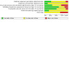
Summary of findings 1. Thrombolytic therapy versus heparin: primary outcome measures for pulmonary embolism.
| Thrombolytic therapy versus heparin: primary outcome measures for pulmonary embolism | |||||
| Patient or population: people with acute PE Setting: hospital Intervention: thrombolytic therapy Comparison: heparin | |||||
| Outcomes | Anticipated absolute effects* (95% CI) | Relative effect (95% CI) | No. of participants (RCTs) | Certainty of the evidence (GRADE) | |
| Risk with heparin | Risk with thrombolytic therapy | ||||
|
Death from all causes (duration of follow‐up: from 7 days to 12 months) |
Study population | OR 0.58 (0.38 to 0.88) | 2319 (19) | ⊕⊕⊝⊝ lowb | |
| 47 per 1000 | 28 per 1000 (19 to 42) | ||||
| Moderatea | |||||
| 49 per 1000 | 29 per 1000 (19 to 43) | ||||
|
Recurrence of pulmonary emboli (duration of follow‐up: from 7 days to 12 months) |
Study population | OR 0.54 (0.32 to 0.91) | 2050 (12) | ⊕⊕⊝⊝ lowb | |
| 39 per 1000 | 21 per 1000 (13 to 36) | ||||
| Moderatea | |||||
| 42 per 1000 | 23 per 1000 (14 to 38) | ||||
|
Major haemorrhagic events (duration of follow‐up: from 7 days to 12 months) |
Study population | OR 2.84 (1.92 to 4.20) | 2101 (15) | ⊕⊕⊕⊝ moderatec | |
| 35 per 1000 | 94 per 1000 (65 to 133) | ||||
| Moderatea | |||||
| 24 per 1000 | 66 per 1000 (46 to 95) | ||||
|
Minor haemorrhagic events (duration of follow‐up: from 7 days to 12 months) |
Study population | OR 2.97 (1.66 to 5.30) | 1757 (13) | ⊕⊕⊝⊝ lowc,d | |
| 96 per 1000 | 239 per 1000 (149 to 359) | ||||
| Moderatea | |||||
| 86 per 1000 | 219 per 1000 (135 to 333) | ||||
| *The basis for the assumed risk (e.g. the median control group risk across studies) is provided in footnotes. The risk in the intervention group (and its 95% confidence interval) is based on the assumed risk in the comparison group and the relative effect of the intervention (and its 95% CI). GRADE Working Group grades of evidence CI: confidence interval; OR: odds ratio; PE: pulmonary embolism; RCT: randomised controlled trial. | |||||
| GRADE Working Group grades of evidence High certainty: We are very confident that the true effect lies close to that of the estimate of the effect Moderate certainty: We are moderately confident in the effect estimate: The true effect is likely to be close to the estimate of the effect, but there is a possibility that it is substantially different Low certainty: Our confidence in the effect estimate is limited: The true effect may be substantially different from the estimate of the effect Very low certainty: We have very little confidence in the effect estimate: The true effect is likely to be substantially different from the estimate of effectGRADE Working Group grades of evidence. | |||||
aMedian control group risk from the studies included in this meta‐analysis. bDowngraded by two levels for very serious risk of bias (due to serious risk of selection, performance and other bias in most included studies). cDowngraded by one level for serious risk of bias (due to serious risk of selection, performance and other bias in some included studies). dDowngraded by one level for inconsistency (due to moderate heterogeneity; I2 = 55%).
Background
Description of the condition
A pulmonary embolus (a blood clot in the artery of the lungs) is a life‐threatening condition known as pulmonary embolism (PE), that is accompanied by significant morbidity and mortality. Massive and submassive PEs are subtypes of PE that are often encountered in the literature, even though the definitions of these subtypes are often vague and can lead to ambiguity (Goldhaber 2002). Because the severity and prognosis of PE vary widely, risk stratification after PE diagnosis is essential. The American Heart Association defines massive PE, submassive PE, and low‐risk PE based on associated clinical deterioration and short‐term mortality (Table 2; Jaff 2011; Sista 2017). Submassive or massive PE has been used interchangeably with the terms intermediate‐risk or high‐risk PE, respectively (Gupta 2018).
1. American Heart Association definitions of massive, submassive, and low‐risk PE.
| Risk classification | Definition | Short‐term mortality |
| Massive PE | Acute PE with haemodynamically‐unstable manifestations such as sustained hypotension (systolic blood pressure < 90 mmHg for at least 15 minutes or requiring inotropic support, not due to a cause other than PE, such as arrhythmia, hypovolaemia, sepsis, or left ventricular dysfunction), lack of pulse, or persistent profound bradycardia (heart rate < 40 beats per minute (bpm) with signs or symptoms of shock) | 25% to 65% |
| Submassive PE | Haemodynamically stable (without systemic hypotension (systolic blood pressure > 90 mmHg)) people who present with either right ventricular dysfunction or myocardial necrosis (RV dysfunction (CT, BPN/proBNP, ECG changes) or myocardial necrosis (elevated troponins)) | 3% |
| Low‐risk PE | Absence of hypotension, RV dysfunction, and myocardial necrosis | < 1% |
BPN: B‐type natriuretic peptide CT: computed tomography ECG: electrocardiography PE: pulmonary embolism RV: right ventricular
Several options are available for the management of PE. Anticoagulation therapy forms the foundation of PE management (Hepburn‐Brown 2019). In massive or high‐risk PE, where restoration of pulmonary arterial flow is urgently required due to right ventricular failure, prompt therapeutic intervention is imperative. In such cases, thrombolysis (peripheral or catheter‐directed) or surgical embolectomy should be considered (Hepburn‐Brown 2019; Konstantinides 2020; Tapson 2017). For people with submassive or intermediate‐risk PE, guidelines recommend that management strategy should be prospectively planned and rescue thrombolytic treatment is necessary if the situation deteriorates (Kearon 2016; Konstantinides 2020).
Description of the intervention
Although the thrombotic origin of PE has been well documented for almost two centuries, anticoagulation (anti‐clotting drugs) as treatment for venous thromboembolism (VTE) dates back less than a century, and thrombolysis was initiated only relatively recently. In 1962, Browse and James reported that streptokinase could lyse (break up) pulmonary emboli in dogs and humans. Four patients treated with different dosage regimens experienced striking clinical improvement (Browse 1962). Additional studies show that patients who had hypotension (low blood pressure) responded quickly to streptokinase therapy, and their lung scans returned almost completely to normal (Bottiger 1994; Browse 1962; Chesterman 1969). However, improvement was less marked in those with associated cardiopulmonary disease and recurrent emboli (Hirsh 1971; Meneveau 2006).
The findings of four clinical studies of urokinase for PE indicate that improvement with urokinase was more apparent than with heparin alone (Genton 1968; Sasahara 1967; Sautter 1967; Tow 1967). Based on this promising experience, the National Heart and Lung Institute organised a multi‐institutional randomised controlled trial (RCT) to evaluate thrombolytic agents for treatment of PE. Results of Phase I (the Urokinase Pulmonary Embolism Trial ‐ UPET) show that a 12‐hour infusion of urokinase followed by heparin and oral anticoagulants, compared to heparin and oral anticoagulants alone, increased the resolution rate of pulmonary thromboemboli (Hyers 1970). Phase II (the Urokinase‐Streptokinase Pulmonary Embolism Trial ‐ USPET), completed in 1973, shows comparable results for two additional thrombolytic regimens: 24 hours of streptokinase and 24 hours of urokinase. Increasing the duration of urokinase administration to 24 hours conferred little benefit, and the distinction between 24 hours of urokinase and 24 hours of streptokinase was not clear (UPET Study Group 1974). These trials did not document actual improvement in survival; however, people with massive embolism did derive major physiological benefit. Thrombolytic agents may therefore be useful for severely‐ill patients with massive embolism or submassive embolism, especially when accompanied by shock.
In the late 1980s, recombinant tissue‐type plasminogen activator (rt‐PA) was introduced for treatment of acute PE, and an RCT reported its faster action and greater safety in comparison with urokinase (Goldhaber 1988). One multicentre study showed that rt‐PA decreased mean pulmonary arterial pressure (Meyer 1992). Effects of intravenous rt‐PA on arterial blood gases and right ventricular function were compared with the effect of heparin treatment in acute PE. Results show that rt‐PA is more effective for acute PE than heparin alone, and that a high dose of rt‐PA leads to rapid improvement in arterial blood gases and lung perfusion images, with no clinical episodes of recurrent PE (Goldhaber 1993; Yamasawa 1992). The collaborative PIOPED study suggested that rt‐PA given over two hours has little effect on angiographic clot burden but may produce some improvement in haemodynamics. However, this treatment is not without risk (Tapson 2017). In the PEITHO trial, people with submassive PE treated with tenecteplase had less haemodynamic decompensation but an increased risk of major haemorrhage and stroke (Meyer 2014). Until now, the effectiveness of thrombolytic therapy in PE remains under discussion (Eberle 2018; Hepburn‐Brown 2019).
Why it is important to do this review
Although good evidence shows that thrombolytic agents are superior to heparin alone in accelerating the lysis of pulmonary emboli, restoring normal pulmonary circulation, and decreasing strain on the right side of the heart, few data are available on their long‐term benefits for PE (Chatterjee 2014). Studies of the long‐term benefit of thrombolytic therapy for people with PE suggest that thrombolytic therapy preserves the normal haemodynamic response to exercise and maintains cardiac output during long‐term follow‐up, possibly preventing recurrence of VTE and development of pulmonary hypertension (Sharma 2000).
Although it is difficult to prove that thrombolytic agents decrease mortality from pulmonary emboli, one large registry shows that thrombolytic treatment was associated with a 50% reduction in death risk among clinically‐stable patients with right ventricular enlargement (Konstantinides 1999), and another prospective RCT showed that thrombolytic therapy reduced the mortality rate of massive acute PE (Jerjes‐Sánchez 1995).
Different thrombolytic agents ‐ rt‐PA (e.g. alteplase), streptokinase, and urokinase ‐ are efficacious in dissolving clots (Stambaugh 1981; Stewart 2020). However, these agents are not without risk, sometimes leading to frequent massive bleeding, including intracranial haemorrhage (Chatterjee 2014; Dalla‐Volta 1992). Other studies show that bleeding and fever were increased in streptokinase‐treated patients, but both were generally controllable, with most bleeding occurring at the puncture site (Goldhaber 1993; Sasahara 1973). Several recent meta‐analyses conducted to assess the efficacy and safety of thrombolytic therapy for treatment of PE show no obvious differences in mortality, nor in risk of PE relapse between the group of people receiving thrombolytic agents and the group not receiving them (Cao 2014; Gao 2015; Liu 2014; Marti 2014; Nakamura 2014). However, they reveal substantial differences between these two groups in the risk of bleeding events (Chatterjee 2014; Gao 2015).
Although most studies agree that thrombolytic agents are superior to heparin alone in accelerating the lysis of pulmonary thromboemboli, their benefits for reduced death rates from PE and influence on survival and risks of associated haemorrhagic complications remain unclear, especially for people with submassive (intermediate‐risk) PE. This review attempts to ascertain the efficacy of thrombolytic agents for treatment of PE. This is the fourth update of a review first published in 2006.
Objectives
To assess the effects of thrombolytic therapy for acute pulmonary embolism.
Methods
Criteria for considering studies for this review
Types of studies
We included all randomised controlled trials (RCTs) that compared thrombolytic therapy (e.g. streptokinase, urokinase, recombinant tissue plasminogen activator (rt‐PA), alteplase) followed by heparin versus heparin alone, heparin plus placebo, or surgical intervention (e.g. embolectomy) for people with acute pulmonary embolism (PE). We did not include trials that compared two different thrombolytic agents or different doses of the same thrombolytic drug.
Types of participants
We included participants who had symptoms or signs of PE, confirmed by pulmonary angiography, ventilation/perfusion lung scan, or another validated measurement.
Types of interventions
We included any type of thrombolytic drug (e.g. streptokinase, urokinase, rt‐PA, alteplase) followed by heparin versus heparin alone, heparin plus placebo, or surgical intervention (e.g. embolectomy).
Types of outcome measures
We analysed the following clinical outcome measures on an intention‐to‐treat (ITT) basis.
Primary outcomes
Death from all causes
Recurrence of pulmonary emboli
-
Haemorrhagic events
Major haemorrhagic events: a decreased haemoglobin concentration > 2 G/dL; retroperitoneal or intracranial bleeding; transfusion of two or more units of blood, which may or may not lead to discontinuation of anticoagulant treatment
Minor haemorrhagic events: other bleeding events not meeting the criteria for major bleeding
Secondary outcomes
Haemodynamic improvement and thrombolysis: immediate clinical, haemodynamic, angiographic, perfusion lung scanning, or echocardiographic outcomes or the rapidity of resolution of PE as judged by the change in total pulmonary resistance (TPR) over the initial hours
Chronic thromboembolic pulmonary hypertension after three months, six months, and one year, and at the end of the follow‐up period
Differences in coagulation parameters over time
Post‐thrombotic syndrome (PTS): complications after deep vein thrombosis (DVT) may include persistent oedema (swelling), pain, purpura (bleeding into the skin), increased skin pigmentation, eczematoid (eczema‐like) dermatitis, pruritus (itchiness), ulceration, and cellulitis (bacterial infection just below the skin). All of these complications result from impaired return of blood through the veins of the lower leg to the heart. This is determined by using any validated measurement for PTS
Escalation of treatment
Hospital stay
Survival time
Composite clinical outcome: sum per participant of mortality, recurrent PE, and major and minor haemorrhagic events
Quality of life (QoL)
Healthcare cost comparison
Search methods for identification of studies
Electronic searches
The Cochrane Vascular Information Specialist conducted systematic searches of the following databases for RCTs and controlled clinical trials without language, publication year, or publication status restrictions.
Cochrane Vascular Specialised Register via the Cochrane Register of Studies (CRS‐Web, searched on 17 August 2020)
Cochrane Central Register of Controlled Trials (CENTRAL), in the Cochrane Library, and Cochrane Register of Studies Online (CRSO; 2020, Issue 8)
MEDLINE (Ovid MEDLINE® Epub Ahead of Print, In‐Process & Other Non‐Indexed Citations, Ovid MEDLINE® Daily, and Ovid MEDLINE®) (searched from 1 January 2017 to 17 August 2020)
Embase Ovid (searched from 1 January 2017 to 17 August 2020)
Cumulative Index to Nursing and Allied Health Literature (CINAHL) Ebsco (searched from 1 January 2017 to 17 August 2020)
Allied and Complementary Medicine Database (AMED) Ovid (searched from 1 January 2017 to 17 August 2020)
The Information Specialist modelled search strategies for other databases on the search strategy designed for CENTRAL. When appropriate, we combined these strategies with adaptations of the highly‐sensitive search strategy designed by Cochrane for identifying randomised controlled trials and controlled clinical trials (as described in the Cochrane Handbook for Systematic Reviews of Interventions, Chapter 6; Lefebvre 2011). We have provided the search strategies used for major databases in Appendix 1.
The Information Specialist searched the following trials registries on 17 August 2020.
World Health Organization International Clinical Trials Registry Platform (who.int/trialsearch);
ClinicalTrials.gov (clinicaltrials.gov).
Searching other resources
For this update, review authors searched all references from included studies.
Data collection and analysis
Selection of studies
Two review authors (ZZ, QH) independently assessed the titles and abstracts of all trial reports identified by the searches. Whenever possible, we obtained the full‐text hard copies for studies that appeared to fulfil the selection criteria. Each review author had a list of selected papers and duplicate sets of the papers for independent analyses. To ascertain that the study met the inclusion criteria, we used a standard form to collect information on type of study, types of participants, and types of interventions, and we resolved disagreements through discussion.
Data extraction and management
Two review authors (ZZ, QH) independently extracted information on participants, methods, interventions, outcomes, and results using a pre‐tested form and resolving disagreements through discussion.
Assessment of risk of bias in included studies
We recorded data about the methodological criteria used by investigators in all included studies. We have presented these in the 'Risk of bias' tables and have discussed them in the text where relevant. Two review authors (ZZ, QH) independently assessed trials for risks of bias in adequate sequence generation; allocation concealment; blinding of participants, personnel, and outcomes assessors; attrition bias (i.e. whether all participants were accounted for in the analysis (intention‐to‐treat, or ITT)); selective reporting; and other potential types of bias. We graded each domain as 'low risk of bias', 'high risk of bias', or 'unclear risk of bias', according to the guidelines provided in the Cochrane Handbook for Systematic Reviews of Interventions (Higgins 2011).
Sequence generation
Examples of randomisation methods falling into each 'Risk of bias' category for generation of the allocation sequence include the following:
Low risk of bias: adequate generation of allocation sequence encompasses randomisation methods such as computer‐generated numbers, a table of random numbers, shuffling of cards or envelopes, coin‐ or dice‐tossing, and drawing of lots;
High risk of bias: inadequate generation of allocation sequence refers to group allocations by case record number; date of birth; day, month, or year of admission; judgement of the clinician or the participant; laboratory test or series of tests; and availability of the intervention;
Unclear risk of bias: study authors reported generation of the allocation sequence unclearly.
Allocation concealment
Examples of methods used for allocation concealment that fall into each category include the following:
Low risk of bias: adequate allocation concealment was achieved through central randomisation (including telephone, web‐based, and pharmacy‐controlled randomisation; sealed opaque containers administered serially to participants);
High risk of bias: inadequate allocation concealment occurred by any procedure that was transparent before allocation;
Unclear risk of bias: trials provided insufficient information to allow a judgement on risk of bias.
Blinding
Double‐blinding methods include masking the clinician (person delivering treatment), the participant, and the outcomes assessor to treatment allocation. We determined risk of bias in line with the following examples:
Low risk of bias: we considered masking of both participants and the results assessor as carrying low risk of performance and detection bias. We did not consider blinding necessary for mortality or other outcomes not influenced by blinding;
High risk of bias: non‐blinded assessment of outcomes such as quality of life (QoL) carry high risk of bias; for objective outcomes (e.g. death), we did not consider this necessary;
Unclear risk of bias: studies did not provide sufficient information for a judgement of 'yes' or 'no'. We considered single‐blinding of the results assessor to carry moderate risk of performance bias, detection bias, or both. If single‐blinding was performed on participants but not on the results assessor, we considered the outcomes to carry high risk of detection bias.
Incomplete outcome data
'Incomplete outcome data' refers to a mismatch between the number of randomised participants and the number included in the main analysis. Examples of the three risk categories include the following:
Low risk of bias: trials are not missing outcome data or note few exclusions and attrition; an ITT analysis is possible;
High risk of bias: the rate of exclusion, attrition, or both is higher than 15%, or there are wide differences in exclusions between intervention group and control group, whichever ITT analysis is used;
Unclear risk of bias or moderate risk of bias: trials report the rate of exclusion or attrition (or both) as higher than 10%, whichever ITT analysis is used.
Selective reporting
If the protocol of the included study was available, we compared outcomes in the protocol versus those in the published report. If the protocol was not available, we compared outcomes listed in the Methods section of the study against those presented in the Results.
Other bias
We assessed potential factors affecting the precision of an estimate of included studies.
All quality criteria met: low risk of bias;
One or more of the quality criteria met in part: unclear risk of bias;
One or more criteria not met: high risk of bias.
We resolved disagreements about whether or not a trial fulfilled certain quality criteria through discussion with a third review author (BD). We have detailed all quality criteria ratings and supporting information in the 'Risk of bias' tables (see Characteristics of included studies).
Measures of treatment effect
We analysed the data using Review Manager 5 (Review Manager 2020). We summarised dichotomous data as an odds ratio (OR) and continuous data as a mean difference (MD), using a 95% confidence interval (CI) throughout.
Unit of analysis issues
For multiple‐arm trials, we included the intervention group of interest according to the objective in our review. We took care to avoid double‐counting of participants when we included multiple‐arm trials. For cross‐over trials, we planned to include the first period of the trial and to exclude the subsequent period to prevent interference with previous drugs, even if the trial reported a washout period. For cluster‐RCTs, we planned to calculate the effective sample size both in the intervention group and in the control group based on the numbers of clusters and participants, and then, when necessary, to use the generic inverse variance method to pool this type of data according to recommendations provided in the Cochrane Handbook for Systematic Reviews of Interventions (Higgins 2011).
Dealing with missing data
We contacted trial authors for missing data. For this review, we analysed outcome measures on an ITT basis.
Assessment of heterogeneity
For detecting heterogeneity across studies, we used the Chi2 test with a 10% level of statistical significance, establishing a P value of 0.1 as the cutoff value to determine statistical significance. We used the I2 statistic to estimate total variation across studies. We considered an I2 value less than 40% to represent low‐level heterogeneity, 40% to 50% as representing a moderate level of heterogeneity, 50% to 90% as showing a substantial level of heterogeneity, and 75% to 100% as indicating considerable heterogeneity (Higgins 2011).
Within each subgroup, we used Chi2 analyses to test for statistical evidence of heterogeneity among studies, and we used I2 to measure the degree of inconsistency across studies. When Chi2 analysis was significant and I2 values were in excess of 50%, we analysed differences in participant selection, baseline values, risk of bias, design, and methods that could possibly explain the heterogeneity.
Assessment of reporting biases
Funnel plots have a limited role when used with small numbers of studies (fewer than 10) in a meta‐analysis. Our review included only a few studies (fewer than 10) in each subgroup, so we did not use this approach to assess reporting bias. In the future, if we can include more studies in a subgroup, we will use a funnel plot to assess the presence of publication bias. However, we did attempt to access the protocols of the included studies to assess selective reporting bias.
Data synthesis
We used a random‐effects model for pooled analysis of heterogeneous data (I2 = 40% to 100%) and a fixed‐effect model for individual study data and pooled analyses of homogeneous data (I2 < 40%). We used the Mantel‐Haenszel method to synthesise dichotomous data and the inverse variance method to synthesise continuous data. When it was not possible to undertake meta‐analyses, we described a systematic approach to synthesising the findings of multiple studies.
Subgroup analysis and investigation of heterogeneity
We analysed subgroups according to the different types of interventions included in the review. We also performed a subgroup analysis according to different types of PE (massive/submassive) for the primary outcomes. We analysed studies of submassive PE that used an ultrasound‐assisted, catheter‐directed thrombolysis system (USAT (rt‐PA)) separately from other studies investigating submassive PE, because USAT (rt‐PA) is a new and different intervention from traditional thrombolytic therapy. For studies that included both massive and other unknown PE types, we categorised participants as 'type of PE unknown'. We used the interaction test (whereby an I2 statistic is computed for heterogeneity across subgroup results) for subgroup differences in Review Manager 2020 as the basis for interpreting subgroup analyses. For future updates, and if the necessary data become available, we plan to analyse subgroups according to different doses and durations of intervention.
Sensitivity analysis
We performed a sensitivity analysis based on the methodological quality of included studies. We excluded very low‐quality studies from the pooled meta‐analysis. In this review, we defined very low‐quality studies as having high risk in two or more 'Risk of bias' domains.
Summary of findings and assessment of the certainty of the evidence
In this review, we included only RCTs. We used GRADEpro software to help us create Table 1 for the comparison 'Thrombolytic therapy versus heparin' (GRADEpro GDT). We reported the primary outcomes of death from all causes; recurrence of pulmonary emboli; and major and minor haemorrhagic events based on an ITT population. We downgraded the evidence from 'high certainty' by one or two levels for serious or very serious study limitations (risk of bias), indirectness and inconsistency of evidence, imprecision of effect estimates, or potential publication bias, according to recommendations provided in the Cochrane Handbook for Systematic Reviews of Interventions (Ryan 2016).
Results
Description of studies
Results of the search
We included three new studies in this update (Ahmed 2018; Sinha 2017; Zhang 2018). Of these, two were identified from database search results (Ahmed 2018; Zhang 2018), and one from reference checking (Sinha 2017). We identified two additional reports for two already included studies (Kline 2014; Meyer 2014). We excluded an additional 11 studies (Allen 2020; Avgerinos 2018; Bin 2019; Cimen 2019; Er 2018; NCT03581877; Petolat 2019; Tapson 2018; Wang 2018; Yilmaz 2019; Zhao 2018). We identified four new ongoing studies (IRCT2014042017343N1; NCT03854266; NCT03988842; NCT04430569) and one additional record for a study previously assessed as ongoing (EudraCT: 2017‐005075‐91‐DK). See Figure 1.
1.

Study flow diagram.
Included studies
We included a total of 21 studies with 2401 participants (Ahmed 2018; Becattini 2010; Dalla‐Volta 1992; Dotter 1979; Fasullo 2011; Goldhaber 1993; Jerjes‐Sánchez 1995; Kline 2014; Konstantinides 2002; Kucher 2014; Levine 1990; Ly 1978; Marini 1988; Meyer 2014; PIOPED 1990; Sharifi 2013; Sinha 2017; Taherkhani 2014; Tibbutt 1974; UPETSG 1970; Zhang 2018). We were able to use 20 of the included trials (2371 participants) in the meta‐analysis; the other study lacked outcome data (Marini 1988).
Design
All included RCTs except for Marini 1988 used a parallel design and included two study arms (Marini 1988 had three arms). Nine were multi‐centre RCTs (Becattini 2010; Dalla‐Volta 1992; Kline 2014; Konstantinides 2002; Kucher 2014; Levine 1990; Meyer 2014; PIOPED 1990; UPETSG 1970); one was a two‐centre study (Tibbutt 1974); and the remainder were single‐centre studies (Ahmed 2018; Dotter 1979; Fasullo 2011; Goldhaber 1993; Jerjes‐Sánchez 1995; Ly 1978; Marini 1988; Sharifi 2013; Sinha 2017; Taherkhani 2014; Zhang 2018).
Participants
All trials focused on adults aged 18 or over. Trials took place in Italy (Becattini 2010; Dalla‐Volta 1992; Fasullo 2011), the United States (Dotter 1979; Goldhaber 1993; Kline 2014; PIOPED 1990; UPETSG 1970), Canada (Levine 1990), China (Zhang 2018), Egypt (Ahmed 2018), Norway (Ly 1978), Germany (Konstantinides 2002), Germany and other European countries (Kucher 2014; Meyer 2014), India (Sinha 2017), Iran (Taherkhani 2014), and the United Kingdom (Tibbutt 1974). Three studies did not describe the study setting or country (Jerjes‐Sánchez 1995; Marini 1988; Sharifi 2013). All trials stated baseline data and analysed comparability. Fourteen trials included participants with submassive PE (Ahmed 2018; Becattini 2010; Dalla‐Volta 1992; Fasullo 2011; Goldhaber 1993; Kline 2014; Konstantinides 2002; Kucher 2014; Levine 1990; Meyer 2014; Sharifi 2013; Sinha 2017; Taherkhani 2014; Zhang 2018), and only one study included only participants with massive PE (Jerjes‐Sánchez 1995). We were unable to identify the type of PE in six studies (Dotter 1979; Ly 1978; Marini 1988; PIOPED 1990; Tibbutt 1974; UPETSG 1970).
Interventions
Studies involved different types of thrombolytics, including alteplase, urokinase, streptokinase, rt‐PA, ultrasound‐assisted catheter‐directed thrombolysis system, and tenecteplase, usually followed by heparin. The control intervention was heparin alone in 13 included trials (Ahmed 2018; Dalla‐Volta 1992; Dotter 1979; Goldhaber 1993; Jerjes‐Sánchez 1995; Kucher 2014; Ly 1978; Marini 1988; Sharifi 2013; Taherkhani 2014; Tibbutt 1974; UPETSG 1970; Zhang 2018). The remaining eight trials used placebo plus heparin (Becattini 2010; Fasullo 2011; Kline 2014; Konstantinides 2002; Levine 1990; Meyer 2014; PIOPED 1990; Sinha 2017). No studies compared thrombolytics versus surgical intervention.
Outcome measures
Investigators reported a variety of outcome measures. Most trials reported overall mortality, recurrence of PE, and haemorrhagic events. Main outcome measures also included perfusion lung scanning, haemodynamic outcomes, and angiographic score. Two trials that performed perfusion lung scanning reported data at several time points (first, third, and seventh days post‐treatment) (Levine 1990; UPETSG 1970). Six trials reported haemodynamic outcomes in 10 subgroups (Meyer 2014; PIOPED 1990; Sinha 2017; Tibbutt 1974; UPETSG 1970; Zhang 2018). Six other trials reported length of hospital stay or hospitalised status of the participant, including rate of rehospitalisation (Ahmed 2018; Kucher 2014; Meyer 2014; Sharifi 2013; Sinha 2017; Taherkhani 2014). Kline 2014 reported on functional capacity and quality of life (using the Venous Insufficiency Epidemiological and Economic Study, or VEINES, questionnaire and score). None of the trials assessed healthcare costs.
See the Characteristics of included studies table for further details.
Ongoing studies
We identified four new ongoing studies for this update (IRCT2014042017343N1; NCT03854266; NCT03988842; NCT04430569). We found an additional report to a study previously listed as ongoing (NCT04088292). The total number of ongoing studies is therefore nine (EudraCT: 2005‐001070‐27; IRCT2014042017343N1; NCT01531829; NCT02604238; NCT03218410; NCT03854266; NCT03988842; EudraCT: 2017‐005075‐91‐DK; NCT04430569). See Characteristics of ongoing studies for further details.
Excluded studies
For this 2020 update, we identified 11 new excluded studies (Allen 2020; Avgerinos 2018; Bin 2019; Cimen 2019; Er 2018; NCT03581877; Petolat 2019; Tapson 2018; Wang 2018; Yilmaz 2019; Zhao 2018), bringing the total number of excluded studies to 66 (Abdelsamad 2011; Agnelli 1997; Alexandru Ion 2017; Allen 2020; Avgerinos 2018; Barrios 2017; Bell 1974; Bell 1976; Bell 1977; Bhardwaj 2010; Bin 2019; Carroll 2018; Charbonnier 1984; Chen 2009; Cimen 2019; Comerota 2009; De Takats 1973; Er 2018; Erkan 2002; Francois 1986; Goldhaber 1989; Goldhaber 1992; Goldhaber 1994; IRCT201104245625N2; Jin 2012; Jing 2018; Konstantinides 1998; Lehnert 2017; Liu 2012; Marder 1978; Meneveau 1997; Meneveau 1998; Meyer 1992; Miller 1971; Muhl 2007; NCT00968929; NCT01956955; NCT03581877; Ohayon 1986; Palla 1997; Pang 2007; Petolat 2019; Prandoni 1985; Research Group on Urokinase and PE 1984; Saponjski 2002; Sasahara 1975; Sharma 2000; Sors 1994; Tapson 2018; Tebbe 1999; Tebbe 2009; UKEP Study Group 1987; UPET Study Group 1974; Verstraete 1988; Wang 2009; Wang 2010; Wang 2018; Wu 2010; Xu 2016; Yang 2007; Yang 2009; Yang 2011; Yilmaz 2019; Yilmazel 2018; Zhao 2018; Zhu 2008). See the Characteristics of excluded studies table for further details. The reasons for exclusion were mainly as follows: not a true RCT, compared different thrombolytic agents and compared different doses or usages of thrombolytics. One previously excluded study (NCT00680628) was reclassified as an additional publication of included study Kline 2014.
Risk of bias in included studies
We judged all included studies to be at low or unclear risk for selection bias (allocation concealment) and reporting bias. Two studies were at high risk of selection bias (random sequence generation) (Jerjes‐Sánchez 1995; Ly 1978), two studies were at high risk of attrition bias (Ly 1978; Tibbutt 1974), three studies were at high risk of performance and detection bias (Ahmed 2018; Goldhaber 1993; Taherkhani 2014), three studies are at high risk of performance bias (Kucher 2014; Sharifi 2013; Tibbutt 1974), and seven studies were at high risk of other bias (Dotter 1979; Jerjes‐Sánchez 1995; Kline 2014; Kucher 2014; Meyer 2014; Taherkhani 2014; Tibbutt 1974). Only Meyer 2014 provided sufficient detail for assessment of all domains as having low or high risk of bias. Marini 1988 did not provide enough information on any domain to allow a clear determination of risk. One study had either unclear or high risk of bias in all domains (Dotter 1979).
We defined studies as being at high risk of overall bias if two or more assessment domains carried a high risk of bias, with seven studies meeting this criterion (Ahmed 2018; Goldhaber 1993; Jerjes‐Sánchez 1995; Kucher 2014; Ly 1978; Taherkhani 2014; Tibbutt 1974). See Figure 2 and Figure 3 for a summary of the general risk of bias of included studies.
2.
Risk of bias graph: review authors' judgements about each risk of bias item presented as percentages across all included studies.
3.

Risk of bias summary: review authors' judgements about each risk of bias item for each included study.
Allocation
Six trials clearly described appropriate random sequence generation (Fasullo 2011; Goldhaber 1993; Kline 2014; Konstantinides 2002; Meyer 2014; UPETSG 1970). Thirteen trials did not clearly describe random sequence generation so were judged to be at unclear risk (Ahmed 2018; Becattini 2010; Dalla‐Volta 1992; Dotter 1979; Kucher 2014; Levine 1990; Marini 1988; PIOPED 1990; Sharifi 2013; Sinha 2017; Taherkhani 2014; Tibbutt 1974; Zhang 2018). Although two studies used the appropriate method to generate the sequence, Ly 1978 did not randomise five included participants (four in the streptokinase group, one in the heparin group); and numbers were unbalanced between the intervention group and the control group at the onset of PE in Jerjes‐Sánchez 1995. We therefore judged these two studies as having high risk of selection bias.
Only three of the 21 trials described adequate allocation concealment (Goldhaber 1993; Meyer 2014; UPETSG 1970). Thirteen trials poorly reported methods, mainly by omitting any mention of allocation concealment (Becattini 2010; Dalla‐Volta 1992; Dotter 1979; Fasullo 2011; Jerjes‐Sánchez 1995; Konstantinides 2002; Kucher 2014; Levine 1990; Marini 1988; PIOPED 1990; Sinha 2017; Taherkhani 2014; Tibbutt 1974). Five trials reported using sealed envelopes, closed envelopes or envelopes during concealment, but their descriptions were not detailed enough (sequential numbering and opaqueness) to allow a definitive judgement (Ahmed 2018; Kline 2014; Ly 1978; Sharifi 2013; Zhang 2018). We contacted trial authors for further clarification but received no response.
Blinding
Seven trials used double‐blinding (Fasullo 2011; Kline 2014; Konstantinides 2002; Levine 1990; Meyer 2014; PIOPED 1990; UPETSG 1970) and so were at low risk of bias for both performance and detection bias. Six trials reported single‐blinding of outcome assessment and so were at low risk of detection bias (Becattini 2010; Dalla‐Volta 1992; Kucher 2014; Ly 1978; Sharifi 2013; Tibbutt 1974). Three of these were at unclear risk of performance bias as insufficient details were provided (Becattini 2010; Dalla‐Volta 1992; Ly 1978), but three were at high risk because they were described as open (Kucher 2014; Sharifi 2013), or because they were unable to blind due to differences in physical characteristics of the intervention (Tibbutt 1974). Five trials did not document blinding and were assessed at unclear risk of both performance and detection bias (Dotter 1979; Jerjes‐Sánchez 1995; Marini 1988; Sinha 2017; Zhang 2018). Two trials were described as non‐blinded and were judged to be at high risk of performance and detection bias (Ahmed 2018; Goldhaber 1993). Taherkhani 2014 reported single‐blinding of participants and the blinding was broken, so we assigned it high risk of bias.
Incomplete outcome data
Apart from Dalla‐Volta 1992 and Dotter 1979, all trials either described the withdrawal rate or provided sufficient information for this to be calculated. Withdrawal rates varied from 0% in Ahmed 2018, Fasullo 2011, Jerjes‐Sánchez 1995, Levine 1990, PIOPED 1990, Sinha 2017, Taherkhani 2014, UPETSG 1970 and Zhang 2018 (low risk of bias), to 45% in Ly 1978 (among participants with an angiographic response to 72 hours of treatment in the heparin group) and 63% in Tibbutt 1974 (for long‐term follow‐up at six months; data unstable between different follow‐up periods), both of which were judged to be at high risk of attrition bias. The remaining two studies described post‐randomisation exclusions well and were at low risk of bias (Goldhaber 1993; Meyer 2014).
Selective reporting
Four studies had low reporting bias according to their study protocols (Becattini 2010; Fasullo 2011; Kline 2014; Meyer 2014). We were unable to access the protocols of the remaining included studies, so they were all at an unclear risk of selective reporting bias.
Other potential sources of bias
We judged seven trials to be at high risk of other bias, and the rest carried unclear risk. Reasons included small sample size, potential conflicts of interest, inconsistent randomisation, and non‐ITT methods of analysing outcome data. All included studies had relatively small sample sizes. The largest sample size in the included studies was 1006 participants (Meyer 2014), and the smallest was eight (Jerjes‐Sánchez 1995). The limited number of participants could introduce a potential source of bias. Similarly, pharmaceutical companies funded some studies, which may constitute a conflict of interest, even though some study authors state there was no influence from these companies during the whole study period (Dotter 1979; Kline 2014; Kucher 2014; Meyer 2014). Taherkhani 2014 included a small sample size, and although 59 participants had submassive pulmonary thromboembolism, only 50 participants were randomised. In the same way, Tibbutt 1974 included a small sample size, and two participants were transferred from the control group to the treatment group; moreover, investigators did not analyse outcome data on an ITT basis. We therefore assessed these studies as having high risk of other potential bias (Dotter 1979; Jerjes‐Sánchez 1995; Kline 2014; Kucher 2014; Meyer 2014; Taherkhani 2014; Tibbutt 1974). All other studies were at an unclear risk of other bias.
Effects of interventions
See: Table 1
We were not able to include in the meta‐analyses one of the 21 included trials, because it provided no data that we could extract (Marini 1988). Our meta‐analyses therefore included up to 20 trials with a total of 2371 participants. We analysed primary outcome measures on an ITT basis. We analysed all participants who dropped out of the study according to their original group, regardless of whether or not they completed or received that treatment.
Primary outcome measures
Death from all causes
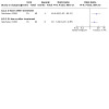
Nineteen trials reported on death from all causes (Becattini 2010; Dalla‐Volta 1992; Dotter 1979; Fasullo 2011; Goldhaber 1993; Jerjes‐Sánchez 1995; Kline 2014; Konstantinides 2002; Kucher 2014; Levine 1990; Ly 1978; Meyer 2014; PIOPED 1990; Sharifi 2013; Sinha 2017; Taherkhani 2014; Tibbutt 1974; UPETSG 1970; Zhang 2018). The 19 trials included in the meta‐analysis reported a total of 87 deaths: 32 in the thrombolytics group and 55 in the heparin group. Pooled analyses showed that across all studies, giving thrombolytics reduced the incidence of death (odds ratio (OR) 0.58, 95% confidence interval (CI) 0.38 to 0.88; 19 studies, 2319 participants, low‐certainty evidence; Analysis 1.1). The analysis showed that statistical heterogeneity between studies was at a low level (I2 = 0%). The test for subgroup differences indicated no clear difference between the types of thrombolytic used for death from all causes (P = 0.73; Analysis 1.1).
1.1. Analysis.

Comparison 1: Thrombolytic therapy versus heparin: primary outcome measures, Outcome 1: Death from all causes
We carried out a sensitivity analysis by excluding studies at high risk of bias (Goldhaber 1993; Jerjes‐Sánchez 1995; Kucher 2014; Ly 1978; Taherkhani 2014; Tibbutt 1974). However, we no longer found clear evidence to support a difference between the two groups for all‐cause mortality (OR 0.71, 95% CI 0.45 to 1.13; 13 studies, 2046 participants; Analysis 2.1). The analysis still shows that statistical heterogeneity between studies was at a low level (I2 = 0%). Because some studies carried a high risk of bias, we downgraded the certainty of the evidence for this outcome from high to low (Table 1). We also performed a subgroup analysis according to different types of PE (massive/submassive/unknown types of PE), and found no clear effects between subgroups (P = 0.30). Only the massive PE subgroup showed that thrombolytic therapy may have an effect on death (Analysis 3.1). We found no clear evidence to support a difference between thrombolytic therapy and heparin for death in the other subgroups.
2.1. Analysis.

Comparison 2: Thrombolytic therapy versus heparin: primary outcome measures (sensitivity analysis according to study quality), Outcome 1: Death from all causes
3.1. Analysis.

Comparison 3: Thrombolytic therapy versus heparin: primary outcome measures (subgroup analysis according to types of PE), Outcome 1: Death from all causes
Meyer 2014 reported long‐term mortality rates for participants with intermediate‐risk PE. Researchers in this study followed about 70% of participants over two years (median 37.8 months) and reported that tenecteplase treatment did not affect long‐term mortality rates compared to placebo and heparin. We were unable to include the data in our meta‐analysis, as the other included studies reported short‐term mortality (follow‐up period less than three months for most studies). Further analyses may be possible in future updates.
Recurrence of pulmonary emboli
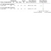
Twelve studies reported on the recurrence of pulmonary emboli (Becattini 2010; Dalla‐Volta 1992; Dotter 1979; Fasullo 2011; Goldhaber 1993; Konstantinides 2002; Levine 1990; Meyer 2014; Sharifi 2013; Sinha 2017; UPETSG 1970; Zhang 2018). Pooled data comparing thrombolytics versus heparin show that the thrombolytics group experienced less recurrence than the heparin group (OR 0.54, 95% CI 0.32 to 0.91; 12 studies, 2050 participants, low‐certainty evidence; Analysis 1.2). Analyses show that statistical heterogeneity between studies was at a low level (I2 = 0%).The test for subgroup differences indicated no clear difference between types of thrombolytic used and recurrence of PE (P = 0.59; Analysis 1.2).
1.2. Analysis.

Comparison 1: Thrombolytic therapy versus heparin: primary outcome measures, Outcome 2: Recurrence of pulmonary emboli
We performed a sensitivity analysis by removing one study at high risk of bias (Goldhaber 1993). The analysis showed no clear effect of thrombolytics on recurrence of PE comparing with heparin (OR 0.60, 95% CI 0.35 to 1.04; 11 studies, 1949 participants; Analysis 2.2). Hence, we downgraded the certainty of the evidence for this outcome from high to low for very serious risk of bias (selection, performance, and detection bias) (Table 1). We also performed a subgroup analysis by different types of PE (submassive/unknown types of PE) and found no conclusive evidence showing a difference between subgroups (test for subgroup differences: P = 0.46). The 'unknown types of PE' subgroup provided no clear evidence to support a difference between thrombolytic therapy and heparin (Analysis 3.2).
2.2. Analysis.

Comparison 2: Thrombolytic therapy versus heparin: primary outcome measures (sensitivity analysis according to study quality), Outcome 2: Recurrence of pulmonary emboli
3.2. Analysis.

Comparison 3: Thrombolytic therapy versus heparin: primary outcome measures (subgroup analysis according to types of PE), Outcome 2: Recurrence of pulmonary emboli
Major and minor haemorrhagic events
Major haemorrhagic events
Fifteen studies reported on major haemorrhagic events (Ahmed 2018; Becattini 2010; Dalla‐Volta 1992; Fasullo 2011; Goldhaber 1993; Kline 2014; Konstantinides 2002; Levine 1990; Ly 1978; Meyer 2014; PIOPED 1990; Sinha 2017; Tibbutt 1974; UPETSG 1970; Zhang 2018). The total number of these events was 136: 99 in the thrombolytics group and 37 in the heparin group. Pooled analyses showed that across 15 studies comparing thrombolytics versus heparin, more major bleeding events occurred after treatment with thrombolytics (OR 2.84, 95% CI 1.92 to 4.20; 15 studies, 2101 participants; moderate‐certainty evidence; Analysis 1.3). The result was not changed even after four studies at high risk of bias (Ahmed 2018; Goldhaber 1993; Ly 1978; Tibbutt 1974), were excluded in a sensitivity analysis (OR 2.91, 95% CI 1.92 to 4.39; 11 studies, 1893 participants; Analysis 2.3). Analysis showed low levels of statistical heterogeneity between studies, both before (I2 = 3%) and after (I2 = 26%) the sensitivity analysis. We downgraded the certainty of the evidence for this outcome from high to moderate for 'Risk of bias' concerns (Table 1).
1.3. Analysis.

Comparison 1: Thrombolytic therapy versus heparin: primary outcome measures, Outcome 3: Major haemorrhagic events
2.3. Analysis.

Comparison 2: Thrombolytic therapy versus heparin: primary outcome measures (sensitivity analysis according to study quality), Outcome 3: Major haemorrhagic events
The test for subgroup differences indicated no clear difference between types of thrombolytic used and major haemorrhagic events (P = 0.05; Analysis 1.3). We also performed a subgroup analysis by different types of PE (submassive/unknown types of PE) and found no subgroup effects between subgroups (P = 0.30; Analysis 3.3).
3.3. Analysis.

Comparison 3: Thrombolytic therapy versus heparin: primary outcome measures (subgroup analysis according to types of PE), Outcome 3: Major haemorrhagic events
Two studies explicitly reported on the occurrence of haemorrhagic stroke after treatment (Meyer 2014; Sinha 2017). Both studies compared tenecteplase plus heparin versus placebo plus heparin, with a total number of events of 12: 11 in the thrombolytic group and 1 in the heparin group. Pooled data show more haemorrhagic stroke occurred in the thrombolytic groups than in the heparin group (OR 7.59, 95% CI 1.38 to 41.72; 2 studies, 1091 participants; Analysis 1.4).
1.4. Analysis.

Comparison 1: Thrombolytic therapy versus heparin: primary outcome measures, Outcome 4: Haemorrhagic stroke
Minor haemorrhagic events
Thirteen studies reported on minor haemorrhagic events (Ahmed 2018; Becattini 2010; Dalla‐Volta 1992; Fasullo 2011; Kucher 2014; Levine 1990; Ly 1978; Meyer 2014; Sinha 2017; Taherkhani 2014; Tibbutt 1974; UPETSG 1970; Zhang 2018). Pooled analyses comparing thrombolytics versus heparin show more minor haemorrhagic events occurred in the thrombolytics group (OR 2.97, 95% CI 1.66 to 5.30; 13 studies, 1757 participants; low‐certainty evidence; Analysis 1.5). Analyses show that statistical heterogeneity between the included studies was at a substantial level (I2 = 55%), so we used a random‐effects model for the pooled analysis. After excluding the five studies at high risk of bias (Ahmed 2018; Kucher 2014; Ly 1978; Taherkhani 2014; Tibbutt 1974), we still observed this difference between the two groups (OR 3.82, 95% CI 2.06 to 7.09; 8 studies, 1541 participants; Analysis 2.4). We downgraded the certainty of the evidence for this outcome from high to low for 'Risk of bias' concerns and inconsistency (large heterogeneity) (Table 1). The test for subgroup differences indicated no clear difference between types of thrombolytic used and minor haemorrhagic events (P = 0.07; Analysis 1.5). We also performed a subgroup analysis by different types of PE (submassive/unknown types of PE) and found a subgroup effect between subgroups (P = 0.02); we found a difference between the two groups in the 'submassive PE' subgroup but not in the 'unknown types of PE' subgroup (Analysis 3.4).
1.5. Analysis.

Comparison 1: Thrombolytic therapy versus heparin: primary outcome measures, Outcome 5: Minor haemorrhagic events
2.4. Analysis.

Comparison 2: Thrombolytic therapy versus heparin: primary outcome measures (sensitivity analysis according to study quality), Outcome 4: Minor haemorrhagic events
3.4. Analysis.

Comparison 3: Thrombolytic therapy versus heparin: primary outcome measures (subgroup analysis according to types of PE), Outcome 4: Minor haemorrhagic events
Secondary outcome measures
Haemodynamic improvement and thrombolysis
Pulmonary arterial systolic pressure improvement
Four studies comparing thrombolytics with heparin show consistent results in the improvement of pulmonary arterial systolic pressure at follow‐up times of 24 hours, 72 hours and 7 days (Sinha 2017; Tibbutt 1974; UPETSG 1970; Zhang 2018). At 24 hours after treatment, UPETSG 1970 compared urokinase versus heparin in 147 participants and Zhang 2018 compared rt‐PA versus heparin in 66 participants; both show that thrombolytic treatment had a small effect on pulmonary arterial systolic pressure improvement (mean difference (MD) −4.41 mmHg, 95% CI −4.62 to −4.20; MD −12.4 mmHg, 95% CI −17.23 to −7.57, respectively; Analysis 4.1). Of the two remaining studies, one compared streptokinase versus heparin in 21 participants at 72 hours (Tibbutt 1974) and the other compared tenecteplase versus heparin in 86 participants at 7 days (Sinha 2017). These also showed a possible effect following thrombolytic treatment (MD −11.60 mmHg, 95% CI −20.81 to −2.39; MD −3.02 mmHg, 95% CI −4.75 to −1.29, respectively; Analysis 4.1). Although not pooled, these results indicate that thrombolytics may decrease pulmonary arterial systolic pressure to a greater extent than heparin, and that the effect is similar for various thrombolytics. However, the small number of overall participants involved and the high risk of bias attached to Tibbutt 1974 warrants caution when interpreting the results.
4.1. Analysis.

Comparison 4: Thrombolytic therapy versus heparin: haemodynamic outcomes, Outcome 1: Pulmonary arterial systolic pressure improvement (mmHg)
Mean pulmonary arterial pressure improvement
Three studies comparing thrombolytics versus heparin showed contradictory results in the improvement in mean pulmonary arterial pressure (PIOPED 1990; Tibbutt 1974; UPETSG 1970). Although rt‐PA versus heparin at 1½ hours showed no clear effect for thrombolytic treatment in PIOPED 1990 (MD −3.00 mmHg, 95% CI −16.91 to 10.91; 13 participants; Analysis 4.2), the two remaining studies reported a small effect on mean pulmonary arterial pressure improvement at 24 and 72 hours in favour of thrombolytic treatment (MD −4.41 mmHg, 95% CI −4.62 to −4.20; 147 participants; MD −7.50 mmHg, 95% CI −12.80 to −2.20; 17 participants, respectively; Analysis 4.2). However, the small number of participants involved warrants caution when interpreting the results.
4.2. Analysis.

Comparison 4: Thrombolytic therapy versus heparin: haemodynamic outcomes, Outcome 2: Mean pulmonary arterial pressure improvement (mmHg)
Right ventricular end‐diastolic pressure improvement
Two studies showed contradictory results for right ventricular end‐diastolic pressure improvement. UPETSG 1970 compared urokinase versus heparin in 142 participants, and after 24 hours noted a small difference in right ventricular end‐diastolic pressure improvement in favour of thrombolytic treatment (MD −2.21 mmHg, 95% CI −2.35 to −2.07; Analysis 4.3). On the other hand, Tibbutt 1974 compared streptokinase versus heparin in 19 participants, observing no clear difference after 72 hours (MD 1.20 mmHg, 95% CI −2.59 to 4.99; Analysis 4.3). However, we judged Tibbutt 1974 to be at high risk of bias in this review and the number of participants involved in this analysis was small, so results must be interpreted with caution.
4.3. Analysis.

Comparison 4: Thrombolytic therapy versus heparin: haemodynamic outcomes, Outcome 3: Right ventricular end‐diastolic pressure improvement (mmHg)
Total pulmonary resistance improvement
UPETSG 1970 compared urokinase versus heparin in 113 participants, finding a small difference in favour of urokinase at 24 hours after treatment (MD −0.33 dyn·s·cm‐5, 95% CI −0.35 to −0.31; Analysis 4.4). Tibbutt 1974 compared streptokinase versus heparin in 12 participants at 72 hours after treatment, finding no clear difference between treatment and control (MD 0.30 dyn·s·cm‐5, 95% CI −0.83 to 1.43; Analysis 4.4). PIOPED 1990 compared rt‐PA versus heparin in 13 participants at 1½ hours after treatment, and although these results appear to favour rt‐PA, no clear difference between the two groups is evident (MD −180.00 dyn·s·cm‐5, 95% CI −883.55 to 523.55; Analysis 4.4). Again, the small number of participants involved and the high risk of bias for Tibbutt 1974 warrants caution when interpreting the results.
4.4. Analysis.

Comparison 4: Thrombolytic therapy versus heparin: haemodynamic outcomes, Outcome 4: Total pulmonary resistance improvement (dyn·s·cm‐5)
Cardiac index improvement (L/min/m²)
Two studies show contradictory results for cardiac index improvement (Tibbutt 1974; UPETSG 1970). Tibbutt 1974 compared streptokinase versus heparin in 13 participants, observing a small difference in cardiac index improvement in favour of heparin (MD −0.60, 95% CI −1.05 to −0.15; Analysis 4.5). UPETSG 1970, which compared urokinase versus heparin in 115 participants, reported a small difference in cardiac index improvement in favour of urokinase (MD 0.20, 95% CI 0.15 to 0.25; Analysis 4.5). Results must be interpreted with caution due to high risk of bias in Tibbutt 1974, and the small number of participants involved.
4.5. Analysis.

Comparison 4: Thrombolytic therapy versus heparin: haemodynamic outcomes, Outcome 5: Cardiac index improvement (L/min/m²)
Other haemodynamic outcomes
UPETSG 1970, with 160 participants, compared urokinase versus heparin at 24 hours after treatment, showing small differences in favour of urokinase in right ventricular systolic pressure (MD −6.90 mmHg, 95% CI −7.25 to −6.55; Analysis 4.6), right arterial mean pressure (MD −1.94 mmHg, 95% CI −2.05 to −1.83; Analysis 4.7), arterial‐venous oxygen difference (MD −0.31 vol %, 95% CI −0.37 to −0.25; Analysis 4.8), and arterial PO₂ (MD 8.45 mmHg, 95% CI 7.84 to 9.06; Analysis 4.9).
4.6. Analysis.

Comparison 4: Thrombolytic therapy versus heparin: haemodynamic outcomes, Outcome 6: Right ventricular systolic pressure improvement (mmHg) at 24 hours
4.7. Analysis.

Comparison 4: Thrombolytic therapy versus heparin: haemodynamic outcomes, Outcome 7: Right arterial mean pressure improvement (mmHg) at 24 hours
4.8. Analysis.

Comparison 4: Thrombolytic therapy versus heparin: haemodynamic outcomes, Outcome 8: Arterial‐venous oxygen difference (vol %) at 24 hours
4.9. Analysis.

Comparison 4: Thrombolytic therapy versus heparin: haemodynamic outcomes, Outcome 9: Arterial PO₂ (mmHg) improvement at 24 hours
Three studies reported haemodynamic decompensation rates among submassive PE participants in the thrombolytics group and the heparin group (Meyer 2014; Sinha 2017; Zhang 2018). Zhang 2018 compared rt‐PA versus heparin in 66 participants, while Meyer 2014 and Sinha 2017 compared tenecteplase versus heparin in 1005 and 86 participants, respectively. The total number of these events was 46: 10 in the thrombolytics group and 36 in the heparin group. Pooled analyses show that fewer haemodynamic decompensation events occurred after thrombolytic therapy (OR 0.26, 95% CI 0.13 to 0.53; 3 studies, 1157 participants; Analysis 4.10).
4.10. Analysis.

Comparison 4: Thrombolytic therapy versus heparin: haemodynamic outcomes, Outcome 10: Haemodynamic decompensation
Perfusion lung scanning
UPETSG 1970 compared urokinase versus heparin, expressing perfusion defects as a percentage of total normal perfusion of both lungs. At days 1 and 2, results show a difference in favour of urokinase (day 1: MD 3.50%, 95% CI 1.32 to 5.68; 142 participants; Analysis 5.1; day 2: MD 3.10%, 95% CI 0.15 to 6.05; 133 participants; Analysis 5.2). Subsequent results include the following: at day 5: MD 2.00% (95% CI −1.60 to 5.60; 126 participants; Analysis 5.3); at day 14: MD 0.20% (95% CI −4.26 to 4.66; 116 participants; Analysis 5.5); and at one year MD −1.10% (95% CI −7.57 to 5.37; 57 participants; Analysis 5.7). These results show that on days 1 and 2 after treatment, either the total normal perfusion of both lungs or the proportion of lung not perfused in those treated with thrombolytics was greater than in those treated with heparin, and on days 5 and 14 and at one year follow‐up there was no clear effect of urokinase. A second study comparing rt‐PA versus heparin (Goldhaber 1993), in which perfusion defects were expressed as the proportion of lung not perfused, also showed a small effect in favour of rt‐PA at day 1 (MD 0.13%, 95% CI 0.05 to 0.21; 101 participants; Analysis 5.1). However, results must be interpreted with caution due to high risk of bias in Goldhaber 1993.
5.1. Analysis.

Comparison 5: Thrombolytic therapy versus heparin: perfusion lung scanning (absolute resolution), Outcome 1: Day 1
5.2. Analysis.

Comparison 5: Thrombolytic therapy versus heparin: perfusion lung scanning (absolute resolution), Outcome 2: Day 2
5.3. Analysis.

Comparison 5: Thrombolytic therapy versus heparin: perfusion lung scanning (absolute resolution), Outcome 3: Day 5
5.5. Analysis.

Comparison 5: Thrombolytic therapy versus heparin: perfusion lung scanning (absolute resolution), Outcome 5: Day 14
5.7. Analysis.

Comparison 5: Thrombolytic therapy versus heparin: perfusion lung scanning (absolute resolution), Outcome 7: Absolute resolution (1‐year follow‐up)
Dalla‐Volta 1992 compared alteplase plus heparin versus heparin alone, showing no clear effect on total lung score between the two groups at day 7 (MD 1.70, 95% CI −1.04 to 4.44; 21 participants; Analysis 5.4); however, results show a small difference in favour of alteplase at day 30 after treatment (MD 2.80, 95% CI 0.35 to 5.25; 22 participants; Analysis 5.6). Comparison of scores by change from baseline in both groups provides no clear evidence to support a difference between the two groups at day 7 or at day 30 (day 7: MD 1.80, 95% CI −0.51 to 4.11; 21 participants; Analysis 5.4; day 30: MD 0.70, 95% CI −1.37 to 2.77; 22 participants; Analysis 5.6). These results show that alteplase plus heparin and heparin alone may improve total lung scores with similar effect, but by day 30 the score in the alteplase‐plus‐heparin group was higher than the score in the heparin‐alone group. Due to the small number of participants involved, the results should be interpreted with caution.
5.4. Analysis.

Comparison 5: Thrombolytic therapy versus heparin: perfusion lung scanning (absolute resolution), Outcome 4: Day 7
5.6. Analysis.

Comparison 5: Thrombolytic therapy versus heparin: perfusion lung scanning (absolute resolution), Outcome 6: Day 30
Levine 1990 compared rt‐PA plus heparin versus placebo plus heparin, showing no clear difference in the number of participants with greater than 50% improvement on lung scan at 24 hours after treatment (OR 3.84, 95% CI 0.94 to 15.73; 57 participants; Analysis 6.1). We could not estimate this in the PIOPED 1990 study.
6.1. Analysis.

Comparison 6: Thrombolytic therapy versus heparin: number of patients with greater than 50% improvement on lung scan, Outcome 1: Day 1
Pulmonary angiogram assessment
Researchers evaluated pulmonary angiograms using the Miller index (Miller 1971). The overall total score for pulmonary angiograms in Dalla‐Volta 1992 shows a small reduction in the alteplase‐plus‐heparin group (MD −3.4, 95% CI −4.72 to −2.08; 36 participants; Analysis 7.1).
7.1. Analysis.

Comparison 7: Thrombolytic therapy versus heparin: pulmonary angiogram assessment, Outcome 1: Change from baseline at 2 hours
Ly 1978 and Tibbutt 1974 compared streptokinase versus heparin, and, when pooled, results show a small difference in angiographic score changes from baseline to 72 hours in favour of streptokinase (MD −9.3, 95% CI −12.81 to −5.78; 47 participants, 2 studies; Analysis 7.2). This indicates that changes in angiographic score from baseline to 72 hours after treatment were greater in participants treated with streptokinase than in those treated with heparin. These results must be interpreted with caution, because both studies carried high risk of bias according to our review criteria.
7.2. Analysis.

Comparison 7: Thrombolytic therapy versus heparin: pulmonary angiogram assessment, Outcome 2: Change from baseline at 72 hours
Echocardiograms
Eight studies performed echocardiograms (Ahmed 2018; Becattini 2010; Fasullo 2011; Goldhaber 1993; Kucher 2014; Sinha 2017; Taherkhani 2014; Zhang 2018). Goldhaber 1993 compared rt‐PA plus heparin versus heparin alone; panellists decided by consensus whether right ventricular wall motion was normal or mildly (1+), moderately (2+), or severely (3+) hypokinetic. Tricuspid regurgitation was visually assessed according to the size of the largest colour doppler jet as absent, mild (1+), moderate (2+), or severe (3+). This study showed that the rt‐PA group had increased numbers of participants with improved right ventricular wall movement (OR 2.90, 95% CI 0.98 to 8.60 at 3 hours; OR 3.20, 95% CI 1.20 to 8.57 at 24 hours; 89 participants; Analysis 8.1) and tricuspid regurgitation (OR 6.35, 95% CI 1.90 to 21.17 at 3 hours; OR 3.20, 95% CI 1.20 to 8.57 at 24 hours; 89 participants; Analysis 8.2). Sinha 2017, comparing tenecteplase plus heparin versus placebo plus heparin, also reported that the thrombolytic group had a higher rate of right ventricular function improvement at 7 days after treatment (OR 3.46, 95% CI 1.42 to 8.42; 86 participants; Analysis 8.1).
8.1. Analysis.

Comparison 8: Thrombolytic therapy versus heparin: echocardiograms, Outcome 1: Right ventricular wall movement improvement
8.2. Analysis.
Comparison 8: Thrombolytic therapy versus heparin: echocardiograms, Outcome 2: Tricuspid regurgitation improvement
Fasullo 2011 compared alteplase plus heparin versus heparin alone, assessing inferior vena cava, doppler acceleration time, paradoxical systolic septal motion, tricuspid annular plane systolic excursion, and B‐type natriuretic peptide (BNP) values (at baseline; at 24, 48, and 72 hours; at six days; at discharge; and at three months and six months). Investigators found earlier improvement in the thrombolytics group compared with the placebo group, with evident differences after 24 hours that lasted throughout hospitalisation and during the follow‐up period. Another study compared USAT (rt‐PA) plus heparin versus heparin alone (Kucher 2014), reporting the right‐to‐left ventricular dimension (RV/LV) ratio at 24 hours and at three months as a primary outcome. Results show a difference between the two groups at 24 hours, but at three months they show no clear effect for the rt‐PA group (P = 0.36). This study also shows that USAT (rt‐PA) had better outcomes at 24 hours than at three months for tricuspid annular systolic excursion, right ventricular‐to‐left ventricular pressure gradient, and minimum inferior vena cava diameter. Taherkhani 2014 compared alteplase or streptokinase plus enoxaparin versus enoxaparin alone. This study reported no clear differences between the two groups in normalisation of the RV. Zhang 2018 compared rt‐PA plus enoxaparin versus enoxaparin alone, and reported that the thrombolytics group had more reduction in RV/LV ratio at 24 hours after treatment.
In this review, we found that after treatment, most echocardiogram parameters in individual studies were better in the thrombolytics group than in the control group. For example, Fasullo 2011 reported the paradoxical systolic septal motion (OR 0.24, 95% CI 0.07 to 0.82 at 24 hours; OR 0.35, 95% CI 0.13 to 0.92 at 48 hours; OR 0.29, 95% CI 0.10 to 0.88 at 72 hours; OR 0.12, 95% CI 0.01 to 2.49 at six days; 72 participants; Analysis 8.3); Fasullo 2011, Kucher 2014 and Zhang 2018 reported right‐to‐left ventricular ratio at 24 hours after treatment (MD −0.16, 95% CI −0.21 to −0.11; 197 participants; 3 studies; Analysis 8.4); and Fasullo 2011 with 72 participants, reported additional time points: 48 hours (MD −0.19, 95% CI −0.20 to −0.18), 72 hours (MD −0.14, 95% CI −0.15 to −0.13), six days (MD −0.22, 95% CI −0.23 to −0.21), discharge (MD −0.33, 95% CI −0.34 to −0.32), three months (MD −0.14, 95% CI −0.34 to 0.05; 131 participants; pooled Fasullo 2011 and Kucher 2014), and six months (MD −0.21, 95% CI −0.22 to −0.20; 72 participants) (see Analysis 8.4).
8.3. Analysis.

Comparison 8: Thrombolytic therapy versus heparin: echocardiograms, Outcome 3: Paradoxical systolic septal motion
8.4. Analysis.

Comparison 8: Thrombolytic therapy versus heparin: echocardiograms, Outcome 4: Right ventricle‐to‐left ventricle ratio
Researchers reported similar time points for tricuspid annular plane systolic excursion: 24 hours (MD 0.45, 95% CI −1.18 to 2.07; 131 participants; pooled Fasullo 2011 and Kucher 2014), 48 hours (MD 1.00, 95% CI −0.13 to 2.13; 1 study, 72 participants), 72 hours (MD 1.80, 95% CI 0.67 to 2.93; 1 study, 72 participants), six days (MD 2.50, 95% CI 1.57 to 3.43; 1 study, 72 participants), discharge (MD 2.00, 95% CI 0.75 to 3.25; 1 study, 72 participants), three months (MD 0.33, 95% CI −3.18 to 3.85; 131 participants, 2 studies; pooled Fasullo 2011 and Kucher 2014), and six months (MD 1.30, 95% CI 0.28 to 2.32; 1 study, 72 participants; see Analysis 8.5). Kucher 2014 reported the right ventricular‐to‐right atrial pressure gradient (MD −6.30, 95% CI −13.06 to 0.46 at 24 hours; MD 3.20, 95% CI −4.77 to 11.17 at three months; Analysis 8.6) and the minimum inferior vena cava diameter (MD −6.60, 95% CI −9.36 to −3.84 at 24 hours; MD −0.50, 95% CI −2.79 to 1.79 at three months; 1 study, 59 participants; Analysis 8.7).
8.5. Analysis.

Comparison 8: Thrombolytic therapy versus heparin: echocardiograms, Outcome 5: Tricuspid annular plane systolic excursion
8.6. Analysis.

Comparison 8: Thrombolytic therapy versus heparin: echocardiograms, Outcome 6: Right ventricular‐to‐right atrial pressure gradient
8.7. Analysis.
Comparison 8: Thrombolytic therapy versus heparin: echocardiograms, Outcome 7: Minimum inferior vena cava diameter
BNP values showed faster reduction in the thrombolytics group than in the placebo group during hospitalisation at six days after admission. Becattini 2010 also reported reduction in echocardiography parameters and found small differences in decreases in both right ventricle end‐diastolic dimension and the right‐to‐left end‐diastolic dimension ratio at 24 hours in favour of tenecteplase, but the difference was not maintained during the seven‐day follow‐up period (data were unavailable). These figures indicate that treatment with thrombolytics plus heparin possibly results in more participants with improved right ventricular wall movement and tricuspid regurgitation than treatment with heparin alone.
Chronic thromboembolic pulmonary hypertension
Six studies compared thrombolytic therapy plus heparin versus heparin alone for pulmonary hypertension (Ahmed 2018; Fasullo 2011; Sharifi 2013; Sinha 2017; Taherkhani 2014; Zhang 2018). Fasullo 2011 and Zhang 2018 reported that participants in the thrombolytics group had lower pulmonary hypertension than participants in the heparin group at 24 hours (MD −11.28, 95% CI −16.80 to −5.76; 2 studies, 138 participants). Fasullo 2011 and Sharifi 2013 reported the outcome at 48 hours (MD −7.37, 95% CI −9.20 to −5.53; 2 studies, 193 participants), and Fasullo 2011 and Ahmed 2018 reported the outcome at 72 hours (MD −7.65, 95% CI −10.03 to −5.28; 2 studies, 124 participants). Fasullo 2011 and Taherkhani 2014 reported at six days (MD −5.69, 95% CI −9.37 to −2.02; 2 studies, 122 participants); Sinha 2017 at seven days (MD −5.33, 95% CI −7.14 to −3.52; 1 study, 86 participants); Fasullo 2011 at discharge (MD −8.00, 95% CI −9.78 to −6.22; 1 study, 72 participants) and at three months (MD −7.00, 95% CI −17.18 to 3.18; 1 study, 72 participants); Fasullo 2011 and Sharifi 2013 at six months (MD −11.95, 95% CI −23.71 to −0.19; 2 studies, 193 participants); and Sharifi 2013 at 28 months (MD −15.00, 95% CI −17.32 to −12.68; 1 study, 128 participants) (see Analysis 8.8).
8.8. Analysis.

Comparison 8: Thrombolytic therapy versus heparin: echocardiograms, Outcome 8: Pulmonary hypertension
Differences in coagulation parameters over time
Fibrinogen
Two studies (Dalla‐Volta 1992; PIOPED 1990) comparing thrombolytic versus heparin treatment at less than three hours after treatment show a small difference in fibrinogen levels in favour of thrombolytics (total MD −2.68 g/L, 95% CI −4.36 to −1.00; 2 studies, 45 participants; Analysis 9.1). However, we found no clear evidence to support a difference between the two groups at 24 hours (MD −1.61 g/L, 95% CI −3.99 to 0.76; 2 studies, 114 participants), nor at 48 hours (MD −0.60 g/L, 95% CI −1.40 to 0.20; 1 study, 83 participants) after treatment (see Analysis 9.1). This may indicate that thrombolytic treatment results in a lower level of fibrinogen than heparin treatment. Levine 1990 also reported this comparison, but we could not extract the data from this study, as it reported changes in mean fibrinogen levels in a figure, showing that the thrombolytics group had a lower level of fibrinogen than the placebo group within 24 hours after treatment. In this update, an additional report of Kline 2014 stated that the fibrinogen levels between the two groups showed no evident difference at three‐month follow‐up (P = 0.862).
9.1. Analysis.

Comparison 9: Thrombolytic therapy versus heparin: haemocoagulation variables, Outcome 1: Fibrinogen (g/L)
D‐dimer
As a molecular marker of haemostatic activation, D‐dimer indicates fibrin turnover both from intravascular fibrin formation and from subsequent lysis of a fibrin clot. Dalla‐Volta 1992 and PIOPED 1990 both reported a difference between participants treated with thrombolytics plus heparin and those treated with heparin alone at two hours after treatment. We used a random‐effects method to pool as heterogeneity was very high (I2 = 86%), which was reflected in the pooled analysis (MD 21.04, 95% CI −4.60 to 46.69; 2 studies, 45 participants; Analysis 9.2). Dalla‐Volta 1992 reported a difference between two groups at 24 hours after treatment (MD 5.30 µg/mL, 95% CI 2.12 to 8.48; 31 participants; Analysis 9.2). These results show that D‐dimer concentrations may be higher in the thrombolytics‐plus‐heparin group than in the heparin‐alone group. However, an additional report from Kline 2014 for this update reports no clear difference in D‐dimer concentrations between the tenecteplase group and the control group at three‐month follow‐up (P = 0.05).
9.2. Analysis.

Comparison 9: Thrombolytic therapy versus heparin: haemocoagulation variables, Outcome 2: D‐dimer (µg/mL)
Plasminogen
Dalla‐Volta 1992 reported a difference in concentrations of plasminogen at two hours (MD −60.30%, 95% CI −71.92 to −48.68; 22 participants) and at 24 hours (MD −36.00%, 95% CI −48.06 to −23.94; 21 participants) after treatment in favour of alteplase (see Analysis 9.3). This shows that treatment with alteplase plus heparin may result in a lower plasminogen concentration than treatment with heparin alone.
9.3. Analysis.

Comparison 9: Thrombolytic therapy versus heparin: haemocoagulation variables, Outcome 3: Plasminogen (%)
Post‐thrombotic syndrome
No data were available for this outcome. If data become available, we will include them in future updates.
Escalation of treatment
Two studies reported the number of participants who needed escalation of treatment after receiving thrombolytics versus heparin (Konstantinides 2002; Taherkhani 2014). Konstantinides 2002 compared alteplase plus heparin versus heparin alone, finding that fewer participants in the alteplase‐plus‐heparin group needed escalation of treatment for in‐hospital events compared with the heparin‐alone group. Pooling these data with data from alteplase or streptokinase plus enoxaparin versus enoxaparin alone (Taherkhani 2014), we found a small difference in favour of alteplase (OR 0.32, 95% CI 0.16 to 0.64; 2 studies, 306 participants; Analysis 10.1).This indicates that fewer participants required rescue thrombolysis in the thrombolytic‐plus‐heparin group than in the heparin‐alone group.
10.1. Analysis.

Comparison 10: Thrombolytic therapy versus heparin: other outcomes, Outcome 1: Escalation of treatment
Hospital stay
Five studies that compared thrombolytics versus heparin reported length of hospital stay (Ahmed 2018; Kucher 2014; Sharifi 2013; Sinha 2017; Taherkhani 2014). Analyses show that statistical heterogeneity between included studies was at a considerable level (I2 = 93%), and given the heterogeneity of the interventions, we used a random‐effects model for the pooled analysis. For delivery technique for the thrombolytic drug, one study used the USAT system (Kucher 2014), and another study delivered a 'safe dose' of rt‐PA through intravenous injection (Sharifi 2013). The other three studies respectively used alteplase or streptokinase (Taherkhani 2014), streptokinase (Ahmed 2018) and tenecteplase (Sinha 2017). Pooled analysis of the five studies yielded clear evidence to support the thrombolytics group having a shorter hospital stay compared with the heparin group (MD −1.40 days, 95% CI −2.69 to −0.11; 5 studies, 368 participants; Analysis 10.2). After we excluded the studies at high risk of bias (Ahmed 2018; Kucher 2014; Taherkhani 2014) as part of the sensitivity analysis, we still found evidence to support a difference between the two groups for hospital stay (MD −2.72 days, 95% CI −2.95 to −2.49; 2 studies, 207 participants; Analysis 11.1).
10.2. Analysis.
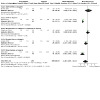
Comparison 10: Thrombolytic therapy versus heparin: other outcomes, Outcome 2: Hospital stay
11.1. Analysis.

Comparison 11: Thrombolytic therapy versus heparin: other outcomes (sensitivity analysis according to study quality), Outcome 1: Hospital stay
Survival time
Duration of follow‐up varied, and researchers reported deaths up to 72 hours (Tibbutt 1974), 10 days (Levine 1990), 14 days (Goldhaber 1993; Ly 1978; UPETSG 1970), 19 days (PIOPED 1990), 30 days (Dalla‐Volta 1992; Konstantinides 2002), and 180 days after randomisation (Fasullo 2011). Fasullo 2011 and Konstantinides 2002 show a beneficial effect of thrombolytics over control in clinical event‐free survival time. The other studies did not report this outcome. However, we could not extract data from the two studies to conduct a pooled analysis because they provided only a figure for survival time. Thus we cannot draw a specific conclusion about survival time nor about thrombolytic effects on death.
Composite clinical outcome
Four studies reported the important composite clinical outcome (Kline 2014; Meyer 2014; Sinha 2017; Zhang 2018). Meyer 2014, Sinha 2017 and Zhang 2018 reported all‐cause death or haemodynamic decompensation. Pooled analysis shows fewer composite events of death or haemodynamic decompensation occurred in the thrombolytics group than in the control group (OR 0.36, 95% CI 0.20 to 0.66; 3 studies, 1157 participants; Analysis 10.3). For this update, we identified an additional report for Meyer 2014, which gave the composite outcome of chronic thromboembolic pulmonary hypertension (CTEPH) or post‐PE impairment (Barco 2019). Kline 2014 reported recurrent venous thromboembolism (VTE), poor functional capacity, and low perception of wellness as measured by the 36‐item Short Form Health Survey (SF‐36). However, we found no clear evidence to support a difference between the two groups for these outcomes. As these composite clinical outcomes are different, it is not appropriate to pool them.
10.3. Analysis.

Comparison 10: Thrombolytic therapy versus heparin: other outcomes, Outcome 3: Composite clinical outcome
Quality of life
One study reported participants' quality of life (QoL) in the follow‐up period. Kline 2014 reported the number of participants who remained in the intensive care unit on day 2 and the QoL as measured by the VEINES QoL score and SF‐36. This study showed that QoL measured by both VEINES QoL and SF‐36 at 90 days follow‐up was similar between the two treatment groups (P = 0.40 and P = 0.67, respectively).
Healthcare cost comparison
None of the included trials reported cost comparisons. If data become available, we will report this information in future updates.
Discussion
Summary of main results
We have described the main results in Table 1.
Outcomes analysed
Overall, results for included outcomes were somewhat unsatisfactory and susceptible to bias, due to the fact that most outcomes in the subgroups were extracted from only one or two studies. Most studies had small sample sizes, and some were of low methodological quality. For this update we found only three studies with four or more 'Risk of bias' domains assessed as being at low risk of bias (Fasullo 2011; Meyer 2014; UPETSG 1970). However, Meyer 2014 received funds from several companies, which may have caused conflicts of interest. In addition, some trials did not assess the most important outcomes defined in this review or did not evaluate the potential complications of thrombolytic therapy.
Clinical interpretation of the data
Thrombolytic agents showed possible benefit for death compared with heparin (OR 0.58, 95% CI 0.38 to 0.88; 19 studies, 2319 participants; low‐certainty evidence) and recurrence of PE (OR 0.54, 95% CI 0.32 to 0.91; 12 studies, 2050 participants; low‐certainty evidence). However, after excluding six studies at high risk of bias, we found no clear evidence to support a difference between the two groups for risk of death (OR 0.71, 95% CI 0.45 to 1.13; 13 studies, 2046 participants). We also found no clear effect of thrombolytics on recurrence of PE comparing with heparin after removing one study at high risk of bias (OR 0.60, 95% CI 0.35 to 1.04; 11 studies, 1949 participants).
Results indicate more major haemorrhagic events in the thrombolytics group than in the control group (OR 2.84, 95% CI 1.92 to 4.20; 15 studies, 2101 participants; moderate‐certainty evidence); and more minor haemorrhagic events in the thrombolytics group than in the control group (OR 2.97, 95% CI 1.66 to 5.30; 13 studies, 1757 participants; low‐certainty evidence). The treatment effect was not changed after exclusion of trials at high risk of bias for these outcomes.
The total number of deaths was 87 among 2319 participants, and the total number of major haemorrhagic events was 136 among 2101 participants over all studies. From the confidence intervals, we can see a relatively precise estimate of treatment effect for recurrence of PE and for major and minor haemorrhagic events. However, the treatment effect for mortality was influenced by exclusion of studies at high risk of bias in a sensitivity analysis, even only slight alterations in the confidence intervals were evident. Thus, additional high‐quality studies are needed before firm conclusions can be drawn.
Two studies reported on haemorrhagic stroke, which occurred more often in the thrombolytics group than in the control group (OR 7.59, 95% CI 1.38 to 41.72) (Meyer 2014; Sinha 2017). Both studies compared tenecteplase with heparin. However, we cannot draw a strong conclusion from this result, due to the limited numbers of included studies and participants involved.
Many of the results for the remaining secondary outcome measures of this review are based on only one or two studies. In addition, studies used different follow‐up periods and interventions, resulting in unavoidable heterogeneity. The very limited results appear to suggest that thrombolytic therapy may be better than heparin for improving haemodynamic outcomes, perfusion lung scanning, pulmonary angiogram assessment, echocardiograms, pulmonary hypertension, coagulation parameters, composite clinical outcomes, need for escalation and survival time to a greater extent than heparin alone. However, the heterogeneity of the studies and the small number of participants involved warrant caution when interpreting results.
Thrombolytic therapy showed a probable advantage over heparin for length of hospital stay. Participants treated with thrombolytics had a shorter stay than with heparin before (MD −1.40 days, 95% CI −2.69 to −0.11) and after (MD −2.72 days, 95% CI −2.95 to −2.49) excluding the study at high risk of bias. However, substantial heterogeneity (I2 = 93%) between the studies and the small number of participants (368 participants) involved in this analysis warrant caution when results are interpreted.
None of the included studies provided data on post‐thrombotic syndrome or on cost comparison. Given the risks of bias in the included studies and the inconsistent results, we cannot draw any strong conclusions about the benefit of thrombolytic agents versus heparin in this review.
Overall completeness and applicability of evidence
We assessed the effectiveness and safety of thrombolytic therapy for people with acute PE. We found that thrombolytic therapy was probably better than comparison treatments in reducing the odds ratio of death and recurrence of PE, and it may improve some composite clinical outcomes and haemodynamic parameters (perfusion lung scanning, pulmonary angiogram assessment, and echocardiograms). All participants in the included studies were adults aged 18 or over, with a mean age of about 60. However, the included trials did not include strict subgroups of elderly participants. Only Meyer 2014 included a subgroup for participants over 75, and results of this study provide no clear evidence to support a difference between the two groups in their composite outcome 'death or haemodynamic decompensation' (OR 0.63, 95% CI 0.24 to 1.66). Possible differences between adults and elderly people therefore need further investigation.
Based on data from Konstantinides 2002, Perlroth 2007 highlighted that the heparin group required treatment escalation approximately three times more often than the thrombolytics group, but the researchers observed no difference in the risk of death from PE. Investigators also found that treatment with heparin alone was more effective and less costly than treatment with alteplase plus heparin among participants with submassive PE and right ventricular dysfunction. However, we were not able to investigate any differences between thrombolytic therapy and the comparison treatment by healthcare costs and post‐thrombotic syndrome, as no included studies reported on these outcomes. In clinical practice, haemodynamic parameters and the age of people with PE must be considered, especially for haemodynamically‐unstable people and those aged over 75.
The included studies involved different types of thrombolytics, including alteplase, urokinase, streptokinase, rt‐PA, ultrasound‐assisted catheter‐directed thrombolysis systems, and tenecteplase, usually followed by heparin. The control intervention was heparin alone or placebo plus heparin. No studies compared thrombolytics versus surgical intervention; surgical thrombectomy in massive PE will probably not lend itself to a prospective analysis (Duffett 2020). The test for subgroup differences indicates no clear difference between the types of thrombolytic used and death from all causes, recurrence of PE, major or minor haemorrhagic events. Pooled data from two studies indicate that tenecteplase may increase the odds ratio of haemorrhagic stroke (OR 7.59, 95% CI 1.38 to 41.72, 1091 participants). Possible differences between different types of thrombolytics need further investigation. Catheter‐directed administration may have theoretical advantages (e.g. low dose and more targeted) (Stewart 2020), but data are too scarce to make any judgement.
In Meyer 2014, researchers followed about 70% of participants over two years (median 37.8 months) and found that tenecteplase treatment did not affect long‐term mortality among participants with intermediate‐risk PE. This report also provided echocardiographic data for only about 30% of participants (144/506 (28.5%) in the tenecteplase group and 146/499 (28.9%) in the control group) with long‐term follow‐up (at least 24 months), showing that tenecteplase treatment did not reveal clear differences in residual pulmonary hypertension nor in RV dysfunction. In addition, another report of Meyer 2014 included in this update revealed that among 219 follow‐up participants with six‐month and 37‐month echocardiographic results, the composite outcome of CTEPH or post‐PE impairment at 37 months in two groups was 14.3% and 12.1% respectively, showing no obvious difference (Barco 2019). We could not use these data in our meta‐analysis because the other included studies reported only short‐term mortality (follow‐up period less than three months for most studies). Using data from this report in the meta‐analysis would have further increased heterogeneity. Future studies should therefore further investigate the effect on mortality of thrombolytic treatment with long‐term follow‐up.
Thrombolytics for massive PE (haemodynamically‐unstable patients)
It is clinically very important to differentiate haemodynamically‐stable and unstable patients (massive PE). However, we have identified only one study to date that compared thrombolytic therapy versus heparin in people with massive PE (Jerjes‐Sánchez 1995). Four studies included both massive and unknown PE types (Dotter 1979; Ly 1978; Tibbutt 1974; UPETSG 1970). The proportion of massive PE varied, at 8% in Ly 1978, 9% in UPETSG 1970, 23% in Tibbutt 1974, and 71% in Dotter 1979. As we did not know the types of PE for all participants included in these studies, we decided to include these four studies in the 'type of PE unknown' group. In PIOPED 1990, all included participants had an unclear PE type. We therefore included this study in the same group.
Jerjes‐Sánchez 1995 shows unbalanced allocation in the two groups at onset of PE, so we judged this study as having high risk of bias due to the high risk of selective reporting and the very small sample size. In this RCT, eight participants received 1,500,000 IU of streptokinase in one hour through a peripheral vein followed by heparin, or heparin alone. The four participants who were randomised to streptokinase (mean time of onset of symptoms of the first event of PE: 2½ hours) improved in the first hour after treatment, survived, and over two years of follow‐up did not present with pulmonary arterial hypertension. All four participants treated with heparin alone (mean time of onset of symptoms for the first event of PE: 34¾ hours) died within one to three hours after arrival at the emergency department. We excluded this study along with another five studies at high risk of bias from the sensitivity analysis (Goldhaber 1993; Kucher 2014; Ly 1978; Taherkhani 2014; Tibbutt 1974), and this appreciably influenced the results for death from all causes between the two groups. Thus, the data from Jerjes‐Sánchez 1995 had an influence on the pooled analysis. Even though we performed a subgroup analysis according to different types of PE (massive/submassive/unknown types of PE) for the primary outcomes, these results cannot be extrapolated to massive PE due to the limitations of Jerjes‐Sánchez 1995.
Quality of the evidence
We have described the main results in Table 1.
Randomisation and blinding
Only three studies reported both appropriate random sequence generation and allocation concealment (Goldhaber 1993; Meyer 2014; UPETSG 1970), and seven reported satisfactory blinding (Fasullo 2011; Kline 2014; Konstantinides 2002; Levine 1990; Meyer 2014; PIOPED 1990; UPETSG 1970). Goldhaber 1993 and Ahmed 2018 reported no blinding, and Taherkhani 2014 indicated that blinding was broken. Although two studies used the appropriate method to generate the random sequence, Ly 1978 included five participants who were not randomised, and Jerjes‐Sánchez 1995 reported unbalanced numbers between intervention and control groups at the onset of PE. The remaining trials did not provide clear details about random sequence generation, allocation concealment, or blinding. Three studies were of high risk of performance bias but low risk of detection bias (Kucher 2014; Sharifi 2013; Tibbutt 1974). One study was unclear in all items for 'Risk of bias' assessment (Marini 1988), and in one study, all 'Risk of bias' domains were at unclear or high risk (Dotter 1979). This could have biased study results in favour of treatment.
Sample size
Many of the outcome measures were only reported in one or two studies, so some effect sizes have wide confidence intervals (such as for incidence of haemorrhagic stroke). Many of the included studies involved small numbers of participants. For some outcomes, we may not have been able to detect any real effects of thrombolytics because of small sample size.
Conflicts of interest
Four included studies were funded by related pharmaceutical companies (Dotter 1979; Kline 2014; Kucher 2014; Meyer 2014), and some study authors worked for these companies (Meyer 2014). This could potentially result in conflicts of interest in drafting and reporting results of the study, even though study authors stated there was no influence over the whole study period.
Heterogeneity
Within each subgroup, we used Chi2 analyses to test for statistical evidence of heterogeneity among studies, and we used I2 to measure the degree of inconsistency across studies. When Chi2 analysis was significant and I2 values were in excess of 50%, we analysed differences in participant selection, baseline values, risk of bias, design, and methods that could possibly explain the heterogeneity.
Heterogeneity in responses did not appear to result from differences in methods used for most of the primary outcomes (including overall mortality, recurrence of pulmonary emboli, and major haemorrhagic events), nor for some of the secondary outcomes (mean pulmonary arterial pressure improvement, pulmonary angiogram assessment (72 hours after treatment), and echocardiograms). We obtained dynamic measurements from studies comparing thrombolytics versus heparin, showing a similar effect on those outcomes listed above. We found no dose‐related or time‐related relationships in these dynamic analyses.
We noted moderate heterogeneity in dynamic analysis of minor bleeding in studies comparing thrombolytics versus heparin (Ahmed 2018; Becattini 2010; Dalla‐Volta 1992; Fasullo 2011; Kucher 2014; Levine 1990; Ly 1978; Meyer 2014; Sinha 2017; Taherkhani 2014; Tibbutt 1974; UPETSG 1970; Zhang 2018), and we found a difference between the two therapies. We took this into consideration for assessing the certainty of the evidence. A great degree of heterogeneity was present in the dynamic analysis for D‐dimer concentration at two hours after treatment (Dalla‐Volta 1992; PIOPED 1990), pulmonary artery systolic pressure (Ahmed 2018; Fasullo 2011; Sharifi 2013; Sinha 2017; Taherkhani 2014; Zhang 2018), and right ventricle‐to‐left ventricle ratio (Fasullo 2011; Kucher 2014; Zhang 2018), showing differences between the two treatment groups. This may be a consequence of the different types of thrombolytics and heparin used in the two studies (see Characteristics of included studies table). We performed subgroup analysis for various thrombolytic agents. However, given the lack of data on different thrombolytic strategies, such as routes, doses and durations, we could not perform further subgroup analysis. Future updates should include subgroup analyses for different treatment strategies once more relevant studies are published.
Due to the above issues, we downgraded the overall certainty of the evidence presented in this review by one or two levels to moderate or low, because of concerns over risks of bias and inconsistency.
Potential biases in the review process
We analysed outcomes on an ITT basis, using a random‐effects model for pooled analysis of heterogeneous data (I2 = 40% to 100%). We did this to reduce the bias of estimated intervention effects.
We classified studies as having high risk of overall bias if two or more 'Risk of bias' domains carried a high risk of bias. We conducted a sensitivity analysis by excluding these studies to assess the effects of this exclusion on the estimated intervention effect.
Agreements and disagreements with other studies or reviews
Individually, almost all studies show that thrombolytic therapy may reduce the primary outcomes defined by the study, especially echocardiographic parameters. Our meta‐analysis of all included studies shows probable beneficial results for thrombolysis for death from all causes. However, the sensitivity analysis, when studies labelled as having high risk of bias were removed, provides no clear evidence to support a difference between the two groups in terms of death (OR 0.71, 95% CI 0.45 to 1.13). Results from Jerjes‐Sánchez 1995 show an apparent benefit for the thrombolytics group, and removing this study from the sensitivity analysis may explain the changes in results. For this update, we added two studies (Sinha 2017; Zhang 2018) with a total of 152 participants to the outcome of all‐cause death, and the result is consistent with the review of Chatterjee 2014. However, Chatterjee 2014 did not use the GRADE criteria to assess the certainty of the evidence.
In the meta‐analysis by Chatterjee 2014, study authors reported that theirs was the first analysis of thrombolysis in PE with sufficient statistical power to detect associations with a meaningful mortality reduction. However, these investigators included the data from Jerjes‐Sánchez 1995, which we labelled as carrying a high risk of bias, Furthermore, Chatterjee 2014 did not perform a sensitivity analysis according to the methodological quality of included studies and did not assess conflicts of interest in the included trials, which may introduce bias to the review process. According to the Cochrane Handbook for Systematic Reviews of Interventions (Higgins 2011), the Peto OR works well when individual odds ratios are close to one (i.e. intervention effects are small), and when events are not particularly common (the ideal event rate is below 1%). In Chatterjee 2014, the range of the OR was 0.03 to 5.80 and the maximum event rate was 100% (4/4) in the control group. Thus, the Peto OR method used by Chatterjee 2014 to pool data may not be the most appropriate way to analyse it.
Our review included different subgroup analyses compared with Chatterjee 2014 and with evidence‐based guidelines (NICE 2012). In our review, we placed studies that included participants with unclear types of PE into the 'type of PE unknown' group, which was more precise than the subgroups used by Chatterjee 2014 and NICE 2012 (namely, unstable/massive and stable/submassive). The National Institute for Health and Clinical Excellence (NICE) guideline did not reveal obvious reductions in overall mortality in the unstable/massive and stable/submassive PE subgroups (OR 0.52, 95% CI 0.24 to 1.15; OR 0.67, 95% CI 0.30 to 1.51, respectively). Four other reviews did not report obvious reductions in overall mortality, especially for participants with submassive PE or haemodynamically‐stable participants (Cao 2014; Liu 2014; Marti 2014; Nakamura 2014). Larger clinical trials focusing on this association are therefore needed.
Authors' conclusions
Implications for practice.
Low‐certainty evidence suggests that thrombolytics may reduce death following acute pulmonary embolism compared with heparin (the effectiveness was mainly driven by one trial with massive PE).
Thrombolytic therapy may be helpful in reducing the recurrence of PE but may cause more major and minor haemorrhagic events, including haemorrhagic stroke. After sensitivity analyses excluding studies with high risk of bias, results did not change for major and minor haemorrhagic events whereas the effect on PE recurrence was weakened, but all were limited due to small participant numbers and heterogeneity.
We note that most of the studies included in this review considered participants with submassive PE. Only one study focused on participants with massive PE, finding an apparent benefit for thrombolysis.
More studies of high methodological quality are needed to assess the safety and cost effectiveness of thrombolytic therapy for people with acute PE.
Implications for research.
Investigators planning future randomised trials should:
use a study design that incorporates double‐blinding and adequate concealment of treatment allocation;
focus their assessment on patient‐important outcomes (e.g. mortality, haemorrhagic events (especially for intracranial haemorrhage), escalation of treatment);
plan and evaluate measures of cost effectiveness and quality of life, functional outcomes (e.g. six‐minute walk test, cardiopulmonary exercise testing) in mid‐term to long‐term search;
define the different types of PE to differentiate clinical subgroups;
compare safety and effectiveness of catheter‐based thrombolysis (CDT or USAT) with heparin or systemic thrombolysis; for such patients expanded examinations of haemorrhagic complications (e.g. plasminogen, anti‐Factor Xa, partial thromboplastin time) might be helpful;
assess differences between different age groups.
What's new
| Date | Event | Description |
|---|---|---|
| 22 April 2021 | Amended | Peer reviewer acknowledgement amended |
History
Protocol first published: Issue 4, 2002 Review first published: Issue 2, 2006
| Date | Event | Description |
|---|---|---|
| 24 September 2020 | New search has been performed | Searches were rerun. Three additional studies were included, and 11 additional studies were excluded. Four new ongoing studies were added. |
| 24 September 2020 | New citation required but conclusions have not changed | Searches were rerun. Three additional studies were included, and 11 additional studies were excluded. Four new ongoing studies were added. A new author joined the team and review text was updated in keeping with current Cochrane standards. No change to conclusions. |
| 20 September 2018 | New search has been performed | Searches were rerun. One article related to an included study was identified. Three new ongoing studies were added, and nine additional studies were excluded |
| 20 September 2018 | New citation required but conclusions have not changed | Searches were rerun. One article related to an included study was identified. Three new ongoing studies were added, and nine additional studies were excluded. No change was made to conclusions |
| 1 September 2015 | New search has been performed | Searches were rerun. Ten additional studies were included, and 15 additional studies were excluded. Three new ongoing studies were added |
| 1 September 2015 | New citation required but conclusions have not changed | Searches were rerun. Ten additional studies were included, and 15 additional studies were excluded. Three new ongoing studies were added. Review text was updated in keeping with Cochrane policy including 'Risk of bias' assessments and 'Summary of findings' tables. Conclusions were not changed |
| 20 April 2010 | New search has been performed | No new trials were found that met the review inclusion criteria. Ten additional trials were excluded. Minor amendments were made, and minor errors were corrected |
| 11 May 2009 | New citation required but conclusions have not changed | Qiukui Hao was added as a review author, and Qin Wang was removed as a review author |
| 8 July 2008 | Amended | Review was converted to new review format |
Notes
The protocol for this review was developed and published under the auspices of the Cochrane Airways Collaborative Review Group. As the scope of treatment of pulmonary embolism was considered to be better suited to the Peripheral Vascular Diseases (PVD) Group, whose scope already included prevention of pulmonary embolism, this review was passed over to the PVD Group. The PVD Group was renamed Cochrane Vascular in July 2015.
The published protocol did not specify methods used for statistical analysis. We have added details of the analysis to the full review. We have also clarified the review objectives.
Acknowledgements
We thank the Cochrane Vascular editorial base and team, including the review group Editors and Co‐ordinating Editors, for their advice during the writing of this review.
The review authors and the Cochrane Vascular editorial base are grateful to the following peer reviewers for their time and comments: Dr Daniel Schimmel, Northwestern Medicine, USA; Dr Ronald Sing, Carolinas Medical Center, Charlotte, NC, USA; Dr Akhilesh K Sista, Division of Vascular and Interventional Radiology, NYU‐Langone School of Medicine, NY, USA; Maedeh Dorri, Iran; Danial Sayyad, Iran.
Appendices
Appendix 1. Database searches
| Source | Search strategy | Hits retrieved |
| VASCULAR REGISTER IN CRSW | #1 PE OR Pulmonary Embolism AND INREGISTER AND #2 (lung or pulmonary) near (embol* or clot*) AND INREGISTER #3 #1 OR #2 #4 Thrombol* AND INREGISTER #5 #3 AND #4 |
17 AUG 2020: 47 |
| CENTRAL via CRSO | #1 MESH DESCRIPTOR Pulmonary Embolism EXPLODE ALL TREES #2 ((lung or pulmonary) near (embol* or clot*) ):TI,AB,KY #3 MESH DESCRIPTOR Thrombolytic Therapy EXPLODE ALL TREES #4 MESH DESCRIPTOR Fibrinolytic Agents EXPLODE ALL TREES #5 MESH DESCRIPTOR Plasminogen Activators EXPLODE ALL TREES #6 MESH DESCRIPTOR Fibrinolysis EXPLODE ALL TREES #7 (streptokinase or urokinase or alteplase ):TI,AB,KY #8 (9 thromboly* or fibrinoly* ):TI,AB,KY #9 (avelizin or awelysin or celiase or distreptase or kabikinase or kabivitrum or streptase or streptodecase or apsac or anistreplase or monteplase or apsac):TI,AB,KY #10 (avelizin or awelysin or celiase or distreptase or kabikinase or kabivitrum or streptase or streptodecase or apsac or anistreplase or monteplase or apsac):TI,AB,KY #11 (activase or saruplase or retavase or abbokinase or abbokinase or renokinase or u‐pa ):TI,AB,KY #12 ((clot* or thrombus) near3 (lyse or lysis or dissolv* or dissolution)):TI,AB,KY #13 (tPA or t‐PA or rtPA or rt‐PA or plasminogen or plasmin or alteplase or actilyse):TI,AB,KY #14 (anistreplase or streptodornase or pro‐urokinase or prourokinase or pro‐uk or lumbrokinase or duteplase or lanoteplase or pamiteplase or reteplase or saruplase or staphylokinase or streptase or tenecteplase or desmoteplase or retevase):TI,AB,KY #15 #1 OR #2 #16 #3 OR #4 OR #5 OR #6 OR #7 OR #8 OR #9 OR #10 OR #11 OR #12 OR #13 OR #14 #17 #15 AND #16 |
17 AUG 2020: 170 |
| Clinicaltrials.gov | Pulmonary Embolism | Thrombolytic Therapy OR Fibrinolytic Agents OR Plasminogen Activators OR Fibrinolysis | 17 AUG 2020: 19 |
| ICTRP Search Portal | Pulmonary Embolism | Thrombolytic Therapy OR Fibrinolytic Agents OR Plasminogen Activators OR Fibrinolysis | 17 AUG 2020: Not available |
| MEDLINE | 1 exp Pulmonary Embolism/ 2 ((lung or pulmonary) adj2 (embol* or clot*)).ti,ab. 3 exp Thrombolytic Therapy/ 4 exp Fibrinolytic Agents/ 5 exp Plasminogen Activators/ 6 exp FIBRINOLYSIS/ 7 (streptokinase or urokinase or alteplase).ti,ab. 8 (thromboly* or fibrinoly*).ti,ab. 9 (avelizin or awelysin or celiase or distreptase or kabikinase or kabivitrum or streptase or streptodecase or apsac or anistreplase or monteplase or apsac).ti,ab. 10 (activase or saruplase or retavase or abbokinase or abbokinase or renokinase or u‐pa).ti,ab. 11 ((clot* or thrombus) adj3 (lyse or lysis or dissolv* or dissolution)).ti,ab. 12 (tPA or t‐PA or rtPA or rt‐PA or plasminogen or plasmin or alteplase or actilyse).ti,ab. 13 (anistreplase or streptodornase or pro‐urokinase or prourokinase or pro‐uk or lumbrokinase or duteplase or lanoteplase or pamiteplase or reteplase or saruplase or staphylokinase or streptase or tenecteplase or desmoteplase or retevase).ti,ab. 14 1 or 2 15 or/3‐13 16 14 and 15 17 randomized controlled trial.pt. 18 controlled clinical trial.pt. 19 randomized.ab. 20 placebo.ab. 21 drug therapy.fs. 22 randomly.ab. 23 trial.ab. 24 groups.ab. 25 or/17‐24 26 exp animals/ not humans.sh. 27 25 not 26 28 16 and 27 |
17 AUG 2020: 466 |
| EMBASE | 1 exp lung embolism/ 2 ((lung or pulmonary) adj2 (embol* or clot*)).ti,ab. 3 1 or 2 4 exp fibrinolytic therapy/ 5 exp fibrinolytic agent/ 6 exp plasminogen activator/ 7 exp fibrinolysis/ 8 (streptokinase or urokinase or alteplase).ti,ab. 9 (thromboly* or fibrinoly).ti,ab. 10 (avelizin or awelysin or celiase or distreptase or kabikinase or kabivitrum or streptase or streptodecase or apsac or anistreplase or monteplase or apsac).ti,ab. 11 (activase or saruplase or retavase or abbokinase or abbokinase or renokinase or u‐pa).ti,ab. 12 ((clot* or thrombus) adj3 (lyse or lysis or dissolv* or dissolution)).ti,ab. 13 (tPA or t‐PA or rtPA or rt‐PA or plasminogen or plasmin or alteplase or actilyse).ti,ab. 14 (anistreplase or streptodornase or pro‐urokinase or prourokinase or pro‐uk or lumbrokinase or duteplase or lanoteplase or pamiteplase or reteplase or saruplase or staphylokinase or streptase or tenecteplase or desmoteplase or retevase).ti,ab. 15 4 or 5 or 6 or 7 or 8 or 9 or 10 or 11 or 12 or 13 or 14 16 3 and 15 17 randomized controlled trial/ 18 controlled clinical trial/ 19 random$.ti,ab. 20 randomization/ 21 intermethod comparison/ 22 placebo.ti,ab. 23 (compare or compared or comparison).ti. 24 ((evaluated or evaluate or evaluating or assessed or assess) and (compare or compared or comparing or comparison)).ab. 25 (open adj label).ti,ab. 26 ((double or single or doubly or singly) adj (blind or blinded or blindly)).ti,ab. 27 double blind procedure/ 28 parallel group$1.ti,ab. 29 (crossover or cross over).ti,ab. 30 ((assign$ or match or matched or allocation) adj5 (alternate or group$1 or intervention$1 or patient$1 or subject$1 or participant$1)).ti,ab. 31 (assigned or allocated).ti,ab. 32 (controlled adj7 (study or design or trial)).ti,ab. 33 (volunteer or volunteers).ti,ab. 34 trial.ti. 35 or/17‐34 36 16 and 35 |
17 AUG 2020: 543 |
| CINAHL | S31 S17 AND S30 S30 S18 OR S19 OR S20 OR S21 OR S22 OR S23 OR S24 OR S25 OR S26 OR S27 OR S28 OR S29 S29 (MH "Random Assignment") S28 (MH "Single‐Blind Studies") or (MH "Double‐Blind Studies") or (MH "Triple‐Blind Studies") S27 (MH "Crossover Design") S26 (MH "Factorial Design") S25 (MH "Placebos") S24 (MH "Clinical Trials") S23 TX "multi‐centre study" OR "multi‐center study" OR "multicentre study" OR "multicenter study" OR "multi‐site study" S22 TX crossover OR "cross‐over" 14,383 S21 AB placebo* S20 TX random* S19 TX trial* S18 TX "latin square" S17 S3 AND S16 S16 S4 OR S5 OR S6 OR S7 OR S8 OR S9 OR S10 OR S11 OR S12 OR S13 OR S14 OR S15 S15 TX anistreplase or streptodornase or pro‐urokinase or prourokinase or pro‐uk or lumbrokinase or duteplase or lanoteplase or pamiteplase or reteplase or saruplase or staphylokinase or streptase or tenecteplase or desmoteplase or retevase S14 TX tPA or t‐PA or rtPA or rt‐PA or plasminogen or plasmin or alteplase or actilyse S13 TX ((clot* or thrombus) n3 (lyse or lysis or dissolv* or dissolution)) S12 TX ((clot* or thrombus) n3 (lyse or lysis or dissolv* or dissolution)) S11 TX activase or saruplase or retavase or abbokinase or abbokinase or renokinase or u‐pa 54 S10 TX avelizin or awelysin or celiase or distreptase or kabikinase or kabivitrum or streptase or streptodecase or apsac or anistreplase or monteplase or apsac S9 TX thromboly* or fibrinoly* S8 TX streptokinase or urokinase or alteplase S7 (MH "Fibrinolysis") S6 (MH "Plasminogen Activators+") S5 (MH "Fibrinolytic Agents+") S4 (MH "Thrombolytic Therapy") S3 S1 OR S2 S2 (lung or pulmonary) n3(embol* or clot*) S1 (MH "Pulmonary Embolism") |
17 AUG 2020: 62 |
| AMED | 1 ((lung or pulmonary) adj2 (embol* or clot*)).ti,ab. 2 exp fibrinolytic agent/ 3 exp fibrinolysis/ 4 (streptokinase or urokinase or alteplase).ti,ab. 5 (thromboly* or fibrinoly).ti,ab. 6 (avelizin or awelysin or celiase or distreptase or kabikinase or kabivitrum or streptase or streptodecase or apsac or anistreplase or monteplase or apsac).ti,ab. 7 (activase or saruplase or retavase or abbokinase or abbokinase or renokinase or u‐pa).ti,ab. 8 ((clot* or thrombus) adj3 (lyse or lysis or dissolv* or dissolution)).ti,ab. 9 (tPA or t‐PA or rtPA or rt‐PA or plasminogen or plasmin or alteplase or actilyse).ti,ab. 10 (anistreplase or streptodornase or pro‐urokinase or prourokinase or pro‐uk or lumbrokinase or duteplase or lanoteplase or pamiteplase or reteplase or saruplase or staphylokinase or streptase or tenecteplase or desmoteplase or retevase).ti,ab. 11 or/2‐10 12 1 and 11 13 exp CLINICAL TRIALS/ 14 RANDOM ALLOCATION/ 15 DOUBLE BLIND METHOD/ 16 Clinical trial.pt. 17 (clinic* adj trial*).tw. 18 ((singl* or doubl* or trebl* or tripl*) adj (blind* or mask*)).tw. 19 PLACEBOS/ 20 placebo*.tw. 21 random*.tw. 22 PROSPECTIVE STUDIES/ 23 or/13‐22 24 12 and 23 |
17 AUG 2020: 0 |
Data and analyses
Comparison 1. Thrombolytic therapy versus heparin: primary outcome measures.
| Outcome or subgroup title | No. of studies | No. of participants | Statistical method | Effect size |
|---|---|---|---|---|
| 1.1 Death from all causes | 19 | 2319 | Odds Ratio (M‐H, Fixed, 95% CI) | 0.58 [0.38, 0.88] |
| 1.1.1 rt‐PA vs heparin | 5 | 359 | Odds Ratio (M‐H, Fixed, 95% CI) | 0.57 [0.16, 2.06] |
| 1.1.2 Streptokinase vs heparin | 4 | 94 | Odds Ratio (M‐H, Fixed, 95% CI) | 0.24 [0.06, 0.93] |
| 1.1.3 Urokinase vs heparin | 1 | 160 | Odds Ratio (M‐H, Fixed, 95% CI) | 0.80 [0.26, 2.50] |
| 1.1.4 Tenecteplase vs heparin | 4 | 1233 | Odds Ratio (M‐H, Fixed, 95% CI) | 0.74 [0.38, 1.45] |
| 1.1.5 Alteplase vs heparin | 3 | 364 | Odds Ratio (M‐H, Fixed, 95% CI) | 0.61 [0.23, 1.65] |
| 1.1.6 USAT (rt‐PA) vs heparin | 1 | 59 | Odds Ratio (M‐H, Fixed, 95% CI) | 0.31 [0.01, 7.96] |
| 1.1.7 Streptokinase or alteplase vs enoxaparin heparin | 1 | 50 | Odds Ratio (M‐H, Fixed, 95% CI) | 0.13 [0.01, 2.58] |
| 1.2 Recurrence of pulmonary emboli | 12 | 2050 | Odds Ratio (M‐H, Fixed, 95% CI) | 0.54 [0.32, 0.91] |
| 1.2.1 rt‐PA vs heparin | 4 | 346 | Odds Ratio (M‐H, Fixed, 95% CI) | 0.18 [0.04, 0.84] |
| 1.2.2 Urokinase vs heparin | 1 | 160 | Odds Ratio (M‐H, Fixed, 95% CI) | 0.72 [0.31, 1.65] |
| 1.2.3 Tenecteplase vs heparin | 3 | 1149 | Odds Ratio (M‐H, Fixed, 95% CI) | 0.55 [0.16, 1.92] |
| 1.2.4 Alteplase vs heparin | 3 | 364 | Odds Ratio (M‐H, Fixed, 95% CI) | 0.76 [0.25, 2.30] |
| 1.2.5 Streptokinase vs heparin | 1 | 31 | Odds Ratio (M‐H, Fixed, 95% CI) | 0.33 [0.01, 8.83] |
| 1.3 Major haemorrhagic events | 15 | 2101 | Odds Ratio (M‐H, Fixed, 95% CI) | 2.84 [1.92, 4.20] |
| 1.3.1 rt‐PA vs heparin | 4 | 238 | Odds Ratio (M‐H, Fixed, 95% CI) | 2.90 [0.43, 19.30] |
| 1.3.2 Streptokinase vs heparin | 3 | 107 | Odds Ratio (M‐H, Fixed, 95% CI) | 1.64 [0.34, 8.07] |
| 1.3.3 Urokinase vs heparin | 1 | 160 | Odds Ratio (M‐H, Fixed, 95% CI) | 2.23 [1.00, 4.99] |
| 1.3.4 Tenecteplase vs heparin | 4 | 1232 | Odds Ratio (M‐H, Fixed, 95% CI) | 4.64 [2.59, 8.30] |
| 1.3.5 Alteplase vs heparin | 3 | 364 | Odds Ratio (M‐H, Fixed, 95% CI) | 0.71 [0.23, 2.16] |
| 1.4 Haemorrhagic stroke | 2 | 1091 | Odds Ratio (M‐H, Fixed, 95% CI) | 7.59 [1.38, 41.72] |
| 1.5 Minor haemorrhagic events | 13 | 1757 | Odds Ratio (M‐H, Random, 95% CI) | 2.97 [1.66, 5.30] |
| 1.5.1 rt‐PA vs heparin | 2 | 124 | Odds Ratio (M‐H, Random, 95% CI) | 14.38 [3.19, 64.78] |
| 1.5.2 Streptokinase vs heparin | 3 | 107 | Odds Ratio (M‐H, Random, 95% CI) | 0.64 [0.17, 2.43] |
| 1.5.3 Urokinase vs heparin | 1 | 160 | Odds Ratio (M‐H, Random, 95% CI) | 1.52 [0.64, 3.63] |
| 1.5.4 Tenecteplase vs heparin | 3 | 1149 | Odds Ratio (M‐H, Random, 95% CI) | 4.49 [1.35, 14.95] |
| 1.5.5 Alteplase vs heparin | 2 | 108 | Odds Ratio (M‐H, Random, 95% CI) | 2.90 [1.26, 6.66] |
| 1.5.6 USAT (rt‐PA) vs heparin | 1 | 59 | Odds Ratio (M‐H, Random, 95% CI) | 3.11 [0.30, 31.79] |
| 1.5.7 Streptokinase or Alteplase vs enoxaparin heparin | 1 | 50 | Odds Ratio (M‐H, Random, 95% CI) | 2.09 [0.18, 24.61] |
Comparison 2. Thrombolytic therapy versus heparin: primary outcome measures (sensitivity analysis according to study quality).
| Outcome or subgroup title | No. of studies | No. of participants | Statistical method | Effect size |
|---|---|---|---|---|
| 2.1 Death from all causes | 13 | 2046 | Odds Ratio (M‐H, Fixed, 95% CI) | 0.71 [0.45, 1.13] |
| 2.1.1 rt‐PA vs heparin | 4 | 258 | Odds Ratio (M‐H, Fixed, 95% CI) | 0.76 [0.18, 3.30] |
| 2.1.2 Streptokinase vs heparin | 1 | 31 | Odds Ratio (M‐H, Fixed, 95% CI) | 0.50 [0.04, 6.17] |
| 2.1.3 Urokinase vs heparin | 1 | 160 | Odds Ratio (M‐H, Fixed, 95% CI) | 0.80 [0.26, 2.50] |
| 2.1.4 Tenecteplase vs heparin | 4 | 1233 | Odds Ratio (M‐H, Fixed, 95% CI) | 0.74 [0.38, 1.45] |
| 2.1.5 Alteplase vs heparin | 3 | 364 | Odds Ratio (M‐H, Fixed, 95% CI) | 0.61 [0.23, 1.65] |
| 2.2 Recurrence of pulmonary emboli | 11 | 1949 | Odds Ratio (M‐H, Fixed, 95% CI) | 0.60 [0.35, 1.04] |
| 2.2.1 rt‐PA vs heparin | 3 | 245 | Odds Ratio (M‐H, Fixed, 95% CI) | 0.26 [0.04, 1.61] |
| 2.2.2 Urokinase vs heparin | 1 | 160 | Odds Ratio (M‐H, Fixed, 95% CI) | 0.72 [0.31, 1.65] |
| 2.2.3 Tenecteplase vs heparin | 3 | 1149 | Odds Ratio (M‐H, Fixed, 95% CI) | 0.55 [0.16, 1.92] |
| 2.2.4 Alteplase vs heparin | 3 | 364 | Odds Ratio (M‐H, Fixed, 95% CI) | 0.76 [0.25, 2.30] |
| 2.2.5 Streptokinase vs heparin | 1 | 31 | Odds Ratio (M‐H, Fixed, 95% CI) | 0.33 [0.01, 8.83] |
| 2.3 Major haemorrhagic events | 11 | 1893 | Odds Ratio (M‐H, Fixed, 95% CI) | 2.91 [1.92, 4.39] |
| 2.3.1 rt‐PA vs heparin | 3 | 137 | Odds Ratio (M‐H, Fixed, 95% CI) | 1.59 [0.05, 47.52] |
| 2.3.2 Urokinase vs heparin | 1 | 160 | Odds Ratio (M‐H, Fixed, 95% CI) | 2.23 [1.00, 4.99] |
| 2.3.3 Tenecteplase vs heparin | 4 | 1232 | Odds Ratio (M‐H, Fixed, 95% CI) | 4.64 [2.59, 8.30] |
| 2.3.4 Alteplase vs heparin | 3 | 364 | Odds Ratio (M‐H, Fixed, 95% CI) | 0.71 [0.23, 2.16] |
| 2.4 Minor haemorrhagic events | 8 | 1541 | Odds Ratio (M‐H, Random, 95% CI) | 3.82 [2.06, 7.09] |
| 2.4.1 rt‐PA vs heparin | 2 | 124 | Odds Ratio (M‐H, Random, 95% CI) | 14.38 [3.19, 64.78] |
| 2.4.2 Urokinase vs heparin | 1 | 160 | Odds Ratio (M‐H, Random, 95% CI) | 1.52 [0.64, 3.63] |
| 2.4.3 Tenecteplase vs heparin | 3 | 1149 | Odds Ratio (M‐H, Random, 95% CI) | 4.49 [1.35, 14.95] |
| 2.4.4 Alteplase vs heparin | 2 | 108 | Odds Ratio (M‐H, Random, 95% CI) | 2.90 [1.26, 6.66] |
Comparison 3. Thrombolytic therapy versus heparin: primary outcome measures (subgroup analysis according to types of PE).
| Outcome or subgroup title | No. of studies | No. of participants | Statistical method | Effect size |
|---|---|---|---|---|
| 3.1 Death from all causes | 19 | 2319 | Odds Ratio (M‐H, Fixed, 95% CI) | 0.58 [0.38, 0.88] |
| 3.1.1 Submassive PE | 12 | 1993 | Odds Ratio (M‐H, Fixed, 95% CI) | 0.61 [0.37, 1.02] |
| 3.1.2 Submassive PE (USAT (rt‐PA) vs heparin) | 1 | 59 | Odds Ratio (M‐H, Fixed, 95% CI) | 0.31 [0.01, 7.96] |
| 3.1.3 Massive PE | 1 | 8 | Odds Ratio (M‐H, Fixed, 95% CI) | 0.01 [0.00, 0.77] |
| 3.1.4 Type of PE unknown | 5 | 259 | Odds Ratio (M‐H, Fixed, 95% CI) | 0.68 [0.28, 1.62] |
| 3.2 Recurrence of pulmonary emboli | 12 | 2050 | Odds Ratio (M‐H, Fixed, 95% CI) | 0.54 [0.32, 0.91] |
| 3.2.1 Submassive PE | 10 | 1859 | Odds Ratio (M‐H, Fixed, 95% CI) | 0.45 [0.22, 0.92] |
| 3.2.2 Type of PE unknown | 2 | 191 | Odds Ratio (M‐H, Fixed, 95% CI) | 0.68 [0.31, 1.52] |
| 3.3 Major haemorrhagic events | 15 | 2101 | Odds Ratio (M‐H, Fixed, 95% CI) | 2.84 [1.92, 4.20] |
| 3.3.1 Submassive PE | 11 | 1873 | Odds Ratio (M‐H, Fixed, 95% CI) | 3.24 [2.01, 5.21] |
| 3.3.2 Type of PE unknown | 4 | 228 | Odds Ratio (M‐H, Fixed, 95% CI) | 2.07 [1.03, 4.18] |
| 3.4 Minor haemorrhagic events | 13 | 1757 | Odds Ratio (M‐H, Random, 95% CI) | 2.97 [1.66, 5.30] |
| 3.4.1 Submassive PE | 9 | 1483 | Odds Ratio (M‐H, Random, 95% CI) | 4.34 [2.46, 7.66] |
| 3.4.2 Submassive PE (USAT (rt‐PA) vs heparin) | 1 | 59 | Odds Ratio (M‐H, Random, 95% CI) | 3.11 [0.30, 31.79] |
| 3.4.3 Type of PE unknown | 3 | 215 | Odds Ratio (M‐H, Random, 95% CI) | 1.18 [0.57, 2.44] |
Comparison 4. Thrombolytic therapy versus heparin: haemodynamic outcomes.
| Outcome or subgroup title | No. of studies | No. of participants | Statistical method | Effect size |
|---|---|---|---|---|
| 4.1 Pulmonary arterial systolic pressure improvement (mmHg) | 4 | Mean Difference (IV, Fixed, 95% CI) | Totals not selected | |
| 4.1.1 Urokinase vs heparin at 24 hours | 1 | Mean Difference (IV, Fixed, 95% CI) | Totals not selected | |
| 4.1.2 Streptokinase vs heparin at 72 hours | 1 | Mean Difference (IV, Fixed, 95% CI) | Totals not selected | |
| 4.1.3 rt‐PA vs heparin at 24 hours | 1 | Mean Difference (IV, Fixed, 95% CI) | Totals not selected | |
| 4.1.4 Tenecteplase vs heparin at 7 days | 1 | Mean Difference (IV, Fixed, 95% CI) | Totals not selected | |
| 4.2 Mean pulmonary arterial pressure improvement (mmHg) | 3 | Mean Difference (IV, Fixed, 95% CI) | Totals not selected | |
| 4.2.1 Urokinase vs heparin at 24 hours | 1 | Mean Difference (IV, Fixed, 95% CI) | Totals not selected | |
| 4.2.2 Streptokinase vs heparin at 72 hours | 1 | Mean Difference (IV, Fixed, 95% CI) | Totals not selected | |
| 4.2.3 rt‐PA vs heparin at 1.5 hours | 1 | Mean Difference (IV, Fixed, 95% CI) | Totals not selected | |
| 4.3 Right ventricular end‐diastolic pressure improvement (mmHg) | 2 | Mean Difference (IV, Fixed, 95% CI) | Totals not selected | |
| 4.3.1 Urokinase vs heparin at 24 hours | 1 | Mean Difference (IV, Fixed, 95% CI) | Totals not selected | |
| 4.3.2 Streptokinase vs heparin at 72 hours | 1 | Mean Difference (IV, Fixed, 95% CI) | Totals not selected | |
| 4.4 Total pulmonary resistance improvement (dyn·s·cm‐5) | 3 | Mean Difference (IV, Fixed, 95% CI) | Totals not selected | |
| 4.4.1 Urokinase vs heparin at 24 hours | 1 | Mean Difference (IV, Fixed, 95% CI) | Totals not selected | |
| 4.4.2 Streptokinase vs heparin at 72 hours | 1 | Mean Difference (IV, Fixed, 95% CI) | Totals not selected | |
| 4.4.3 rt‐PA vs heparin at 1.5 hours | 1 | Mean Difference (IV, Fixed, 95% CI) | Totals not selected | |
| 4.5 Cardiac index improvement (L/min/m²) | 2 | Mean Difference (IV, Fixed, 95% CI) | Totals not selected | |
| 4.5.1 Streptokinase vs heparin at 72 hours | 1 | Mean Difference (IV, Fixed, 95% CI) | Totals not selected | |
| 4.5.2 Urokinase vs heparin at 24 hours | 1 | Mean Difference (IV, Fixed, 95% CI) | Totals not selected | |
| 4.6 Right ventricular systolic pressure improvement (mmHg) at 24 hours | 1 | Mean Difference (IV, Fixed, 95% CI) | Totals not selected | |
| 4.7 Right arterial mean pressure improvement (mmHg) at 24 hours | 1 | Mean Difference (IV, Fixed, 95% CI) | Totals not selected | |
| 4.8 Arterial‐venous oxygen difference (vol %) at 24 hours | 1 | Mean Difference (IV, Fixed, 95% CI) | Totals not selected | |
| 4.9 Arterial PO₂ (mmHg) improvement at 24 hours | 1 | Mean Difference (IV, Fixed, 95% CI) | Totals not selected | |
| 4.10 Haemodynamic decompensation | 3 | 1157 | Odds Ratio (M‐H, Fixed, 95% CI) | 0.26 [0.13, 0.53] |
| 4.10.1 rt‐PA vs heparin at 3 months | 1 | 66 | Odds Ratio (M‐H, Fixed, 95% CI) | 0.13 [0.01, 2.62] |
| 4.10.2 Tenecteplase vs heparin at 7 days | 2 | 1091 | Odds Ratio (M‐H, Fixed, 95% CI) | 0.28 [0.13, 0.57] |
Comparison 5. Thrombolytic therapy versus heparin: perfusion lung scanning (absolute resolution).
| Outcome or subgroup title | No. of studies | No. of participants | Statistical method | Effect size |
|---|---|---|---|---|
| 5.1 Day 1 | 2 | Mean Difference (IV, Fixed, 95% CI) | Totals not selected | |
| 5.1.1 Urokinase vs heparin | 1 | Mean Difference (IV, Fixed, 95% CI) | Totals not selected | |
| 5.1.2 rt‐PA vs heparin | 1 | Mean Difference (IV, Fixed, 95% CI) | Totals not selected | |
| 5.2 Day 2 | 1 | Mean Difference (IV, Fixed, 95% CI) | Totals not selected | |
| 5.2.1 Urokinase vs heparin | 1 | Mean Difference (IV, Fixed, 95% CI) | Totals not selected | |
| 5.3 Day 5 | 1 | Mean Difference (IV, Fixed, 95% CI) | Totals not selected | |
| 5.3.1 Urokinase vs heparin | 1 | Mean Difference (IV, Fixed, 95% CI) | Totals not selected | |
| 5.4 Day 7 | 1 | Mean Difference (IV, Fixed, 95% CI) | Totals not selected | |
| 5.4.1 Alteplase vs heparin (total lung score) | 1 | Mean Difference (IV, Fixed, 95% CI) | Totals not selected | |
| 5.4.2 Change from baseline | 1 | Mean Difference (IV, Fixed, 95% CI) | Totals not selected | |
| 5.5 Day 14 | 1 | Mean Difference (IV, Fixed, 95% CI) | Totals not selected | |
| 5.5.1 Urokinase vs heparin | 1 | Mean Difference (IV, Fixed, 95% CI) | Totals not selected | |
| 5.6 Day 30 | 1 | Mean Difference (IV, Fixed, 95% CI) | Totals not selected | |
| 5.6.1 Alteplase vs heparin (total lung score) | 1 | Mean Difference (IV, Fixed, 95% CI) | Totals not selected | |
| 5.6.2 Change from baseline | 1 | Mean Difference (IV, Fixed, 95% CI) | Totals not selected | |
| 5.7 Absolute resolution (1‐year follow‐up) | 1 | Mean Difference (IV, Fixed, 95% CI) | Totals not selected | |
| 5.7.1 Urokinase vs heparin | 1 | Mean Difference (IV, Fixed, 95% CI) | Totals not selected |
Comparison 6. Thrombolytic therapy versus heparin: number of patients with greater than 50% improvement on lung scan.
| Outcome or subgroup title | No. of studies | No. of participants | Statistical method | Effect size |
|---|---|---|---|---|
| 6.1 Day 1 | 1 | Odds Ratio (M‐H, Fixed, 95% CI) | Totals not selected | |
| 6.1.1 rt‐PA vs heparin | 1 | Odds Ratio (M‐H, Fixed, 95% CI) | Totals not selected | |
| 6.2 Day 7 | 1 | Odds Ratio (M‐H, Fixed, 95% CI) | Totals not selected | |
| 6.2.1 rt‐PA vs heparin | 1 | Odds Ratio (M‐H, Fixed, 95% CI) | Totals not selected |
6.2. Analysis.

Comparison 6: Thrombolytic therapy versus heparin: number of patients with greater than 50% improvement on lung scan, Outcome 2: Day 7
Comparison 7. Thrombolytic therapy versus heparin: pulmonary angiogram assessment.
| Outcome or subgroup title | No. of studies | No. of participants | Statistical method | Effect size |
|---|---|---|---|---|
| 7.1 Change from baseline at 2 hours | 1 | Mean Difference (IV, Fixed, 95% CI) | Totals not selected | |
| 7.1.1 Alteplase vs heparin (overall total score) | 1 | Mean Difference (IV, Fixed, 95% CI) | Totals not selected | |
| 7.1.2 Alteplase vs heparin (left lung) | 1 | Mean Difference (IV, Fixed, 95% CI) | Totals not selected | |
| 7.1.3 Alteplase vs heparin (right lung) | 1 | Mean Difference (IV, Fixed, 95% CI) | Totals not selected | |
| 7.2 Change from baseline at 72 hours | 2 | Mean Difference (IV, Fixed, 95% CI) | Subtotals only | |
| 7.2.1 Streptokinase vs heparin | 2 | 47 | Mean Difference (IV, Fixed, 95% CI) | ‐9.30 [‐12.81, ‐5.78] |
Comparison 8. Thrombolytic therapy versus heparin: echocardiograms.
| Outcome or subgroup title | No. of studies | No. of participants | Statistical method | Effect size |
|---|---|---|---|---|
| 8.1 Right ventricular wall movement improvement | 3 | Odds Ratio (M‐H, Fixed, 95% CI) | Totals not selected | |
| 8.1.1 3 hours after treatment | 1 | Odds Ratio (M‐H, Fixed, 95% CI) | Totals not selected | |
| 8.1.2 24 hours after treatment | 1 | Odds Ratio (M‐H, Fixed, 95% CI) | Totals not selected | |
| 8.1.3 7 days after treatment | 2 | Odds Ratio (M‐H, Fixed, 95% CI) | Totals not selected | |
| 8.2 Tricuspid regurgitation improvement | 1 | Odds Ratio (M‐H, Fixed, 95% CI) | Totals not selected | |
| 8.2.1 3 hours after treatment | 1 | Odds Ratio (M‐H, Fixed, 95% CI) | Totals not selected | |
| 8.2.2 24 hours after treatment | 1 | Odds Ratio (M‐H, Fixed, 95% CI) | Totals not selected | |
| 8.3 Paradoxical systolic septal motion | 1 | Odds Ratio (M‐H, Fixed, 95% CI) | Totals not selected | |
| 8.3.1 24 hours after treatment | 1 | Odds Ratio (M‐H, Fixed, 95% CI) | Totals not selected | |
| 8.3.2 48 hours after treatment | 1 | Odds Ratio (M‐H, Fixed, 95% CI) | Totals not selected | |
| 8.3.3 72 hours after treatment | 1 | Odds Ratio (M‐H, Fixed, 95% CI) | Totals not selected | |
| 8.3.4 6 days after treatment | 1 | Odds Ratio (M‐H, Fixed, 95% CI) | Totals not selected | |
| 8.4 Right ventricle‐to‐left ventricle ratio | 3 | Mean Difference (IV, Random, 95% CI) | Subtotals only | |
| 8.4.1 24 hours after treatment | 3 | 197 | Mean Difference (IV, Random, 95% CI) | ‐0.16 [‐0.21, ‐0.11] |
| 8.4.2 48 hours after treatment | 1 | 72 | Mean Difference (IV, Random, 95% CI) | ‐0.19 [‐0.20, ‐0.18] |
| 8.4.3 72 hours after treatment | 1 | 72 | Mean Difference (IV, Random, 95% CI) | ‐0.14 [‐0.15, ‐0.13] |
| 8.4.4 6 days after treatment | 1 | 72 | Mean Difference (IV, Random, 95% CI) | ‐0.22 [‐0.23, ‐0.21] |
| 8.4.5 Discharge after treatment | 1 | 72 | Mean Difference (IV, Random, 95% CI) | ‐0.33 [‐0.34, ‐0.32] |
| 8.4.6 3 months after treatment | 2 | 131 | Mean Difference (IV, Random, 95% CI) | ‐0.14 [‐0.34, 0.05] |
| 8.4.7 6 months after treatment | 1 | 72 | Mean Difference (IV, Random, 95% CI) | ‐0.21 [‐0.22, ‐0.20] |
| 8.5 Tricuspid annular plane systolic excursion | 2 | Mean Difference (IV, Random, 95% CI) | Subtotals only | |
| 8.5.1 24 hours after treatment | 2 | 131 | Mean Difference (IV, Random, 95% CI) | 0.45 [‐1.18, 2.07] |
| 8.5.2 48 hours after treatment | 1 | 72 | Mean Difference (IV, Random, 95% CI) | 1.00 [‐0.13, 2.13] |
| 8.5.3 72 hours after treatment | 1 | 72 | Mean Difference (IV, Random, 95% CI) | 1.80 [0.67, 2.93] |
| 8.5.4 6 days after treatment | 1 | 72 | Mean Difference (IV, Random, 95% CI) | 2.50 [1.57, 3.43] |
| 8.5.5 Discharge after treatment | 1 | 72 | Mean Difference (IV, Random, 95% CI) | 2.00 [0.75, 3.25] |
| 8.5.6 3 months after treatment | 2 | 131 | Mean Difference (IV, Random, 95% CI) | 0.33 [‐3.18, 3.85] |
| 8.5.7 6 months after treatment | 1 | 72 | Mean Difference (IV, Random, 95% CI) | 1.30 [0.28, 2.32] |
| 8.6 Right ventricular‐to‐right atrial pressure gradient | 1 | Mean Difference (IV, Fixed, 95% CI) | Totals not selected | |
| 8.6.1 24 hours after treatment | 1 | Mean Difference (IV, Fixed, 95% CI) | Totals not selected | |
| 8.6.2 3 months after treatment | 1 | Mean Difference (IV, Fixed, 95% CI) | Totals not selected | |
| 8.7 Minimum inferior vena cava diameter | 1 | Mean Difference (IV, Fixed, 95% CI) | Totals not selected | |
| 8.7.1 24 hours after treatment | 1 | Mean Difference (IV, Fixed, 95% CI) | Totals not selected | |
| 8.7.2 3 months after treatment | 1 | Mean Difference (IV, Fixed, 95% CI) | Totals not selected | |
| 8.8 Pulmonary hypertension | 6 | Mean Difference (IV, Random, 95% CI) | Subtotals only | |
| 8.8.1 24 hours after treatment | 2 | 138 | Mean Difference (IV, Random, 95% CI) | ‐11.28 [‐16.80, ‐5.76] |
| 8.8.2 48 hours after treatment | 2 | 193 | Mean Difference (IV, Random, 95% CI) | ‐7.37 [‐9.20, ‐5.53] |
| 8.8.3 72 hours after treatment | 2 | 124 | Mean Difference (IV, Random, 95% CI) | ‐7.65 [‐10.03, ‐5.28] |
| 8.8.4 6 days after treatment | 2 | 122 | Mean Difference (IV, Random, 95% CI) | ‐5.69 [‐9.37, ‐2.02] |
| 8.8.5 7 days after treatment | 1 | 86 | Mean Difference (IV, Random, 95% CI) | ‐5.33 [‐7.14, ‐3.52] |
| 8.8.6 Discharge after treatment | 1 | 72 | Mean Difference (IV, Random, 95% CI) | ‐8.00 [‐9.78, ‐6.22] |
| 8.8.7 3 months after treatment | 1 | 72 | Mean Difference (IV, Random, 95% CI) | ‐7.00 [‐17.18, 3.18] |
| 8.8.8 6 months after treatment | 2 | 193 | Mean Difference (IV, Random, 95% CI) | ‐11.95 [‐23.71, ‐0.19] |
| 8.8.9 28 months after treatment | 1 | 121 | Mean Difference (IV, Random, 95% CI) | ‐15.00 [‐17.32, ‐12.68] |
Comparison 9. Thrombolytic therapy versus heparin: haemocoagulation variables.
| Outcome or subgroup title | No. of studies | No. of participants | Statistical method | Effect size |
|---|---|---|---|---|
| 9.1 Fibrinogen (g/L) | 3 | Mean Difference (IV, Random, 95% CI) | Subtotals only | |
| 9.1.1 Less than 3 hours after treatment | 2 | 45 | Mean Difference (IV, Random, 95% CI) | ‐2.68 [‐4.36, ‐1.00] |
| 9.1.2 24 hours after treatment | 2 | 114 | Mean Difference (IV, Random, 95% CI) | ‐1.61 [‐3.99, 0.76] |
| 9.1.3 48 hours after treatment | 1 | 83 | Mean Difference (IV, Random, 95% CI) | ‐0.60 [‐1.40, 0.20] |
| 9.2 D‐dimer (µg/mL) | 2 | Mean Difference (IV, Random, 95% CI) | Subtotals only | |
| 9.2.1 Less than 3 hours after treatment | 2 | 45 | Mean Difference (IV, Random, 95% CI) | 21.04 [‐4.60, 46.69] |
| 9.2.2 24 hours after treatment | 1 | 31 | Mean Difference (IV, Random, 95% CI) | 5.30 [2.12, 8.48] |
| 9.3 Plasminogen (%) | 1 | Mean Difference (IV, Fixed, 95% CI) | Totals not selected | |
| 9.3.1 2 hours after treatment | 1 | Mean Difference (IV, Fixed, 95% CI) | Totals not selected | |
| 9.3.2 24 hours after treatment | 1 | Mean Difference (IV, Fixed, 95% CI) | Totals not selected |
Comparison 10. Thrombolytic therapy versus heparin: other outcomes.
| Outcome or subgroup title | No. of studies | No. of participants | Statistical method | Effect size |
|---|---|---|---|---|
| 10.1 Escalation of treatment | 2 | 306 | Odds Ratio (M‐H, Fixed, 95% CI) | 0.32 [0.16, 0.64] |
| 10.2 Hospital stay | 5 | 368 | Mean Difference (IV, Random, 95% CI) | ‐1.40 [‐2.69, ‐0.11] |
| 10.2.1 USAT (rt‐PA) vs heparin | 1 | 59 | Mean Difference (IV, Random, 95% CI) | 0.30 [‐1.57, 2.17] |
| 10.2.2 rt‐PA vs heparin | 1 | 121 | Mean Difference (IV, Random, 95% CI) | ‐2.70 [‐2.94, ‐2.46] |
| 10.2.3 Streptokinase or alteplase vs enoxaparin heparin | 1 | 50 | Mean Difference (IV, Random, 95% CI) | 0.00 [‐0.83, 0.83] |
| 10.2.4 Streptokinase vs heparin | 1 | 52 | Mean Difference (IV, Random, 95% CI) | ‐1.10 [‐1.96, ‐0.24] |
| 10.2.5 Tenecteplase vs heparin | 1 | 86 | Mean Difference (IV, Random, 95% CI) | ‐3.00 [‐3.98, ‐2.02] |
| 10.3 Composite clinical outcome | 4 | Odds Ratio (M‐H, Fixed, 95% CI) | Subtotals only | |
| 10.3.1 All‐cause death or haemodynamic decompensation | 3 | 1157 | Odds Ratio (M‐H, Fixed, 95% CI) | 0.36 [0.20, 0.66] |
| 10.3.2 Recurrent VTE and poor functional capacity and low perception of wellness | 1 | 83 | Odds Ratio (M‐H, Fixed, 95% CI) | 0.35 [0.01, 8.84] |
| 10.3.3 Poor functional capacity and low perception of wellness | 1 | 83 | Odds Ratio (M‐H, Fixed, 95% CI) | 0.19 [0.02, 1.75] |
| 10.3.4 Recurrent VTE and low perception of wellness | 1 | 83 | Odds Ratio (M‐H, Fixed, 95% CI) | 0.20 [0.01, 4.40] |
| 10.3.5 Chronic thromboembolic pulmonary hypertension or post‐PE impairment | 1 | 219 | Odds Ratio (M‐H, Fixed, 95% CI) | 1.21 [0.55, 2.64] |
Comparison 11. Thrombolytic therapy versus heparin: other outcomes (sensitivity analysis according to study quality).
| Outcome or subgroup title | No. of studies | No. of participants | Statistical method | Effect size |
|---|---|---|---|---|
| 11.1 Hospital stay | 2 | 207 | Mean Difference (IV, Random, 95% CI) | ‐2.72 [‐2.95, ‐2.49] |
| 11.1.1 rt‐PA vs heparin | 1 | 121 | Mean Difference (IV, Random, 95% CI) | ‐2.70 [‐2.94, ‐2.46] |
| 11.1.2 Tenecteplase vs heparin | 1 | 86 | Mean Difference (IV, Random, 95% CI) | ‐3.00 [‐3.98, ‐2.02] |
Characteristics of studies
Characteristics of included studies [ordered by study ID]
Ahmed 2018.
| Study characteristics | ||
| Methods |
Study design: single‐centre, prospective, controlled, randomised, open study Method of randomisation: through opening a closed envelope Blinding: not described Duration: January 2017 to December 2017 Exclusions post‐randomisation: not stated Losses to follow‐up: not described |
|
| Participants |
Country: Egypt Setting: hospital (the Chest Department, Mansoura University Hospitals, and Cardiology Department, Specialized Internal Medicine Hospital, Faculty of Medicine, Mansoura University) No. of participants: 52: 24 in streptokinase group, 28 in heparin group Age (mean ± SD): 42.33 ± 10.11 streptokinase group, 47.21 ± 11.18 heparin group Sex: 14 men, 10 women in streptokinase group; 10 men, 18 women in heparin group Inclusion criteria: adults with PE confirmed by CTPA, fulfil the criteria of submassive PE (which are the presence of RVD with elevated cardiac biomarkers in absence of persistent hypotension) Exclusion criteria: onset of symptoms > 14 days, older than 75 years, parenteral anticoagulation > 24 h, systemic arterial BP ≥ 200/100 mmHg, and those with contraindications to thrombolytic therapy |
|
| Interventions | All participants received an intravenous bolus of UFH in a dose of 5000 U and, thereafter, IV infusion at a dose of 18 U/kg/h continuing until total investigations were completed and until the transfer of participants from the emergency hospital to respiratory or cardiac ICU, after which the participants were classified into 2 groups. Warfarin was started at admission in control group and after the end of thrombolytic therapy in treatment group Treatment group: streptokinase 1.5 million IU, then UFH 18 U/kg/h IV for 24 h, finally SC enoxaparin 1 mg/kg every 12 h Control group: SC enoxaparin 1 mg/kg every 12 h Length of follow‐up: 72 h after starting treatment (echocardiogram), in hospital |
|
| Outcomes |
|
|
| Funding | Not supported or funded by any drug company | |
| Declaration of interests | Authors have no conflict of interests | |
| Notes | — | |
| Risk of bias | ||
| Bias | Authors' judgement | Support for judgement |
| Random sequence generation (selection bias) | Unclear risk | No information provided |
| Allocation concealment (selection bias) | Unclear risk | Through opening a closed envelope (no information about whether the envelope was sealed or opaque) |
| Blinding of participants and personnel (performance bias) All outcomes | High risk | Open study, no other detailed information provided |
| Blinding of outcome assessment (detection bias) All outcomes | High risk | Open study, no other detailed information provided |
| Incomplete outcome data (attrition bias) All outcomes | Low risk | No missing data |
| Selective reporting (reporting bias) | Unclear risk | No information provided |
| Other bias | Unclear risk | Small sample size may be a source of other bias |
Becattini 2010.
| Study characteristics | ||
| Methods |
Study design: multi‐centre, double‐blind, randomised, placebo‐controlled study Method of randomisation: not described Blinding: blind for assessment of efficacy of tenecteplase Duration: July 2006 to December 2006 Exclusions post‐randomisation: not clearly stated Losses to follow‐up: 7 participants |
|
| Participants |
Country: Italy Setting: 15 Italian centres No. of participants: 58: 28 in tenecteplase + heparin group, 30 in placebo + heparin group Age (mean ± SD): 72.1 ± 1.2 tenecteplase + heparin group, 64.5 ± 2.5 placebo + heparin group Sex: 13 men, 15 women in tenecteplase + heparin group; 10 men, 20 women in placebo + heparin group Inclusion criteria: aged between 18 and 85 years with objective diagnosis of PE and onset of symptoms no more than 10 days before randomisation, normal blood pressure (SBP ≥ 100 mmHg), and RVD at echocardiography performed within 24 hours from the diagnosis of PE. The diagnosis of PE was to be done by multi‐detector CT scan, pulmonary angiography, or lung scan Exclusion criteria: chronic pulmonary hypertension, severe COPD, hypertension (SBP > 180 mmHg, DBP > 110 mmHg, or both), clinically relevant bleeding within the last 6 months, a haemorrhagic diathesis, active peptic ulcer, arterial aneurysm, arterial/venous malformation, cancer at increased risk for bleeding, history of stroke, intracranial or spinal surgery. Major surgery, biopsy, or trauma in the 2 months preceding admission were additional criteria for exclusion. Patients were excluded if they had received therapeutic doses of heparin (unfractionated or low‐molecular‐weight heparin) for longer than 72 hours before randomisation, thrombolytic treatment within the previous 4 days, or glycoprotein IIb/IIIa antagonists within the preceding 7 days; if they were on oral anticoagulation or had prolonged cardiopulmonary resuscitation (> 10 minutes) in the last 2 weeks. Severe hepatic or renal failure and subacute bacterial endocarditis were additional criteria for exclusion. Women were excluded in case of pregnancy, lactation, or delivery in the 30 days before randomisation |
|
| Interventions | All participants received UFH and VKA Treatment group: Tenecteplase was given as an IV weight‐adjusted bolus at a dose ranging from 30 to 50 mg, with a 5‐mg step every 10 kg from < 60 to ≥ 90 kg. Maximum bolus dose allowed was of 5000 IU (4000 IU for participants with bodyweight < 67 kg) Control group: placebo instead of tenecteplase Length of follow‐up: follow‐up at 24 hours, 7 days after inclusion; at or before hospital discharge for the outcome echocardiography assessment; at 7 days or before discharge for the outcome clinical deterioration; at 30 days from randomisation for the outcome recurrence of PE and death; at 7 days from randomisation or before discharge for the outcome adverse events |
|
| Outcomes |
|
|
| Funding | This work has been supported by a grant in aid from Boehringer Ingelheim, Italy to the Clinical Research Unit of the University of Perugia | |
| Declaration of interests | The authors have disclosed no conflicts of interest | |
| Notes | — | |
| Risk of bias | ||
| Bias | Authors' judgement | Support for judgement |
| Random sequence generation (selection bias) | Unclear risk | No information provided |
| Allocation concealment (selection bias) | Unclear risk | No information provided |
| Blinding of participants and personnel (performance bias) All outcomes | Unclear risk | Blinding for assessment of efficacy of tenecteplase; no other detailed information provided |
| Blinding of outcome assessment (detection bias) All outcomes | Low risk | Blinding for assessment of efficacy of tenecteplase; no other detailed information provided |
| Incomplete outcome data (attrition bias) All outcomes | Unclear risk | About 12% of participants lost to follow‐up |
| Selective reporting (reporting bias) | Low risk | Reported all outcomes |
| Other bias | Unclear risk | Small sample size may be a source of other bias |
Dalla‐Volta 1992.
| Study characteristics | ||
| Methods |
Study design: multicentre, open, randomised, controlled, parallel study Method of randomisation: not described Blinding: single‐blind for evaluation of angiography and lung scan Duration: October 1988 to November 1990 Exclusions post‐randomisation: not clearly stated Losses to follow‐up: not stated |
|
| Participants |
Country: Italy Setting: hospital No. of participants: 36: 20 in alteplase + heparin group, 16 in heparin‐alone group Age (mean ± SD): 65.7 ± 10.9 in alteplase + heparin group, 63.4 ± 14.5 in heparin‐alone group Sex: 7 men, 13 women in alteplase group; 5 men, 11 women in heparin‐alone group Inclusion criteria: aged 18 to 80 years, clinical signs and symptoms indicating PE, within 10 days of onset; pulmonary angiogram showing vascular obstruction > 30% corresponding to Miller index score > 11 Exclusion criteria: cardiogenic shock defined by SBP < 90 mmHg and urinary output < 20 mL/h; surgical procedure or organ biopsy in previous 7 days; gastrointestinal or genitourinary bleeding during previous 3 months; stroke or transient ischaemic attack in previous 3 months; puncture of non‐compressible vessels; uncontrolled hypertension; haematological disorders and contraindication to use of heparin; severe hepatic or renal insufficiency; pregnancy or lactation |
|
| Interventions |
Treatment group: alteplase 100 mg (10 mg bolus + 50 mg IV for 1 hour + 40 mg in 2 hours), then heparin IV (continuous) Control group: heparin 1750 IU/h IV for 7 to 10 days Length of follow‐up: at 2 hours and 24 hours after end of infusion (blood coagulation tests), at 7 to 30 days (lung scan) |
|
| Outcomes |
|
|
| Funding | No details were provided | |
| Declaration of interests | No details were provided | |
| Notes | — | |
| Risk of bias | ||
| Bias | Authors' judgement | Support for judgement |
| Random sequence generation (selection bias) | Unclear risk | No information provided |
| Allocation concealment (selection bias) | Unclear risk | No information provided |
| Blinding of participants and personnel (performance bias) All outcomes | Unclear risk | No information provided |
| Blinding of outcome assessment (detection bias) All outcomes | Low risk | Single‐blind for evaluation of angiography and lung scan, no other detailed information provided for other outcomes assessment |
| Incomplete outcome data (attrition bias) All outcomes | Unclear risk | No information provided |
| Selective reporting (reporting bias) | Unclear risk | No information provided |
| Other bias | Unclear risk | Small sample size may be a source of other bias |
Dotter 1979.
| Study characteristics | ||
| Methods |
Study design: randomised parallel study Method of randomisation: not described Blinding: not described Duration: not described Exclusions post‐randomisation: not clearly stated Losses to follow‐up: not stated |
|
| Participants |
Country: USA Setting: hospital (University of Oregon Medical School Clinical Research Center or intensive care unit) No. of participants: 31: 15 in streptokinase group, 16 in heparin‐alone group Age (range): 18 to 85 years old Gender: 12 men, 19 women Inclusion criteria: positive pulmonary angiogram (following initial diagnosis on the basis of chest x‐ray, ECG, lung scan, and laboratory test) Exclusion criteria: haemorrhagic diathesis, severe systemic hypertension (grade Ⅲ or Ⅳ), streptococcal infection, active tuberculosis, serious liver disease with bleeding, cerebrovascular accident in previous 6 months, suspected carotid artery thrombosis, atrial fibrillation, major surgery in previous 10 days, pregnancy at any stage or the postpartum period, hepatic or renal biopsy in previous 10 days, translumbar aortography in previous 2 weeks |
|
| Interventions |
Treatment group: streptokinase administered by constant peripheral vein infusion as a loading dose of 250,000 IU over a 20‐ to 30‐minute period, followed by maintenance dosage of not less than 100,000 IU/h for 18 to 72 hours. The rate of infusion of the maintenance dose was adjusted on the basis of the TT at periodic intervals during treatment, followed by heparin and oral anticoagulants (1200 ± 300 units/h by infusion) Control group: Loading dose of 1500 U of heparin per kg of body weight followed by a similar constant rate infusion, monitored by Lee‐White clotting times or activated clotting times at 4‐hour intervals during the first 24 hours, and subsequently at 12‐hour intervals Length of follow‐up: not clearly stated; may be 2 hours, 4 hours, 8 hours, 12 hours, 16 hours, 20 hours, 24 hours, 72 hours, and 7 days |
|
| Outcomes |
|
|
| Funding | This study was supported in part by Hoechst‐Roussel Pharmaceuticals, Inc., the George Alfred Cook Memorial Fund through the Medical Research Foundation of Oregon, and United States Public Health Service grants HL 03275 and 05828 | |
| Declaration of interests | No details were provided | |
| Notes | This study provides just a comparison, with no report of the method of randomisation used, how treatment allocation was concealed, analysis methods used, or numbers of post‐randomisation dropouts or withdrawals. This study had previously been excluded and was reassessed and included in this update according to strict criteria for included studies in the Cochrane Handbook for Systematic Reviews of Interventions | |
| Risk of bias | ||
| Bias | Authors' judgement | Support for judgement |
| Random sequence generation (selection bias) | Unclear risk | No information provided |
| Allocation concealment (selection bias) | Unclear risk | No information provided |
| Blinding of participants and personnel (performance bias) All outcomes | Unclear risk | No information provided |
| Blinding of outcome assessment (detection bias) All outcomes | Unclear risk | No information provided |
| Incomplete outcome data (attrition bias) All outcomes | Unclear risk | No information provided |
| Selective reporting (reporting bias) | Unclear risk | No information provided |
| Other bias | High risk | Small sample size may be a source of other bias. This study was supported in part by Hoechst‐Roussel Pharmaceuticals, Inc. |
Fasullo 2011.
| Study characteristics | ||
| Methods |
Study design: double‐blind, randomised, placebo‐controlled study Method of randomisation: preliminary computer algorithm Blinding: blinded for assessment of outcomes and assignment of all participants Duration: January 2005 to June 2009 Exclusions post‐randomisation: not clearly stated Losses to follow‐up: none |
|
| Participants |
Country: Italy Setting: hospital No. of participants: 72: 37 in alteplase + heparin group, 35 in placebo + heparin‐alone group Age (mean ± SD): 55 ± 16.7 in alteplase + heparin group, 57 ± 15.5 in placebo ± heparin‐alone group Sex: 21 men, 16 women in alteplase + heparin group; 20 men, 15 women in placebo + heparin‐alone group Inclusion criteria: symptom onset within previous 6 hours for first episode of acute SPE; normal blood pressure SBP > 100 mmHg; RVD at echocardiogram; positive lung spiral computed tomography and dyspnoea; chest pain; tachypnoea; hypoxaemia PO₂ ≤ 75 mmHg; PCO₂ < 40 mmHg; oxygen saturation < 90% in room air; D‐dimer elevation; electrocardiography with S1‐Q3‐T3 pattern; inversion of T waves in V1 to V4; right bundle branch block or right axis deviation Exclusion criteria: active internal bleeding, recent intracranial bleeding, intracranial tumour or seizure history, ischaemic stroke in previous 2 months, neurosurgery during last month, surgery in previous 10 days, puncture of incompressible vessel in previous 10 days, trauma in previous 15 days, uncontrolled hypertension (SBP > 180 mmHg and DBP > 110 mmHg), haemorrhagic disorder of thrombocytopaenia (< 100,000), severe impaired hepatic or renal function, gastrointestinal bleeding in previous 10 days, pregnancy, > 75 years of age. Also excluded were those with arterial aneurysm or arterial/venous malformation and cancer at increased risk for bleeding and patients with chronic pulmonary hypertension or severe COPD; those who had received therapeutic doses of heparin (UFH or LMWH) for longer than 72 hours before randomisation, thrombolytic treatment within the previous 4 days, or glycoprotein IIb/IIIa antagonists within the preceding 7 days; and those taking oral anticoagulation |
|
| Interventions |
Before randomisation: IV bolus of 5000 IU of UFH Treatment group: alteplase 100 mg (Actilyse as a 10‐mg bolus, followed by a 90‐mg IV infusion over a period of 2 hours), then heparin and warfarin (continuous) Control group: matching placebo, heparin, and warfarin Length of follow‐up: every week for the first month, every 2 weeks for the subsequent 3 months, and every month successively for the next 6 months (recurrence of PE). At every follow‐up, clinical, ECG, echocardiographic, and laboratory assessments were performed. In addition, spiral CT and lower abdominal CT and Doppler echography of the inferior limbs were repeated 3 and 6 months after thrombolytic treatment |
|
| Outcomes |
|
|
| Funding | No details were provided | |
| Declaration of interests | No details were provided | |
| Notes | — | |
| Risk of bias | ||
| Bias | Authors' judgement | Support for judgement |
| Random sequence generation (selection bias) | Low risk | Preliminary computer algorithm |
| Allocation concealment (selection bias) | Unclear risk | No information provided |
| Blinding of participants and personnel (performance bias) All outcomes | Low risk | Blinding of all participants |
| Blinding of outcome assessment (detection bias) All outcomes | Low risk | Blinding for assessment of outcomes |
| Incomplete outcome data (attrition bias) All outcomes | Low risk | No missing data |
| Selective reporting (reporting bias) | Low risk | Reported all outcomes |
| Other bias | Unclear risk | Small sample size may be a source of other bias |
Goldhaber 1993.
| Study characteristics | ||
| Methods |
Study design: single‐centre, randomised controlled trial Method of randomisation: consecutively‐numbered sealed envelopes generated by permuted block random‐number sequences Blinding: non‐blinded Duration: November 1988 to July 1991 Exclusions post‐randomisation: 1 Losses to follow‐up: 1 |
|
| Participants |
Country: USA Setting: hospital No. of participants: 101: 46 in rt‐PA group, 55 in heparin group Age (mean): 58 in alteplase group, 59 in heparin‐only group Sex: 16 men, 30 women in rt‐PA group; 28 men, 27 women in heparin group Inclusion criteria: aged 18 years or over; symptoms and signs of PE within 14 days; PE confirmed by high‐probability ventilation‐perfusion lung scans, pulmonary angiograms, or both within 24 hours of randomisation; abnormal but not high‐probability scans were considered eligible if angiograms demonstrated pulmonary arterial thrombus Exclusion criteria: major internal bleeding in previous 6 months; intracranial or intraspinal disease; operation or biopsy in previous 10 days (or open heart surgery within previous 14 days); occult blood in stool; platelet count < 100,000/µL; SBP > 200 mmHg or DBP > 110 mmHg; severe impairment in hepatic function; pregnancy; active infective endocarditis; haemorrhagic retinopathy; or any concurrent condition considered to limit survival to within 1 month |
|
| Interventions |
Treatment group: rt‐PA 100 mg by infusion over 2 hours (50 mg/h), then administered 1000 IU/h heparin, when PTT or TT was < 2 times control. Subsequent heparin dose achieved PTT = 1.5 to 2.5 times the upper limit of normal Control group: heparin, initial dose 5000 IU bolus followed by 1000 IU/h continuous IV, 4 hours after the dose of heparin according to PTT Target PTT = 1.5 to 2.5 times upper limit of normal Participants received heparin for at least 5 days and were given oral anticoagulants Length of follow‐up: echocardiography at 3 hours and 24 hours; perfusion lung scan at 24 hours after treatment started. Adverse events followed up for 14 days or longer |
|
| Outcomes |
|
|
| Funding | This study was supported, in part, by a grant from Genetech Inc, South San Francisco, California | |
| Declaration of interests | No details were provided | |
| Notes | — | |
| Risk of bias | ||
| Bias | Authors' judgement | Support for judgement |
| Random sequence generation (selection bias) | Low risk | Generated by permuted block random‐number sequences |
| Allocation concealment (selection bias) | Low risk | Consecutively‐numbered sealed envelopes |
| Blinding of participants and personnel (performance bias) All outcomes | High risk | Non‐blinded |
| Blinding of outcome assessment (detection bias) All outcomes | High risk | Non‐blinded |
| Incomplete outcome data (attrition bias) All outcomes | Low risk | Intention‐to‐treat; 1 loss to follow‐up |
| Selective reporting (reporting bias) | Unclear risk | No information provided |
| Other bias | Unclear risk | Small sample size may be a source of other bias |
Jerjes‐Sánchez 1995.
| Study characteristics | ||
| Methods |
Study design: single‐centre, prospective, randomised controlled trial Method of randomisation: "withdrawal of a sealed envelope from a closed box that initially contained 40 envelopes numbered consecutively from 1 to 40; even numbers were assigned to SK + heparin and odd numbers to heparin"; no information provided for concealment of the sequence Blinding: not described Duration: not described Exclusions post‐randomisation: none Losses to follow‐up: none |
|
| Participants |
Country: not described Setting: not described No. of participants: 8: 4 in streptokinase group, 4 in heparin‐alone group Age (mean ± SD): 51 ± 22.89 in streptokinase group, 46.5 ± 10.28 in heparin‐alone group Sex: 3 men, 1 woman in streptokinase group; 2 men, 2 women in heparin‐alone group Inclusion criteria: age ≥ 15 years; previously healthy; PE diagnosis sustained by high clinical suspicion; PE proven by high‐probability V/Q lung scan, suggestive echocardiogram, or radionuclide venogram; massive PE, defined as > 9 obstructed segments on V/Q lung scan with or without cardiogenic shock, < 9 obstructed segments on V/Q lung scan but with RVD, extensive DVT, or both; symptoms or signs of PE within 14 days after onset of symptoms Exclusion criteria: previous PE; < 3 segmental defects on V/Q lung scan, with normal echocardiogram and without DVT; absolute contraindication for thrombolytic therapy |
|
| Interventions |
Treatment group: streptokinase group received 1,500,000 IU of SK over 1 hour by the peripheral vein, followed by a bolus of 10,000 IU of heparin, then a constant infusion of 1000 IU/h of heparin titrated to a partial thromboplastin time of 2.0 to 2.5 times control Control group: heparin group followed the same regimen, but without streptokinase Length of follow‐up: no information provided |
|
| Outcomes | Mortality | |
| Funding | No details were provided | |
| Declaration of interests | No details were provided | |
| Notes | This study had previously been excluded and was reassessed and included in this update according to strict criteria for included studies provided in the Cochrane Handbook for Systematic Reviews of Interventions | |
| Risk of bias | ||
| Bias | Authors' judgement | Support for judgement |
| Random sequence generation (selection bias) | High risk | Quote: "The patients were randomised to streptokinase followed by heparin or to heparin alone by withdrawal of a sealed envelope from a closed box that initially contained 40 envelopes numbered consecutively from 1 to 40; even numbers were assigned to SK plus heparin and odd numbers to heparin" |
| Allocation concealment (selection bias) | Unclear risk | No information provided |
| Blinding of participants and personnel (performance bias) All outcomes | Unclear risk | No information provided |
| Blinding of outcome assessment (detection bias) All outcomes | Unclear risk | No information provided |
| Incomplete outcome data (attrition bias) All outcomes | Low risk | No missing data |
| Selective reporting (reporting bias) | Unclear risk | No information provided |
| Other bias | High risk | Small sample size; baseline imbalanced (especially for onset of PE ‐ 2½ hours in thrombolytics group vs 34¾ hours in heparin group) |
Kline 2014.
| Study characteristics | ||
| Methods |
Study design: multicentre, randomised, placebo‐controlled trial Method of randomisation: study statistician prepared the sequence of randomisation and linked it to a unique study ID number; sequence was concealed with a sealed envelope Blinding: double‐blind; investigator and outcomes assessors were blinded Duration: June 2008 to October 2012 Exclusions post‐randomisation: none Losses to follow‐up: 7 participants at 3 months |
|
| Participants |
Country: USA Setting: 8 academic medical centres No. of participants: 83: 40 in single‐bolus tenecteplase group, 43 in placebo group Age (mean ± SD): 57 ± 14 tenecteplase group, 54 ± 14 placebo group Sex: 20 men, 20 women in tenecteplase group; 29 men, 14 women in placebo group Inclusion criteria: age > 17 years; PE diagnosed on CTPA performed within 24 hours; normal arterial SBP with evidence of right ventricular strain Exclusion criteria: systolic hypotension (< 90 mmHg), inability to walk, contraindications to fibrinolysis, end‐stage conditions |
|
| Interventions | All participants were treated with full‐dose LMWH. Research pharmacist prepared placebo or tenecteplase in 0.9% saline in an opaque syringe. A site investigator injected the syringe contents as soon as practical. Decisions about long‐term anticoagulant therapy were made at the discretion of the clinical care team Treatment group: tiered‐dose tenecteplase (Genentech Inc., San Francisco, CA, USA) + LMWH Control group: placebo + LMWH Length of follow‐up: 5 days and 3 months |
|
| Outcomes |
Primary outcomes:
Secondary outcomes:
|
|
| Funding | The study was funded by Genentech, Inc | |
| Declaration of interests | No details were provided | |
| Notes | The trial was terminated early because the principal investigator relocated to a new hospital, which led to insoluble problems in transferring contracts. This study was funded by an investigator‐initiated grant from Genentech, Inc | |
| Risk of bias | ||
| Bias | Authors' judgement | Support for judgement |
| Random sequence generation (selection bias) | Low risk | The sequence of randomisation was prepared by the study statistician and was linked to a unique study ID number |
| Allocation concealment (selection bias) | Unclear risk | Quote: "concealment was conducted using sealed envelopes" |
| Blinding of participants and personnel (performance bias) All outcomes | Low risk | Double‐blinded: investigator and outcomes assessors were blinded |
| Blinding of outcome assessment (detection bias) All outcomes | Low risk | Double‐blinded: investigator and outcomes assessors were blinded |
| Incomplete outcome data (attrition bias) All outcomes | Unclear risk | About 8.4% of participants lost to follow‐up |
| Selective reporting (reporting bias) | Low risk | All outcomes reported |
| Other bias | High risk | The study was funded by an investigator‐initiated grant from Genentech, Inc |
Konstantinides 2002.
| Study characteristics | ||
| Methods |
Study design: multicentre, randomised, controlled trial Method of randomisation: adequate according to a standard randomisation programme Blinding: double‐blind Duration: September 1997 to August 2001 Exclusions post‐randomisation: not stated Losses to follow‐up: not described |
|
| Participants |
Country: Germany Setting: 49 medical centres No. of participants: 256: 118 in heparin + alteplase group, 138 in heparin + placebo group Age (mean ± SD): 61.2 ± 10.1 men and 64.4 ± 9.5 women in alteplase group, 60.5 ± 9.7 men and 62.2 ± 12.4 women in placebo group Sex: 54 men, 64 women in alteplase group; 68 men, 70 women in placebo group Inclusion criteria: acute PE and pulmonary hypertension or RVD detected by ECG, precapillary pulmonary hypertension based on catheterisation of right side of heart followed by confirmation of PE, electrocardiographic signs of right ventricular strain followed by confirmation of PE Exclusion criteria: aged over 80 years, haemodynamic instability defined as persistent arterial hypotension (SBP below 90 mmHg) with or without signs of cardiogenic shock, onset of symptoms more than 96 hours before diagnosis, thrombolytic treatment, major surgery or biopsy within previous 7 days, major trauma within previous 10 days, stroke, transient ischaemic attack, craniocerebral trauma, neurological surgery within previous 6 months, gastrointestinal bleeding within previous 3 months, uncontrolled hypertension, known bleeding disorder, intolerance to alteplase, diabetic retinopathy, current oral anticoagulant therapy, pregnancy or lactation, life expectancy less than 6 months, planned use of thrombolytic agents for DVT |
|
| Interventions |
Treatment group: 100 mg alteplase given as 10‐mg bolus followed by 90‐mg IV infusion over 2 hours Control group: matching placebo Both groups also received IV heparin at 1000 IU/h, and rate was adjusted to maintain APTT of 2.0 to 2.5 times the upper limit of normal Oral anticoagulation was started on day 3 after randomisation for all participants Duration of treatment: mean duration in hospital 16.7 ± 8.4 days (range 2 to 70) Length of follow‐up: up to 30 days |
|
| Outcomes |
|
|
| Funding | Supported by Boehringer Ingelheim Pharma (Ingelheim, Germany) | |
| Declaration of interests | No details were provided | |
| Notes | — | |
| Risk of bias | ||
| Bias | Authors' judgement | Support for judgement |
| Random sequence generation (selection bias) | Low risk | According to a standard randomisation programme |
| Allocation concealment (selection bias) | Unclear risk | No information provided |
| Blinding of participants and personnel (performance bias) All outcomes | Low risk | Double‐blinded |
| Blinding of outcome assessment (detection bias) All outcomes | Low risk | Double‐blinded |
| Incomplete outcome data (attrition bias) All outcomes | Unclear risk | ITT |
| Selective reporting (reporting bias) | Unclear risk | No information provided |
| Other bias | Unclear risk | Small sample size may be a source of other bias |
Kucher 2014.
| Study characteristics | ||
| Methods |
Study design: randomised, parallel study Method of randomisation: not described for concealment; randomisation performed in blocks of 3 without stratification Blinding: open‐label and outcomes assessors were blinded Duration: November 2010 to January 2013 Exclusions post‐randomisation: not clearly stated Losses to follow‐up: 4 participants |
|
| Participants |
Countries: Germany and Switzerland Setting: 8 tertiary‐care hospitals No. of participants: 59: 30 in USAT group, 29 in heparin‐alone group Age (mean ± SD): 63 ± 14 years Sex: 53% were women Inclusion criteria: acute symptomatic PE confirmed by contrast‐enhanced CT with embolus located in at least 1 main or proximal lower lobe pulmonary artery; RV/LV ratio ≥ 1 obtained from the echocardiographic apical 4‐chamber view Exclusion criteria: age < 18 or > 80 years; index PE symptom duration > 14 days; insufficient echocardiographic image quality in the apical 4‐chamber view that prohibited measurement of the RV/LV ratio; known significant bleeding risk; administration of thrombolytic agents within previous 4 days; active bleeding; known bleeding diathesis; known coagulation disorder; platelet count < 100,000/µL; previous use of VKA with INR > 2.5 on admission; history of any intracranial or intraspinal surgery or trauma or intracranial/intraspinal bleeding; intracranial neoplasm, arteriovenous malformation, or aneurysm; gastrointestinal bleeding < 3 months; internal eye surgery or haemorrhagic retinopathy < 3 months; major surgery, cataract surgery, trauma, obstetrical delivery, cardiopulmonary resuscitation, or other invasive procedure < 10 days; allergy, hypersensitivity, or thrombocytopaenia from heparin, rt‐PA; severe contrast allergy to iodinated contrast; known right‐to‐left cardiac shunt; large (> 10 mm) right atrial or right ventricular thrombus; haemodynamic decompensation defined as the need for cardiopulmonary resuscitation, or systolic blood pressure < 90 mmHg for at least 15 minutes, or drop in systolic blood pressure by at least 40 mmHg for at least 15 minutes with signs of end‐organ hypoperfusion, or need for catecholamine administration to maintain adequate organ perfusion and systolic blood pressure > 90 mmHg; severe hypertension on repeated readings; pregnancy, lactation, or parturition < 30 days; participation in any other investigational drug or device study; life expectancy < 90 days; inability to comply with study assessments |
|
| Interventions |
Treatment group: all participants were treated via USAT, called EkoSonic MACH4e Endovascular Systems (EKOS Corporation, Bothell, WA, USA). A continuous infusion of rt‐PA at 1 mg/h and saline coolant at 35 mL/h per catheter and intravascular ultrasound delivery were then initiated. After 5 hours of treatment, the infusion rate of rt‐PA was reduced to 0.5 mg/h per catheter for 10 hours. Maximum rt‐PA dose was 20 ± 1 mg for participant with bilateral device placement and 10 ± 0.5 mg for participant with unilateral device placement. At 15 ± 1 hour, rt‐PA infusion and ultrasound delivery were discontinued Control group: UFH was administered immediately after randomisation as an intravenous bolus of 80 IU/kg, followed by an infusion of 18 IU/kg/h (with a maximum initial infusion rate of 1800 IU/h). The intervention for participants already receiving UFH, LMWH, or fondaparinux was adjusted according to different conditions and APTT. The minimum suggested duration of anticoagulation therapy was 3 months Length of follow‐up: 90 days |
|
| Outcomes |
|
|
| Funding | The ULTIMA trial was funded by EKOS Corporation, Bothell, WA | |
| Declaration of interests | Dr Kucher reports being a consultant for EKOS Corp and having received honoraria from Sanof‐Aventis, Boehringer Ingelheim, Pfzer, Bristol‐Myers Squibb, and Bayer. The other authors report no conflicts | |
| Notes | The outcome was analysed based on per‐protocol population. This study was funded by EKOS Corporation (Bothell, WA, USA). Dr Kucher reports being a consultant for EKOS Corp and having received honoraria from Sanofi‐Aventis, Boehringer Ingelheim, Pfizer, Bristol‐Myers Squibb, and Bayer. | |
| Risk of bias | ||
| Bias | Authors' judgement | Support for judgement |
| Random sequence generation (selection bias) | Unclear risk | No details provided |
| Allocation concealment (selection bias) | Unclear risk | No details provided |
| Blinding of participants and personnel (performance bias) All outcomes | High risk | Open‐label, but outcome assessors were blinded |
| Blinding of outcome assessment (detection bias) All outcomes | Low risk | Open‐label, but outcome assessors were blinded |
| Incomplete outcome data (attrition bias) All outcomes | Unclear risk | 6.7% of participants lost to follow‐up |
| Selective reporting (reporting bias) | Unclear risk | No information provided |
| Other bias | High risk | Small sample size may be a source of other bias; possible conflicts of interest |
Levine 1990.
| Study characteristics | ||
| Methods |
Study design: multicentre, randomised controlled trial Method of randomisation: unclear Blinding: double‐blind Duration: not described Exclusions post‐randomisation: none Losses to follow‐up: none |
|
| Participants |
Country: Canada Setting: hospital No. of participants: 58: 33 in rt‐PA group, 25 in placebo group Age (mean ± SD): 61.5 ± 2.7 years in rt‐PA group, 59.6 ± 3.6 years (range 56 to 63) in placebo group Sex: 18 men, 15 women in rt‐PA group; 11 men, 14 women in placebo group Inclusion criteria: acute symptomatic PE documented by pulmonary angiography or ventilation perfusion lung scan, plus DVT confirmed by venography or B‐mode ultrasonography Exclusion criteria: active bleeding process, active peptic ulcer disease, bleeding diathesis, platelet count < 100,000 µL, recent cerebrovascular accident (within previous 2 months), major surgery within previous 10 days, obstetrical delivery or organ biopsy, severe hypertension (SBP > 200 mmHg), pregnancy, clinical symptoms suggestive of PE or longer than 2 weeks in duration, received parenteral heparin for longer than 72 hours, massive PE with hypotension and haemodynamic instability |
|
| Interventions |
Treatment group: rt‐PA 0.6 mg/kg of ideal body weight reconstituted in 50 mL sterile water as bolus injection over 2 minutes Control group: placebo (saline solution) following the same procedure as for treatment group Both groups received initial heparin bolus of 5000 IU, then 30,000 IU for first 24 hours continuous infusion, interrupted only for the duration of the study drug infusion Duration of study period: 10 days Length of follow‐up: 24 hours and 7 days post‐treatment |
|
| Outcomes |
|
|
| Funding | No details were provided | |
| Declaration of interests | No details were provided | |
| Notes | — | |
| Risk of bias | ||
| Bias | Authors' judgement | Support for judgement |
| Random sequence generation (selection bias) | Unclear risk | No information provided |
| Allocation concealment (selection bias) | Unclear risk | No information provided |
| Blinding of participants and personnel (performance bias) All outcomes | Low risk | Double‐blind for primary outcome measure |
| Blinding of outcome assessment (detection bias) All outcomes | Low risk | Double‐blind for primary outcome measure |
| Incomplete outcome data (attrition bias) All outcomes | Low risk | No missing data |
| Selective reporting (reporting bias) | Unclear risk | No information provided |
| Other bias | Unclear risk | Small sample size may be a source of other bias |
Ly 1978.
| Study characteristics | ||
| Methods |
Study design: single‐centre, randomised, controlled trial Method of randomisation: sealed envelopes on the basis of random numbers Blinding: single‐blind for interpretation of pulmonary angiographs Duration: not described Exclusions post‐randomisation: 1 (heparin group) Losses to follow‐up: not described |
|
| Participants |
Country: Norway Setting: Ulleval Hospital No. of participants: 25: 14 in streptokinase group, 11 in heparin group Mean age (range): 56 (23 to 70) in heparin group, 51 (37 to 68) in streptokinase group Sex: 3 men, 8 women in streptokinase group; 8 men, 6 women in heparin group Inclusion criteria: symptoms (< 5 days) of acute major PE, confirmed by angiography Exclusion criteria: minor embolism affecting < 1 lobar artery, known bleeding tendency or recent gastrointestinal or urogenital bleeding, major surgery within last 10 days, recent cerebrovascular episodes, severe hypertension, severe renal or hepatic insufficiency, pregnancy, recent delivery or known malignant disease, aged > 70 years |
|
| Interventions |
Treatment group: streptokinase (Streptase) 250,000 IU loading dose + 0.9% saline 20 mL IV in 20 minutes, then 100,000 IU/h maintenance dose continuous IV for 72 hours, then oral warfarin. If TT < 2 times normal control value, heparin 10,000 to 30,000 IU/d Control group: heparin (Apotekernes Laboratorium, Oslo, Norway) 15,000 IU initial dose IV followed by 30,000 IU/day continuous IV. Dose of heparin was adjusted by TT. Daily dose varied from 30,000 to 60,000 IU Length of follow‐up: not stated |
|
| Outcomes |
Angiographic scores were analysed with and without the 5 non‐randomised participants (see Notes) |
|
| Funding | No details were provided | |
| Declaration of interests | No details were provided | |
| Notes | 5 included participants were NOT randomised (4 in streptokinase group, 1 in heparin group). The decision to include these participants was made before the start of treatment. Of streptokinase‐treated participants, 1 with massive PE was transferred from another hospital for fibrinolytic treatment; 1 was considered to be a candidate for pulmonary embolectomy but the physician decided to treat him with streptokinase instead; 1 had a history of 10 days (angiographic score 24) and 1 probably had the first of 2 embolic episodes 3 weeks earlier (angiographic score 16) Participant allocated to heparin had ulcerative colitis (angiographic score 20) |
|
| Risk of bias | ||
| Bias | Authors' judgement | Support for judgement |
| Random sequence generation (selection bias) | High risk | Random numbers. 5 included participants were not randomised (4 in streptokinase group, 1 in heparin group). The decision to include these participants was made before the start of treatment |
| Allocation concealment (selection bias) | Unclear risk | Used sealed envelopes |
| Blinding of participants and personnel (performance bias) All outcomes | Unclear risk | Single‐blind for interpretation of pulmonary angiographs, no other information provided |
| Blinding of outcome assessment (detection bias) All outcomes | Low risk | Single‐blind for interpretation of pulmonary angiographs |
| Incomplete outcome data (attrition bias) All outcomes | High risk | 45% of participants in the heparin group were withdrawn with an angiographic response to 72 hours of treatment |
| Selective reporting (reporting bias) | Unclear risk | No information provided |
| Other bias | Unclear risk | Small sample size may be a source of other bias |
Marini 1988.
| Study characteristics | ||
| Methods |
Study design: single‐centre, prospective, randomised, controlled trial Method of randomisation: not described Blinding: not described Duration: not described Exclusions post‐randomisation: none Losses to follow‐up: none |
|
| Participants |
Country: not described Setting: not described No. of participants: 30: 10 in urokinase 800,000 IU/12 h for 3 days group; 10 in heparin 30,000 IU/12 h for 7 days group; 10 in urokinase single dose of 3,300,000 IU in 12 hours group Mean age: 52 in urokinase 800,000 IU/12 h for 3‐days group; 47 years in heparin 30,000 IU/12 h for 7‐days group; 60 years in urokinase single dose of 3,300,000 IU in 12‐hours group Sex: 5 men in urokinase 800,000 IU/12 h for 3‐days group; 7 men in heparin 30,000 IU/12 h for 7‐days group; 6 men in urokinase single dose of 3,300,000 IU in 12‐hours group Inclusion criteria: younger than 72 years; > 9 unperfused lung segments; clinically identified embolic episode within previous 7 days; fibrinogen plasma concentration, Lee‐White clotting time, platelet count, PTT within normal plasma level Exclusion criteria: contraindication for thrombolytic therapy, angiographic procedure, or both |
|
| Interventions |
Treatment group: Urokinase 800,000 IU/12 h a day for 3 days or single dose of 3,300,000 IU in 12 hours Control group: Heparin 30,000 IU in a day for 7‐days group Length of follow‐up: 24 hours; 3, 7, and 30 days; 6 and 12 months |
|
| Outcomes |
|
|
| Funding | No details were provided | |
| Declaration of interests | No details were provided | |
| Notes | No data could be extracted. This study was therefore not included in the meta‐analysis. This study had previously been excluded and was reassessed and included in this update according to strict criteria for included studies in the Cochrane Handbook for Systematic Reviews of Interventions | |
| Risk of bias | ||
| Bias | Authors' judgement | Support for judgement |
| Random sequence generation (selection bias) | Unclear risk | No information provided |
| Allocation concealment (selection bias) | Unclear risk | No information provided |
| Blinding of participants and personnel (performance bias) All outcomes | Unclear risk | No information provided |
| Blinding of outcome assessment (detection bias) All outcomes | Unclear risk | No information provided |
| Incomplete outcome data (attrition bias) All outcomes | Unclear risk | No information provided |
| Selective reporting (reporting bias) | Unclear risk | No information provided |
| Other bias | Unclear risk | Small sample size may be a source of other bias |
Meyer 2014.
| Study characteristics | ||
| Methods |
Study design: multicentre, randomised, placebo‐controlled trial Method of randomisation: computer‐generated randomisation scheme; concealment of the sequence was sufficient (via the Internet), and treatment allocation was concealed from all investigators Blinding: double‐blind; participants and all investigators were blinded Duration: November 2007 to July 2012 Exclusions post‐randomisation: 4 in tenecteplase group, 2 in placebo group Losses to follow‐up: none |
|
| Participants |
Country: 12 countries: Austria, Belgium, France, Germany, Greece, Hungary, Israel, Italy, Poland, Portugal, Slovenia, Spain Setting: hospital No. of participants: 1006: 506 in tenecteplase group, 500 in placebo group Age (mean ± SD): 66.5 ± 14.7 in tenecteplase group, 65.8 ± 15.9 in placebo group Sex: 47.8% men in tenecteplase group, 46.3% men in placebo group Inclusion criteria: aged ≥ 18, objectively confirmed acute PE with onset of symptoms 15 days or less before randomisation, RVD confirmed by echocardiography or spiral CT of the chest, myocardial injury confirmed by a positive test for troponin I or troponin T Exclusion criteria: haemodynamic collapse at presentation; known significant bleeding risk; administration of thrombolytic agents within previous 4 days; vena cava filter insertion or pulmonary thrombectomy within previous 4 days; uncontrolled hypertension; treatment with an investigational drug under another study protocol in the previous 7 days (or more, according to local requirements); previous enrolment in this study; known hypersensitivity to tenecteplase, alteplase, unfractionated heparin, or any of the excipients; pregnancy, lactation, or parturition within previous 30 days; known coagulation disorder; any other condition that the investigator feels would place the patient at increased risk if investigational therapy is initiated |
|
| Interventions | All participants received UFH, except participants who had already received LMWH or fondaparinux Treatment group: a single weight‐based intravenous bolus (given over a period of 5 to 10 seconds) of the fibrinolytic agent tenecteplase. Doses ranged from 30 to 50 mg, depending on body weight. Control group: placebo; a single intravenous bolus of the same volume and appearance as the bolus of tenecteplase Length of follow‐up: 7, 30, and 180 days |
|
| Outcomes |
Primary outcomes: Clinical composite of death from any cause or haemodynamic decompensation (or collapse) within 7 days after randomisation Secondary outcomes:
|
|
| Funding | Supported by grants from Programme Hospitalier de Recherche Clinique (AOM 03063, AOM 08231, and AOM 10171) in France, the Federal Ministry of Education and Research (01KG0802 and 01EO1003) in Germany, and Boehringer Ingelheim | |
| Declaration of interests | Some study authors were employed by or received funds or personal fees from related companies | |
| Notes | ‐ | |
| Risk of bias | ||
| Bias | Authors' judgement | Support for judgement |
| Random sequence generation (selection bias) | Low risk | Computer‐generated randomisation scheme |
| Allocation concealment (selection bias) | Low risk | Via the Internet; treatment allocation concealed from all investigators |
| Blinding of participants and personnel (performance bias) All outcomes | Low risk | Double‐blind: participants and all investigators were blinded |
| Blinding of outcome assessment (detection bias) All outcomes | Low risk | Double‐blind: participants and all investigators were blinded |
| Incomplete outcome data (attrition bias) All outcomes | Low risk | Only 6 participants were excluded in the period post‐randomisation |
| Selective reporting (reporting bias) | Low risk | All outcomes reported |
| Other bias | High risk | Although study authors state: "None of the trial funders had any role in the design or conduct of the trial, the analysis of the data, or the preparation of the manuscript", some study authors were employed by or received funds or personal fees from related companies |
PIOPED 1990.
| Study characteristics | ||
| Methods |
Study design: multicentre, randomised, controlled trial Method of randomisation: not described Blinding: double‐blind Duration: November 1986 to June 1987 Exclusions post‐randomisation: none Losses to follow‐up: none |
|
| Participants |
Country: USA Setting: 6 participating hospitals No. of participants: 13: 9 in rt‐PA group, 4 in placebo group Age (range): 20 to 78 years Sex: 9 men, 4 women Inclusion criteria: acute PE Exclusion criteria: not reported |
|
| Interventions |
Treatment group: rt‐PA 40 to 80 mg administered IV at a rate of approximately 1 mg/min, in combination with heparin Control group: matching placebo + heparin Length of follow‐up: 1½ hours, 3 hours, 7 days |
|
| Outcomes |
|
|
| Funding | This study was supported by contracts from the National Heart, Lung, and Blood Institute | |
| Declaration of interests | No details were provided | |
| Notes | — | |
| Risk of bias | ||
| Bias | Authors' judgement | Support for judgement |
| Random sequence generation (selection bias) | Unclear risk | No information provided |
| Allocation concealment (selection bias) | Unclear risk | No information provided |
| Blinding of participants and personnel (performance bias) All outcomes | Low risk | Double‐blind |
| Blinding of outcome assessment (detection bias) All outcomes | Low risk | Double‐blind |
| Incomplete outcome data (attrition bias) All outcomes | Low risk | No missing data |
| Selective reporting (reporting bias) | Unclear risk | No information provided |
| Other bias | Unclear risk | Small sample size may be a source of other bias |
Sharifi 2013.
| Study characteristics | ||
| Methods |
Study design: single‐centre, open, prospective, randomised, controlled trial Method of randomisation: generation of randomised sequence not described; sequence concealed by sealed envelopes Blinding: outcomes assessors Duration: May 2008 to March 2010 Exclusions post‐randomisation: none Losses to follow‐up: 3 in tPA group, 4 in control group |
|
| Participants |
Country: not described Setting: not described No. of participants: 121: 61 in tPA group, 60 in control group Age (mean ± SD): 58 ± 9 in tPA group, 59 ± 10 in control group Sex: 46% men in tPA group, 45% men in control group Inclusion criteria: adults presenting with signs and symptoms suggestive of PE plus imaging documentation on computed tomographic angiography or ventilation/perfusion scanning; patients meeting the criteria of 'moderate' PE with ≥ 2 new signs and symptoms Exclusion criteria: onset of symptoms > 10 days; > 8 hours since the start of parenteral anticoagulation; systemic arterial SBP < 95 or ≥ 200/100 mmHg; eligibility for full‐dose thrombolysis; contraindication to UFH or LMWH; severe thrombocytopaenia; major bleeding within < 2 months requiring transfusion; surgery or major trauma within < 2 weeks; brain mass; neurological surgery, intracerebral haemorrhage, or subdural haematoma within < 1 year; end‐stage illness and conditions; inability to perform echocardiography |
|
| Interventions | All participants received either UFH or subcutaneous enoxaparin Treatment group: tPA + anticoagulation The dose of tPA was 50% of the standard dose (100 mg) commonly used for treatment of PE, termed 'safe dose' thrombolysis Control group: anticoagulation alone Length of follow‐up: 28 ± 5 days |
|
| Outcomes |
Primary outcomes:
Secondary outcomes:
|
|
| Funding | No details were provided | |
| Declaration of interests | The authors have disclosed no conflicts of interest | |
| Notes | — | |
| Risk of bias | ||
| Bias | Authors' judgement | Support for judgement |
| Random sequence generation (selection bias) | Unclear risk | No information provided |
| Allocation concealment (selection bias) | Unclear risk | Quote: "concealment was conducted using sealed envelope" |
| Blinding of participants and personnel (performance bias) All outcomes | High risk | Open study, only outcome assessors were blinded |
| Blinding of outcome assessment (detection bias) All outcomes | Low risk | Outcome assessors were blinded |
| Incomplete outcome data (attrition bias) All outcomes | Unclear risk | 5.5% losses in follow‐up |
| Selective reporting (reporting bias) | Unclear risk | No information provided |
| Other bias | Unclear risk | Small sample size may be a source of other bias |
Sinha 2017.
| Study characteristics | ||
| Methods |
Study design: single‐centre, prospective, randomised study Method of randomisation: not described Blinding: not described Duration: January 2012 to July 2015 Exclusions post‐randomisation: not stated Losses to follow‐up: not described |
|
| Participants |
Country: India Setting: hospital (Department of Cardiology, LPS Institute of Cardiology, G.S.V.M. Medical College, Kanpur, Uttar Pradesh) No. of participants: 86: 45 in tenecteplase group, 41 in placebo group Age (mean ± SD): 54.35 ± 12.1 tenecteplase group, 55.12 ± 11.7 placebo group Sex: 31 men, 14 women in tenecteplase group; 29 men, 12 women in placebo group Inclusion criteria: age 18 ‐ 75 years, acute PE (symptoms ≤ 14 days before presentation) with evidence of RVD as diagnosed by echocardiography and/or biochemical evidence of myocardial injury and confirmed by contrast‐enhanced computed tomography pulmonary angiogram (CECT) Exclusion criteria: symptoms ≥ 14 days; age ≤ 18 years or ≥ 75 years; known hypersensitivity to streptokinase, tenecteplase; uncontrolled hypertension (SBP > 180 mmHg and/or DBP > 110 mmHg at presentation; SBP < 90 mmHg for > 15 min at presentation; known coagulation disorder (including use of vitamin K antagonist), platelet count < 100,000/mm3; and known intracranial neoplasm, past history of haemorrhagic stroke, ischaemic stroke within 1 year, active peptic ulcer or active bleeding (except menstruation) |
|
| Interventions |
Treatment group: single weight‐adjusted bolus dose of intravenous tenecteplase over a period of 5 ‐ 10s Control group: same volume of placebo (normal saline) UFH was administered in both groups:treatment group received only infusion while control group received initial bolus followed by infusion to maintain an APTT 2 ‐ 2½ times the upper limit of the normal range Length of follow‐up: 7 days after randomisation (all‐cause death, haemodynamic decompensation, recurrent PE and echocardiography), 30 days after randomisation (death and rehospitalisation) |
|
| Outcomes |
|
|
| Funding | Stated no funding | |
| Declaration of interests | The authors have disclosed no conflicts of interest | |
| Notes | — | |
| Risk of bias | ||
| Bias | Authors' judgement | Support for judgement |
| Random sequence generation (selection bias) | Unclear risk | No information provided |
| Allocation concealment (selection bias) | Unclear risk | No information provided |
| Blinding of participants and personnel (performance bias) All outcomes | Unclear risk | No information provided |
| Blinding of outcome assessment (detection bias) All outcomes | Unclear risk | No information provided |
| Incomplete outcome data (attrition bias) All outcomes | Low risk | No missing outcome data |
| Selective reporting (reporting bias) | Unclear risk | No information provided |
| Other bias | Unclear risk | Small sample size may be a source of other bias |
Taherkhani 2014.
| Study characteristics | ||
| Methods |
Study design: single‐centre, randomised, controlled trial Method of randomisation: via a computerised system; randomisation performed in blocks Blinding: single‐blind Duration: April 2011 to November 2013 Exclusions post‐randomisation: not stated Losses to follow‐up: not described |
|
| Participants |
Country: Iran Setting: Loghman Hakim Hospital No. of participants: 50: 25 in enoxaparin + alteplase or streptokinase group, 25 in enoxaparin‐alone group Age (mean ± SD): 54.8 ± 14.1 in enoxaparin + alteplase or streptokinase group, 56.6 ± 10.5 in enoxaparin‐alone group Sex: 10 men, 15 women in enoxaparin + alteplase or streptokinase group; 10 men, 15 women in enoxaparin‐alone group Inclusion criteria: people with submassive pulmonary thromboembolism whose diagnosis was confirmed by multi‐slice computed tomography angiography; patients fulfilling at least 1 of the following criteria: echocardiographically detected RV dysfunction or RV enlargement without left ventricular or mitral valve disease and echocardiographically‐detected pulmonary artery hypertension, defined as a tricuspid regurgitant jet velocity > 2.8 m/s Exclusion criteria: age > 80 or < 18 years; haemodynamic instability, defined as persistent arterial hypotension (i.e. systolic pressure below 90 mmHg), with or without signs of cardiogenic shock; major surgery or biopsy within preceding 7 days; major trauma within preceding 10 days; stroke, transient ischaemic attack, craniocerebral trauma, or neurological surgery within preceding 6 months; gastrointestinal bleeding within preceding 3 months; uncontrolled hypertension; known bleeding disorder; current therapy with an oral anticoagulant; current pregnancy or lactation; life expectancy less than 6 months because of underlying disease; planned use of thrombolytic agents for extensive DVT |
|
| Interventions |
Treatment group (enoxaparin + alteplase or streptokinase group): enoxaparin (1 mg/kg SC twice a day) + alteplase (100 mg/90 min) or streptokinase (1,500,000 IU/2 h) Control group (enoxaparin‐alone group): enoxaparin (1 mg/kg SC twice a day) Both groups also received oral anticoagulant therapy, starting on day 3 after randomisation, and the warfarin dosage was adjusted to maintain an international normalised ratio of 2.5 to 3.5 Length of follow‐up: 1 month |
|
| Outcomes |
|
|
| Funding | No details were provided | |
| Declaration of interests | No details were provided | |
| Notes | — | |
| Risk of bias | ||
| Bias | Authors' judgement | Support for judgement |
| Random sequence generation (selection bias) | Unclear risk | According to a computerised system; randomisation performed in blocks (no details of the blocks) |
| Allocation concealment (selection bias) | Unclear risk | No information provided |
| Blinding of participants and personnel (performance bias) All outcomes | High risk | Participants were single‐blinded; blinding was broken |
| Blinding of outcome assessment (detection bias) All outcomes | High risk | Participants were single‐blinded; blinding was broken |
| Incomplete outcome data (attrition bias) All outcomes | Low risk | No missing data |
| Selective reporting (reporting bias) | Unclear risk | No information provided |
| Other bias | High risk | Small sample size. In this study, 59 participants had submassive pulmonary thromboembolism, but the researcher selected only 50 participants for randomisation |
Tibbutt 1974.
| Study characteristics | ||
| Methods |
Study design: 2‐centre, randomised, controlled trial Method of randomisation: not described Blinding: single‐blind analysis of angiographs; not possible to use double‐blinding due to recognisable physical characteristics of streptokinase in solution Duration: not described Exclusions post‐randomisation: 2 in streptokinase group, 5 in heparin group Losses to follow‐up: 10 in streptokinase group, 9 in heparin group |
|
| Participants |
Country: UK Setting: Brompton Hospital (n = 21) and Radcliffe Infirmary (n = 9) No. of participants: 30: 13 in streptokinase group, 17 in heparin group Age (range): mean 51 (29 to 71) in streptokinase group, 47 (25 to 63) in heparin group Sex: 4 men, 9 women in streptokinase group; 11 men, 6 women in heparin group Inclusion criteria: acute or progressive life‐threatening PE verified by angiography Exclusion criteria: recent surgery, gastrointestinal disease, malignant hypertension, recent cerebrovascular episode, pregnancy, recent delivery |
|
| Interventions |
Treatment group: loading dose of streptokinase (600,000 IU in 100 mL normal saline or 5% glucose solution) + 100 mg hydrocortisone infused over 30 minutes through pulmonary artery catheter. Followed by 100,000 IU/h streptokinase IV for 72 hours Control group: 5000 IU heparin (in 100 mL normal saline or 5% glucose solution) + 100 mg hydrocortisone infused over 30 minutes through pulmonary artery catheter. Followed by 2500 IU for 72 hours At 60 hours from start of infusion, warfarin given at 25 mg and continued with laboratory control for next 6 months Dose adjusted according to coagulation test (protamine heparin titration, fibrinogen titre, PTT) Length of follow‐up: 72 hours and 6 months |
|
| Outcomes |
|
|
| Funding | Kabi Pharmaceuticals Limited for financial support | |
| Declaration of interests | No details were provided | |
| Notes | 2 participants transferred from control group to treatment group | |
| Risk of bias | ||
| Bias | Authors' judgement | Support for judgement |
| Random sequence generation (selection bias) | Unclear risk | No information provided |
| Allocation concealment (selection bias) | Unclear risk | No information provided |
| Blinding of participants and personnel (performance bias) All outcomes | High risk | Not possible to use double‐blinding due to recognisable physical characteristics of streptokinase in solution |
| Blinding of outcome assessment (detection bias) All outcomes | Low risk | Single‐blind analysis for angiographs |
| Incomplete outcome data (attrition bias) All outcomes | High risk | 63% losses in long‐term follow‐up |
| Selective reporting (reporting bias) | Unclear risk | No information provided |
| Other bias | High risk | 2 participants transferred from control group to treatment group and data were not analysed as ITT; small sample size may be a source of other bias |
UPETSG 1970.
| Study characteristics | ||
| Methods |
Study design: multicentre, randomised controlled trial Method of randomisation: telephone to Drug Assignment Center Blinding: double‐blind Duration: October 1968 to August 1970 Exclusions post‐randomisation: 1 Losses to follow‐up: 1 |
|
| Participants |
Country: USA Setting: 14 centres No. of participants: 160: 82 in urokinase group, 78 in heparin group Age (years): not clearly stated Sex: 47 men, 35 women in urokinase group; 45 men, 33 women in heparin group Inclusion criteria: well‐documented clinical episode suggesting PE had occurred within 5 days of start of therapy Exclusion criteria: recent operation, contraindication to use of anticoagulant or thrombolytic therapy |
|
| Interventions |
Treatment group: 2 brands of urokinase ‐ Urokinase (Abbott) and Winkinase (Sterling‐Winthrop) ‐ were given randomly by 12‐hour infusion. Loading dose of 2000 CTA IU/lb in 10 minutes, then 2000 CTA IU/lb for 12 hours via infusion pump Control group: heparin; loading dose of 75 IU/lb, then 10 IU/lb/h for 12‐hour infusion Both groups then received heparin for a minimum of 5 days, followed by heparin or warfarin therapy for a total of 14 days. Dosage not clear Follow‐up: 6 to 18 hours after completion of test drug infusion, then at 2 weeks and 3, 6, and 12 months |
|
| Outcomes |
|
|
| Funding | No details were provided | |
| Declaration of interests | No details were provided | |
| Notes | — | |
| Risk of bias | ||
| Bias | Authors' judgement | Support for judgement |
| Random sequence generation (selection bias) | Low risk | Drug Assignment Center |
| Allocation concealment (selection bias) | Low risk | Telephone to Drug Assignment Center |
| Blinding of participants and personnel (performance bias) All outcomes | Low risk | Double‐blind |
| Blinding of outcome assessment (detection bias) All outcomes | Low risk | Double‐blind |
| Incomplete outcome data (attrition bias) All outcomes | Low risk | No missing data |
| Selective reporting (reporting bias) | Unclear risk | No information provided |
| Other bias | Unclear risk | Small sample size may be a source of other bias |
Zhang 2018.
| Study characteristics | ||
| Methods |
Study design: single‐centre, randomised, controlled trial Method of randomisation: generation of randomised sequence not described, sequence concealed by envelopes Blinding: not described Duration: June 2014 to June 2017 Exclusions post‐randomisation: none Losses to follow‐up: none |
|
| Participants |
Country: China Setting: hospital (Department of Respiratory Medicine, The First College of Clinical Medical Science of China Three Gorges University & Yichang Central People’s Hospital) No. of participants: 66: 33 in rt‐PA + LMWH group, 33 in LMWH group Age (mean ± SD): 60.5 ± 12.8 in rt‐PA + LMWH group, 58.6 ± 11.4 in LMWH group Sex: 18 men, 15 women in rt‐PA + LMWH group; 14 men, 19 women in LMWH group Inclusion criteria: acute symptomatic PE confirmed by CTPA with an embolus located in at least 1 main or proximal lower‐lobe pulmonary artery and RV/LV ratio ≥ 0.9 obtained on echocardiographic examination Exclusion criteria: age < 18 or > 80 years; index PE symptom duration > 14 days; uncontrolled hypertension (SBP > 180 mmHg and/or DBP > 110 mmHg at presentation); SBP < 90 mmHg for more than 15 min at presentation, with or without signs of cardiogenic shock; known significant bleeding risk; active bleeding; known coagulation disorder; gastrointestinal bleeding within the preceding 3 months; history of any intracranial or intraspinal surgery or trauma or intracranial/intraspinal bleeding; arteriovenous malformation, or aneurysm; major surgery, cataract surgery, trauma, obstetric delivery, cardiopulmonary resuscitation, or other invasive procedure < 10 days; pregnancy, lactation, or parturition < 30 days; participation in any other investigational drug or device study; life expectancy < 3 months; and inability to comply with study assessments |
|
| Interventions |
Treatment group: 30 mg rt‐PA in continuous intravenous infusion for 2 hours with concomitant LMWH anticoagulation Control group: LMWH anticoagulation alone The activated partial thromboplastin time was determined after the rt‐PA infusion. If the value was < 80 seconds, a SC injection of LMWH (enoxaparin, 1 mg/kg) was given every 12 hours. Overlapping oral anticoagulant therapy (warfarin) was started from day 1 after enoxaparin injection. Enoxaparin was stopped after the INR was stabilised at 2.0 to 3.0 for at least 2 days. Warfarin was continuously used for at least 3 months, and subsequent doses were adjusted to maintain the INR within the 2.0 to 3.0 range, targeting a value of 2½ Follow‐up: 24 hours after treatment, during hospitalisation, 3 months |
|
| Outcomes |
|
|
| Funding | The work was not supported or funded by any drug company | |
| Declaration of interests | Authors have no conflict of interests | |
| Notes | — | |
| Risk of bias | ||
| Bias | Authors' judgement | Support for judgement |
| Random sequence generation (selection bias) | Unclear risk | No information provided |
| Allocation concealment (selection bias) | Unclear risk | Randomly assigned by envelopes (no information about whether the envelope was sealed or opaque) |
| Blinding of participants and personnel (performance bias) All outcomes | Unclear risk | No information provided |
| Blinding of outcome assessment (detection bias) All outcomes | Unclear risk | No information provided |
| Incomplete outcome data (attrition bias) All outcomes | Low risk | No missing data |
| Selective reporting (reporting bias) | Unclear risk | Reported all outcomes |
| Other bias | Unclear risk | Small sample size may be a source of other bias |
APTT: activated partial thromboplastin time. BP: blood pressure. COPD: chronic obstructive pulmonary disease. CT: computed tomography. CTA: Committee on Thrombolytic Agents. CTPA: computed tomographic pulmonary angiography. DBP: diastolic blood pressure. DVT: deep vein thrombosis. ECG: electrocardiogram. ICU: intensive care unit. INR: international normalised ratio. ITT: intention‐to‐treat. IU: international units. IV: intravenous. LMWH: low‐molecular‐weight heparin. LV: left ventricle. PASP: pulmonary artery systolic pressure. PE: pulmonary embolism. PTT: prothrombin time. rt‐PA: recombinant tissue plasminogen activator. RVD: right ventricle dysfunction. RV/LV: right‐to‐left ventricular dimension ratio. SBP: systolic blood pressure. SC: subcutaneous. SD: standard deviation. SK: streptokinase. SPE: submassive pulmonary embolism. TT: thrombin time. U: units. UFH: unfractionated heparin. USAT: ultrasound‐assisted catheter‐directed thrombolysis system. VKA: vitamin K antagonists. V/Q: ventilation/perfusion (lung scan). VTE: venous thromboembolism.
Characteristics of excluded studies [ordered by study ID]
| Study | Reason for exclusion |
|---|---|
| Abdelsamad 2011 | Compared different doses of streptokinase |
| Agnelli 1997 | Compared 2 kinds of rt‐PA |
| Alexandru Ion 2017 | Not a true RCT |
| Allen 2020 | Compared 2 methods of thrombolysis |
| Avgerinos 2018 | Not a true RCT |
| Barrios 2017 | Not a true RCT |
| Bell 1974 | Compared 2 thrombolytics |
| Bell 1976 | Compared streptokinase to urokinase |
| Bell 1977 | Compared 3 doses of thrombolytic |
| Bhardwaj 2010 | Not a true RCT |
| Bin 2019 | Compared 2 thrombolytics |
| Carroll 2018 | Not a true RCT |
| Charbonnier 1984 | Compared defibrase to heparin |
| Chen 2009 | Compared different doses of rt‐PA |
| Cimen 2019 | Not a true RCT |
| Comerota 2009 | Study focuses on acute DVT |
| De Takats 1973 | Not randomised |
| Er 2018 | Not a true RCT |
| Erkan 2002 | Thrombolytic drugs were given to all groups. Thrombolytics were compared |
| Francois 1986 | Compared 2 thrombolytics |
| Goldhaber 1989 | Compared rt‐PA to streptokinase |
| Goldhaber 1992 | Compared rt‐PA to urokinase |
| Goldhaber 1994 | Compared different doses of the same thrombolytic drug |
| IRCT201104245625N2 | Compared different types of thrombolytic regimens |
| Jin 2012 | Compared different thrombolytic regimens |
| Jing 2018 | Not a true RCT |
| Konstantinides 1998 | Not a true RCT |
| Lehnert 2017 | Not a true RCT |
| Liu 2012 | Not a true RCT |
| Marder 1978 | Compared biochemical effects of 2 types of urokinase (tissue culture urokinase with urinary source urokinase); no comparison with heparin or placebo |
| Meneveau 1997 | Compared rt‐PA to streptokinase |
| Meneveau 1998 | Compared streptokinase to alteplase |
| Meyer 1992 | Compared streptokinase to alteplase |
| Miller 1971 | No mention of method of randomisation; only the last 8 participants were randomly allocated to either streptokinase or heparin |
| Muhl 2007 | Compared 2 thrombolytics |
| NCT00968929 | Compared different types of thrombolytic regimens |
| NCT01956955 | Compared LMWH and UFH in combination with thrombolytic treatment |
| NCT03581877 | Compared 2 thrombolytic methods |
| Ohayon 1986 | Compared different doses of urokinase and compared urokinase vs streptokinase |
| Palla 1997 | Not a true RCT |
| Pang 2007 | Not a true RCT |
| Petolat 2019 | Compared ultrasound‐assisted catheter‐directed thrombolysis to UFH |
| Prandoni 1985 | Not a true RCT |
| Research Group on Urokinase and PE 1984 | Compared different doses of urokinase |
| Saponjski 2002 | Not randomised |
| Sasahara 1975 | Compared 2 thrombolytics |
| Sharma 2000 | Not a true RCT |
| Sors 1994 | Compared bolus versus infusion of alteplase |
| Tapson 2018 | Compared different doses of tissue plasminogen activator |
| Tebbe 1999 | Compared reteplase versus alteplase |
| Tebbe 2009 | Compared 2 thrombolytics |
| UKEP Study Group 1987 | Compared 2 doses of thrombolytic |
| UPET Study Group 1974 | Compared 3 doses of thrombolytic |
| Verstraete 1988 | Compared intrapulmonary versus intravenous administration of rt‐PA |
| Wang 2009 | Compared different thrombolytic regimens |
| Wang 2010 | Compared different times of urokinase and different doses of rt‐PA |
| Wang 2018 | Compared reteplase to urokinase |
| Wu 2010 | Compared different doses of rt‐PA |
| Xu 2016 | Not a true RCT |
| Yang 2007 | Comparison of local and systemic thrombolytics |
| Yang 2009 | Compared 2 thrombolytics |
| Yang 2011 | Compared 2 thrombolytics |
| Yilmaz 2019 | It is an oral presentation and only abstract can be obtained. We asked the author for raw data of the study by e‐mail, but no response |
| Yilmazel 2018 | Compared 2 thrombolytics |
| Zhao 2018 | Compared urokinase to rt‐PA |
| Zhu 2008 | Not randomised |
DVT: deep vein thrombosis. LMWH: low‐molecular‐weight heparin. RCT: randomised controlled trial. rt‐PA: recombinant tissue plasminogen activator. UFH: unfractionated heparin.
Characteristics of ongoing studies [ordered by study ID]
EudraCT: 2005‐001070‐27.
| Study name | Open, randomised, mono‐site pilot trial for comparison of thrombolytic efficacy of tenecteplase and alteplase in patients with acute PE |
| Methods | Randomised controlled pilot trial with open‐label and parallel‐group design |
| Participants | N = 20; people with acute PE and indication for thrombolytic therapy are included in the study |
| Interventions | Tenecteplase and alteplase |
| Outcomes | — |
| Starting date | 7 July 2005 |
| Contact information | — |
| Notes | No further details on trial methods available |
EudraCT: 2017‐005075‐91‐DK.
| Study name | Low dose thrombolysis, ultrasound assisted thrombolysis or heparin for intermediate high risk pulmonary embolism (STRATIFY) |
| Methods | Randomised, parallel‐assignment, single‐blinded (outcomes assessor), clinical trial. 3 treatment arms randomised with 1:1:1 allocation |
| Participants | 210 participants (age: 18 years and older) Inclusion criteria:
Exclusion criteria:
Relative contraindications do not preclude randomisation. Relative contraindications include transient ischaemic attack in preceding 6 months, oral anticoagulant therapy, pregnant or within 1 week postpartum, non‐compressible puncture site, traumatic resuscitation, refractory hypertension (SBP > 180 mmHg), advanced liver disease, infective endocarditis, active peptic ulcer |
| Interventions |
Treatment group: USAT + low‐dose thrombolysis: USAT with low‐dose thrombolysis (20 mg of rt‐PA, Alteplase) over 6 hours plus UFH or LMWH within 12 hours of randomisation Low dose thrombolysis: Intravenous low‐dose thrombolysis (20 mg of rt‐PA, Alteplase) over 6 hours plus UFH or LMWH Control group: UFH or LMWH (with option for conventional thrombolysis according to local protocols for haemodynamic deterioration) |
| Outcomes |
Primary outcomes measures:
Secondary outcomes measures:
|
| Starting date | 07 September 2018 |
| Contact information | Jesper Kjærgaard, Rigshospitalet, Denmark Lia Bang, Copenhagen University Hospital Rigshospitalet |
| Notes | An additional report of this was identified from the most recent search (NCT04088292) |
IRCT2014042017343N1.
| Study name | Thrombolytic therapy plus anticoagulant versus anticoagulant alone in submassive pulmonary thromboembolism: (a randomized clinical trial) |
| Methods | Randomised, single‐blinded, parallel‐group design |
| Participants | N = 50 Inclusion criteria:
Exclusion criteria:
|
| Interventions |
Treatment group: Enoxaparin 1 milligram per kilogram 2 times a day for 7 days plus thrombolytic( Streptokinase 1500000 unit or Ateplase 100 milligram) in 2 hours Control group: only Enoxaparine 1 milligram per kilogram 2 times a day for 1 week |
| Outcomes | Primary outcomes: in‐hospital death, clinical deterioration that required an escalation of treatment Secondary outcomes: major bleeding, right ventricular size and pulmonary artery pressure |
| Starting date | 4 April 2011 |
| Contact information | Dr Maryam Taherkhani |
| Notes | This trial was retrospectively registered. The recruitment status is complete, but we can find no clinical study report |
NCT01531829.
| Study name | Low dose rt‐PA for acute normotensive PE with RVD |
| Methods | Randomised controlled trial with open‐label and parallel‐group design |
| Participants | N = 460; both sexes; ≥ 18 and ≤ 75 years of age |
| Interventions |
Treatment group: low‐dose (50 mg/2 h) rt‐PA + LMWH regimen Control group: LMWH |
| Outcomes |
Primary outcome measures:
Secondary outcome measures:
|
| Starting date | July 2009 |
| Contact information | Chen Wang, PhD, MD |
| Notes | No further recent details available |
NCT02604238.
| Study name | Efficacy and safety of half dose alteplase added to heparin in patients with moderate PE (MONALYSE) |
| Methods | Prospective, randomised, open‐label, controlled trial (parallel assignment) |
| Participants | 130 participants with PE (age: 18 to 65 years old) Inclusion criteria:
Exclusion criteria:
|
| Interventions |
Treatment group: administered a "safe dose" of Alteplase (IV infusion of 40 mg within 2 hours (for participants weighing < 50 kg, a loading dose of bolus of 0.5 mg in 1 minute, followed by IV infusion of 40 mg in 2 hours); infusion will be adjusted to maintain the value of APTT between 50 and 70 seconds (1½ to 2½ times the reference value). All participants are treated with LMWH according to Guidelines ESC 2014 heparin; in addition, participants are administered a "safe dose" of Alteplase Control group: all participants are treated with LMWH according to Guidelines ESC 2014 heparin; no treatment added |
| Outcomes | Primary outcome: pulmonary hypertension reduction documented on ECG Secondary outcomes: incidence of recurrent PE fatal or non‐fatal; incidence of haemodynamic shock; incidence of hospital death from all causes Other outcomes: bleeding extracranial minor and major |
| Starting date | Not yet recruiting |
| Contact information | Alberto Conti or Lorella Magnani |
| Notes | ‐ |
NCT03218410.
| Study name | Surgical pulmonary embolectomy versus catheter‐directed thrombolysis in the treatment of PE: a non‐inferiority study (Lungembolism) |
| Methods | Monocentric, randomised, open‐label, controlled, clinical, non‐inferiority trial |
| Participants | Acute symptomatic PE (18 years to 80 years) Inclusion criteria:
Exclusion criteria:
|
| Interventions |
Treatment group: surgical pulmonary embolectomy Control group: CDT |
| Outcomes |
Primary outcome: Difference in RV/LV ratio by contrast‐enhanced chest CT Secondary outcome: Difference in pulmonary occlusion score by contrast‐enhanced chest CT |
| Starting date | 2017 |
| Contact information | Lars Englberger |
| Notes | ‐ |
NCT03854266.
| Study name | Low dose catheter directed thrombolysis for acute pulmonary embolism (BETULA) |
| Methods | Randomised, parallel‐assignment, single‐blinded (outcomes assessor), clinical trial |
| Participants | 60 participants with acute intermediary‐high risk PE (age: 18 years to 80 years) Inclusion criteria:
Exclusion criteria:
|
| Interventions |
Treatment group: Low‐dose alteplase (4/8 milligram) will be infused over 2 hours locally in the pulmonary arteries at the site of the thrombus through an infusion catheter with sideholes (Unifuse, AngioDynamics, US) Control group: Initial dose of UFH is 80 international units per kilo (IU/kg) bolus followed by 18 IU/kg as continuous infusion with dose adjustment every 6 hours and once daily when APTT is 1.5 ‐ 2.3 times control value |
| Outcomes |
Primary outcome measures:
Secondary outcome measures:
Other outcome measures:
|
| Starting date | 10 March 2020 |
| Contact information | Asger Andersen or Jens Erik Nielsen‐Kudsk |
| Notes | ‐ |
NCT03988842.
| Study name | Standard‐dose apixaban AFtEr very low‐dose thromboLYSis for acute Intermediate‐high risk acute pulmonary Embolism (SAFE‐LYSE) |
| Methods | Randomised, parallel‐assignment, quadruple‐blinded (participant, care provider, investigator, outcomes assessor),clinical trial |
| Participants | 40 participants (age: 18 years to 75 years) Inclusion criteria:
Exclusion criteria:
|
| Interventions |
Treatment group: Alteplase + UFH + Apixaban Alteplase 24 mg intravenous infusion for 20 minutes followed by UFH intravenous infusion over 24 hours followed by apixaban 10 mg tablet twice‐daily for 1 week followed by apixaban 5 mg tablet twice‐daily for at least 6 months Control group: Placebo + UFH + apixaban Alteplase placebo solution 24 mg intravenous infusion for 20 minutes followed by UFH intravenous infusion over 24 hours followed by apixaban 10 mg tablet twice‐daily for 1 week followed by apixaban 5 mg tablet twice‐daily for at least 6 months |
| Outcomes |
Primary outcome measures:
Secondary outcome measures:
|
| Starting date | Not yet recruiting |
| Contact information | Joseph Meza or Niree Hindoyan |
| Notes | ‐ |
NCT04430569.
| Study name | Pulmonary Embolism International THrOmbolysis study‐3 (PEITHO‐3) |
| Methods | Randomised, placebo‐controlled, double blind, multicenter, multinational clinical trial |
| Participants | 650 participants (age: 18 years and older) Inclusion criteria:
Exclusion criteria:
|
| Interventions |
Treatment group: Alteplase single intravenous infusion of 0.6 mg/kg of estimated body weight with a maximum of 50 mg given over 15 minutes Control group: Placebo single intravenous infusion of 0.6 mg/kg of estimated bodyweight with a maximum of 50 mg given over 15 minutes |
| Outcomes |
Primary outcome measures:
Secondary outcomes measures:
|
| Starting date | Not yet recruiting |
| Contact information | Guy Meyer or Yvann Frigout |
| Notes | ‐ |
6MWT: 6 Minute Walk Test. AD: atrial diameter. AIDS: acquired immune deficiency syndrome. APTT: activated partial thromboplastin time. ASA: acetylsalicylic acid. BNP: B‐type natriuretic peptide. CDT: catheter‐directed thrombolysis. COPD: chronic obstructive pulmonary disorder. CPR: cardiopulmonary resuscitation. CT: computed tomography. CTEPH: chronic thromboembolic pulmonary hypertension. DVT: deep‐vein thrombosis. ECG: echocardiogram. ESC: European Society of Cardiology. FiO2: Fraction of inspiration O2. GUSTO: Global Use of Strategies to Open Occluded Coronary Arteries. HAS‐BLED: Hypertension, Abnormal renal/liver function, Stroke, Bleeding history or predisposition, Labile international normalized ratio, Elderly, Drugs/alcohol concomitantly. hCG: human chorionic gonadotropin. HIT: heparin‐induced thrombocytopenia. HIV: human immunodeficiency virus. INR: international normalised ratio. IV: intravenous. IVC: inferior vena cava. LMWH: low‐molecular‐weight heparin. LV: left ventricle. NOAC:novel oral anticoagulant. NTproBNP: N‐terminal pro‐brain natriuretic peptide. PAD: pulmonary arterial disease. PE: pulmonary embolism. ptl: platelets. rt‐PA: recombinant tissue plasminogen activator. RV: right ventricle. RVD: right ventricle dysfunction. RVSP: right ventricular systolic pressure. SBP: systolic blood pressure. SF‐36: 36‐Item Short Form Health Survey. sPESI: simplified pulmonary embolism severity index. SpO2: Pulse Oxygen Saturation. TAPSE: tricuspid annular plane systolic excursion. TIA: transient ischaemic attack. tPA: tissue‐type plasminogen activator. TTE: trans‐thoracic echocardiography. UFH: unfractionated heparin. ULN: upper limit of normal. USAT: ultrasound‐assisted thrombolysis. VD: ventricular diameter. VTE: venous thromboembolism.
Differences between protocol and review
2018 version
We revised the methods of 'Assessment of risk of bias in included studies' according to the latest version of the Cochrane Handbook for Systematic Reviews of Interventions (Higgins 2011). We added the secondary outcomes of 'Escalation of treatment' and 'Hospital stay' because of their clinical importance in the treatment of acute pulmonary embolism with thrombolytics. We reordered the outcomes of survival time, composite clinical outcome, QoL, and healthcare cost comparison from primary to secondary outcomes due to clinical importance. We added a subgroup analysis for massive/submassive PE because of its clinical importance in the treatment of acute pulmonary embolism.
We used GRADE to assess the overall certainty of the evidence according to instructions provided in the Cochrane Handbook for Systematic Reviews of Interventions (Higgins 2011), and we summarised this information in a 'Summary of findings' table.
Contributions of authors
Zhiliang Zuo: screened studies for eligibility, assessed the quality of trials, extracted and analysed data, and drafted the review. Jirong Yue: searched the reference lists of included studies, and assessed the quality of trials. Bi Rong Dong: developed the protocol and assessed the quality of trials. Taixiang Wu: ensured that the correct method was used during data extraction and analysis. Guan Jian Liu: assisted with data extraction and analysis. Qiukui Hao: screened studies for eligibility, assessed the quality of trials, extracted and analysed data, and modified the review.
Sources of support
Internal sources
Chinese Cochrane Center, West China Hospital of Sichuan University, Chinese Medical Board of New York (CMB), China
External sources
-
Chief Scientist Office, Scottish Government Health Directorates, The Scottish Government, UK
The Cochrane Vascular editorial base is supported by the Chief Scientist Office.
Declarations of interest
Zhiliang Zuo: none known. Jirong Yue: none known. Birong Dong: none known. Taixiang Wu: none known. Guan Jian Liu: none known. Qiukui Hao: none known.
Edited (no change to conclusions)
References
References to studies included in this review
Ahmed 2018 {published data only}
- Ahmed MA, Abdelsalam SI, Elmorsy RA. Value of thrombolytic therapy for submassive pulmonary embolism patients. Egyptian Journal of Chest Diseases and Tuberculosis 2018;67:413-8. [Google Scholar]
Becattini 2010 {published data only}
- Becattini C, Agnelli G, Salvi A, Grifoni S, Pancaldi LG, Enea I, et al. Bolus tenecteplase for right ventricle dysfunction in hemodynamically stable patients with pulmonary embolism. Thrombosis Research 2010;125(3):e82-6. [DOI] [PubMed] [Google Scholar]
Dalla‐Volta 1992 {published data only}
- Dalla-Volta S, Palla A, Santolicandro A, Giuntini C, Pengo V, Visioli O, et al. PAIMS2: Alteplase combined with heparin versus heparin in the treatment of acute pulmonary embolism. Plasminogen activator Italian multicenter study 2. Journal of the American College of Cardiology 1992;20(3):520-6. [DOI] [PubMed] [Google Scholar]
Dotter 1979 {published data only}
- Dotter CT, Seamon AJ, Rosch J, Porter J. Streptokinase and heparin in the treatment of major pulmonary embolism: a randomised comparison. Vascular Surgery 1979;13(1):42-52. [Google Scholar]
Fasullo 2011 {published data only}
- Fasullo S, Scalzo S, Maringhini G, Ganci F, Cannizzaro S, Basile I, et al. Six-month echocardiographic study in patients with submassive pulmonary embolism and right ventricle dysfunction: comparison of thrombolysis with heparin. American Journal of the Medical Sciences 2011;341(1):33-9. [DOI] [PubMed] [Google Scholar]
Goldhaber 1993 {published data only}
- Goldhaber SZ, Haire WD, Fekdstein ML, Miller M, Toltzis R, Smith JL, et al. Alteplase versus heparin in acute pulmonary embolism: randomised trial assessing right-ventricular function and pulmonary perfusion. Lancet 1993;341(8844):507-11. [DOI] [PubMed] [Google Scholar]
Jerjes‐Sánchez 1995 {published data only}
- Jerjes-Sánchez C, Ramírez-Rivera A, De Lourdes-García M, Arriaga-Nava R, Valencia S, Rosado-Buzzo A, et al. Streptokinase and heparin versus heparin alone in massive pulmonary embolism: a randomized controlled trial. Journal of Thrombosis and Thrombolysis 1995;2(3):227-9. [DOI] [PubMed] [Google Scholar]
Kline 2014 {published data only}
- Kline J, Hernandez J, Kabrhel C, Courtney DM, Jones AE, Nordenholtz K, et al. Randomized trial of tenecteplase or placebo with low molecular weight heparin for acute submassive pulmonary embolism: assessment of patient-oriented cardiopulmonary outcomes at three months. Journal of the American College of Cardiology 2013;61(10 Suppl 1):E2074. [Google Scholar]
- Kline JA, Hernandez J, Hogg MM, Jones AE, Courtney DM, Kabrhel C, et al. Rationale and methodology for a multicentre randomised trial of fibrinolysis for pulmonary embolism that includes quality of life outcomes. Emergency Medicine Australasia 2013;25(6):515-26. [DOI: 10.1111/1742-6723.12159] [DOI] [PubMed] [Google Scholar]
- Kline JA, Hernandez J, Kabrhel C, Courtney DM, Nordenholtz KE, Diercks DB, et al. Randomized trial of tenecteplase or placebo with low molecular weight heparin for acute submassive pulmonary embolism: assessment of patient-oriented cardiopulmonary outcomes at three months. Academic Emergency Medicine 2013;20(5 Suppl 1):S7. [Google Scholar]
- Kline JA, Nordenholz KE, Courtney DM, Kabrhel C, Jones AE, Rondina MT, et al. Treatment of submassive pulmonary embolism with tenecteplase or placebo: cardiopulmonary outcomes at 3 months: multicenter double-blind, placebo-controlled randomized trial. Journal of Thrombosis and Haemostasis 2014;12(4):459-68. [NCT00680628] [DOI] [PubMed] [Google Scholar]
- NCT00680628. Randomized trial of tenecteplase to treat severe submassive pulmonary embolism. clinicaltrials.gov/ct2/show/NCT00680628?cond=pulmonary+embolism&rank=42 (first received 20 May 2008).
- Stewart LK, Nordenholz KE, Courtney M, Kabrhel C, Jones AE, Rondina MT, et al. Comparison of acute and convalescent biomarkers of inflammation in patients with acute pulmonary embolism treated with systemic fibrinolysis vs. placebo. Blood Coagulation & Fibrinolysis 2017;28(8):675-80. [DOI] [PubMed] [Google Scholar]
Konstantinides 2002 {published data only}
- Konstantinides S, Geibel A, Heusel G, Heinrich F, Kasper W. Heparin plus alteplase compared with heparin alone in patients with submassive pulmonary embolism. New England Journal of Medicine 2002;347(15):1143-50. [DOI] [PubMed] [Google Scholar]
Kucher 2014 {published data only}
- Kucher N, Boekstegers P, Muller O, Kupatt C, Beyer-Westendorf J, Heitzer T, et al. Randomized controlled trial of ultrasound-assisted catheter-directed thrombolysis for acute intermediate-risk pulmonary embolism. Circulation 2014;129(4):479-86. [NCT01166997] [DOI] [PubMed] [Google Scholar]
Levine 1990 {published data only}
- Levine M, Hirsh J, Weitz J, Cruickshank M, Neemeh J, Turpie AG, et al. A randomized trial of a single bolus dosage regimen of recombinant tissue plasminogen activator in patients with acute pulmonary embolism. Chest 1990;98(6):1473-9. [DOI] [PubMed] [Google Scholar]
Ly 1978 {published data only}
- Arnesen BL, Eie H, Hol R. A prospective study of streptokinase and heparin in the treatment of major pulmonary embolism. Acta Medica Scandinavica Supplement 1978;621:15. [DOI] [PubMed]
- Ly B, Arneson H, Eie H, Hol R. A controlled clinical trial of streptokinase and heparin in the treatment of major pulmonary embolism. Acta Medica Scandinavica 1978;203(6):465-70. [DOI] [PubMed] [Google Scholar]
Marini 1988 {published data only}
- Giuntini C, Marini C, Di Ricco G, Palla R, Giacomelli V, Rindi M. A controlled clinical trial on the effect of heparin infusion and two regimens of urokinase in acute pulmonary embolism. Giornale Italiano di Cardiologia 1984;14(Suppl 1):26-9. [PubMed] [Google Scholar]
- Marini C, Di Ricco G, Rossi G, Rindi M, Palla R, Giuntini C. Fibrinolytic effects of urokinase and heparin in acute pulmonary embolism: a randomized clinical trial. Respiration 1988;54(3):162-73. [DOI] [PubMed] [Google Scholar]
Meyer 2014 {published data only}
- Barco S, Russo M, Vicaut E, Becattini C, Bertoletti L, Beyer-Westendorf J, et al. Incomplete echocardiographic recovery at 6 months predicts long-term sequelae after intermediate-risk pulmonary embolism. A post-hoc analysis of the Pulmonary Embolism Thrombolysis (PEITHO) trial. Clinical Research in Cardiology 2019;108(7):772-8. [DOI] [PMC free article] [PubMed] [Google Scholar]
- Konstantinides SV, Vicaut E, Danays T, Becattini C, Bertoletti L, Beyer-Westendorf J, et al. Impact of thrombolytic therapy on the long-term outcome of intermediate-risk pulmonary embolism. Journal of the American College of Cardiology 2017;69(12):1536-44. [DOI] [PubMed] [Google Scholar]
- Meyer G, Vicaut E, Danays T, Agnelli G, Becattini C, Beyer-Westendorf J, et al. Fibrinolysis for patients with intermediate-risk pulmonary embolism. New England Journal of Medicine 2014;370(15):1402-11. [NCT00639743] [DOI] [PubMed] [Google Scholar]
- NCT00639743. PEITHO Pulmonary Embolism Thrombolysis Study. clinicaltrials.gov/ct2/show/NCT00639743?term=thrombolysis&rank=14 (first received 20 March 2008).
- PEITHO Steering Committee. Single-bolus tenecteplase plus heparin compared with heparin alone for normotensive patients with acute pulmonary embolism who have evidence of right ventricular dysfunction and myocardial injury: rationale and design of the Pulmonary Embolism Thrombolysis (PEITHO) trial. American Heart Journal 2012;163(1):33-8. [NCT00639743] [DOI] [PubMed]
PIOPED 1990 {published data only}
- PIOPED Investigation Committee. Tissue plasminogen activator for the treatment of acute pulmonary embolism. A collaborative study by the PIOPED investigators. Chest 1990;97(3):528-33. [DOI] [PubMed] [Google Scholar]
Sharifi 2013 {published data only}
- Sharifi M, Bay C, Skrocki L, Rahimi F, Mehdipour M. Moderate pulmonary embolism treated with thrombolysis (from the "mOPETT" Trial). American Journal of Cardiology 2013;111(2):273-7. [DOI] [PubMed] [Google Scholar]
Sinha 2017 {published data only}
- Sinha SK, Sachan M, Goel A, Singh K, Mishra V, Jha MJ, et al. Efficacy and safety of thrombolytic therapy in acute submassive pulmonary embolism: follow-up study. Journal of Clinical Medicine Research 2017;9(2):163-9. [DOI] [PMC free article] [PubMed] [Google Scholar]
Taherkhani 2014 {published data only}
- Taherkhani M, Taherkhani A, Hashemi SR, Langroodi TF, Sadeghi R, Beyranvand M. Thrombolytic-plus-anticoagulant therapy versus anticoagulant-alone therapy in submassive pulmonary thromboembolism (TVASPE study): a randomized clinical trial. Journal of Tehran University Heart Center 2014;9(3):104-8. [PMC free article] [PubMed] [Google Scholar]
Tibbutt 1974 {published data only}
- Tibbutt DA, Davies JA, Anderson JA, Fletcher EW, Hamill J, Thomas ML, et al. Comparison by controlled clinical trial of streptokinase and heparin in treatment of life-threatening pulmonary embolism. British Medical Journal 1974;1(904):343-7. [DOI] [PMC free article] [PubMed] [Google Scholar]
UPETSG 1970 {published data only}
- Sasahara AA, Hyers TM, Cole CM, Ederer F, Murray JA, Wenger NK, et al. The urokinase pulmonary embolism trial. Circulation 1973;47(2 Suppl):7-21. [Google Scholar]
- Sharma GV, Burleson VA, Sasahara AA. Effect of thrombolytic therapy on pulmonary-capillary blood volume in patients with pulmonary embolism. New England Journal of Medicine 1980;303(15):842-5. [DOI] [PubMed] [Google Scholar]
- UPET. Urokinase pulmonary embolism trial. Phase 1 results: a co-operative study. JAMA 1970;214(12):2163-71. [PubMed] [Google Scholar]
Zhang 2018 {published data only}
- Zhang LY, Gao BA, Jin Z, Xiang GM, Gong Z, Zhang TT, et al. Clinical efficacy of low dose recombinant tissue type plasminogen activator for the treatment of acute intermediate-risk pulmonary embolism. Saudi Medical Journal 2018;39(11):1090-5. [DOI] [PMC free article] [PubMed] [Google Scholar]
References to studies excluded from this review
Abdelsamad 2011 {published data only}
- Abdelsamad AA, El-Morsi AS, Mansour AE. Efficacy and safety of high dose versus low dose streptokinase for treatment of submassive pulmonary embolism. Egyptian Heart Journal 2011;63:67-72. [Google Scholar]
Agnelli 1997 {published data only}
- Agnelli G, Iorio A, Parise P, Goldhaber SZ, Levine MN. Fibrinogenolysis and thrombin generation after reduced dose bolus or conventional rt-PA for pulmonary embolism. The Coagulation Project Investigators of the Bolus Alteplase Pulmonary Embolism Group. Blood Coagulation & Fibrinolysis 1997;8(4):216-22. [DOI] [PubMed] [Google Scholar]
Alexandru Ion 2017 {published data only}
- Alexandru Ion A, Andrei C, Raducan M, Sinescu C. The bleeding risk in thrombolysed patients with intermediary-high risk pulmonary embolism. European Journal of Heart Failure 2017;19(Suppl 1):34-5. [Google Scholar]
Allen 2020 {published data only}
- Allen S, Porcaro K, Chan L, Masic D, Morris S, Haines J, et al. Comparison of outcomes in catheter-directed versus ultrasound-assisted thrombolysis for treatment of pulmonary embolism. Journal of the American College of Cardiology 2020;75(11):2266. [Google Scholar]
Avgerinos 2018 {published data only}
- Avgerinos E, Ali AA, Toma C, Wu B, Saadeddin Z, McDaniel B, et al. Catheter-directed thrombolysis versus suction thrombectomy in the management of acute pulmonary embolism. Journal of Vascular Surgery 2018;67(2):e28. [DOI] [PubMed] [Google Scholar]
Barrios 2017 {published data only}
- Barrios D, Chavant J, Rosa-Salazar V, Muriel A, Viallon A, Yusen RD, et al. Treatment of right heart thrombi associated with acute pulmonary embolism. American Journal of Medicine 2017;130:588-95. [DOI] [PubMed] [Google Scholar]
Bell 1974 {published data only}
- Bell WR, Simon TL, Stengle JM, Sherry S. The urokinase-streptokinase pulmonary embolism trial (phase II) results. Circulation 1974;50(6):1070-1. [DOI] [PubMed] [Google Scholar]
Bell 1976 {published data only}
- Bell WR. Streptokinase and urokinase in the treatment of pulmonary thromboemboli; from a national co-operative study. Thrombosis and Haemostasis 1976;35(1):57-69. [PubMed] [Google Scholar]
Bell 1977 {published data only}
- Bell WR. Thrombolytic therapy: differences between streptokinase and urokinase. Vascular Surgery 1977;11(6):378-83. [DOI] [PubMed] [Google Scholar]
Bhardwaj 2010 {published data only}
- Bhardwaj S, Krishna CK, Mukherjee N, Sambi RS, Aggarwal KK, Kapoor N, et al. Treatment of massive pulmonary embolism by tenecteplase. Indian Heart Journal 2010;62(5):454-5. [PubMed] [Google Scholar]
Bin 2019 {published data only}
- Bin H, Binxia S, Xufeng C, Lin L, Jinsong Z. Effects of the combination of alteplase or urokinase with low molecular weight heparin anticoagulant therapy in the treatment of elderly patients with acute submassive pulmonary embolism. Acta Medica Mediterranea 2019;35(3):1287-92. [Google Scholar]
Carroll 2018 {published data only}
- Carroll BJ, Goldhaber SZ, Liu PY, Piazza G. Catheter-directed, ultrasound-facilitated fibrinolysis in obese patients with massive and submassive pulmonary embolism. Journal of Thrombosis and Thrombolysis 2018;45(2):257-63. [DOI] [PubMed] [Google Scholar]
Charbonnier 1984 {published data only}
- Charbonnier B, Raynaud P, Griguier P, Brochier M. Potentializing effects of a defibrillating agent on therapeutic thrombolysis in pulmonary embolism [Effets potentialisateurs d'un agent defibrinant sur la thrombolyse therapeutique dans l'embolie pulmonaire]. Archives des Maladies du Coeur et des Vaisseaux 1984;77:1426.
Chen 2009 {published data only}
- Chen Z. Evaluation of effect of rt-PA therapy and different dose in acute pulmonary embolism [Abstract]. American Thoracic Society 2009 International Conference; 2009 May 15-20; San Diego. In: American Journal of Respiratory and Critical Care Medicine. Vol. 179 (Meeting Abstracts). 2009:A3294.
Cimen 2019 {published data only}
- Cimen P, Alizoroglu D, Unlu M, Kirakli C, Ediboglu O, Erbaycu AE. Safety and effectiveness of thrombolytic therapy compared with standard anticoagulation in subjects with submassive pulmonary embolism. Erciyes Medical Journal 2019;41(2):175-9. [Google Scholar]
Comerota 2009 {published data only}
- Comerota AJ. The ATTRACT trial: rationale for early intervention for iliofemoral DVT. Perspectives in Vascular Surgery and Endovascular Therapy 2009;21(4):221-4. [DOI] [PubMed] [Google Scholar]
De Takats 1973 {published data only}
- De Takats G. The urokinase pulmonary embolism trial. American Journal of Surgery 1973;126(3):311-3. [DOI] [PubMed] [Google Scholar]
Er 2018 {published data only}
- Er M. Recombinant tissue plasminogen activator treatment of pulmonary embolism also improves deep venous thrombosis. Journal of Investigative Medicine 2018;66(7):1045-9. [DOI] [PubMed] [Google Scholar]
Erkan 2002 {published data only}
- Erkan L, Findik S, Atici AG, Uzun O. Thrombolytic therapy in massive pulmonary thromboembolism. European Respiratory Journal 2002;20:237s.
Francois 1986 {published data only}
- Francois G, Charbonnier B, Raynaud P, Garnier LF, Griguer P, Brochier M. Treatment of acute pulmonary embolism with urokinase compared with the combination plasminogen-urokinase. 67 cases [Traitemente de l'embolie pulmonaire aigue par urokinase comparee a l'association plasminogene-urokinase. A propos de 67 cas]. Archives des Maladies du Coeur et des Vaisseaux 1986;79(4):435-42. [PubMed] [Google Scholar]
Goldhaber 1989 {published data only}
- Goldhaber SZ. Tissue plasminogen activator in acute pulmonary embolism. Chest 1989;95(5 Suppl):282S-9S. [DOI] [PubMed] [Google Scholar]
- Goldhaber SZ. TPA versus urokinase in acute pulmonary embolism: results of a randomized controlled trial. Vasa - Supplementum 1989;27:292-4. [PubMed] [Google Scholar]
Goldhaber 1992 {published data only}
- Goldhaber SZ, Kessler CM, Heit JA, Elliot CG. Recombinant tissue-type plasminogen activator versus a novel dosing regimen of urokinase in acute pulmonary embolism: a randomized controlled multicenter trial. Journal of the American College of Cardiology 1992;20(1):24-30. [DOI] [PubMed] [Google Scholar]
Goldhaber 1994 {published data only}
- Goldhaber SZ, Agnelli G, Levine MN. Reduced dose bolus alteplase versus conventional alteplase infusion for pulmonary embolism thrombolysis: an international multicenter randomised trial. Chest 1994;106(3):718-24. [DOI] [PubMed] [Google Scholar]
IRCT201104245625N2 {published data only}
- IRCT201104245625N2. Comparative efficacy and complications of two-hour versus 24-hour regimen of streptokinase in acute pulmonary embolism In Tehran Heart Center. en.irct.ir/trial/6153 (first posted 28 August 2011).
Jin 2012 {published data only}
- Jin J, Ding WB, Yuan RF. Thrombolytic treatment of acute pulmonary thromboembolism: comparison between catheter-directed thrombolysis and venous thrombolysis. Journal of Interventional Radiology 2012;21:667-71. [Google Scholar]
Jing 2018 {published data only}
- Jing X, Zhang G, Zhang B, Dai L, Wang X, Jia L, et al. Efficacy and safety of low-dose urokinase for the treatment of hemodynamically stable AECOPD patients with acute pulmonary thromboembolism. Clinical Respiratory Journal 2018;12(5):1882-90. [DOI] [PubMed] [Google Scholar]
Konstantinides 1998 {published data only}
- Konstantinides S, Tiede N, Geibel A, Olschewski M, Just H, Kasper W. Comparison of alteplase versus heparin for resolution of major pulmonary embolism. American Journal of Cardiology 1998;82(8):966-70. [DOI] [PubMed] [Google Scholar]
Lehnert 2017 {published data only}
- Lehnert P, Moller CH, Mortensen J, Kjaergaard J, SkovOlsen P, Carlsen J, et al. Surgical embolectomy compared to thrombolysis in acute pulmonary embolism: morbidity and mortality. European Journal of Cardio-thoracic Surgery 2017;51(2):354-61. [DOI] [PubMed] [Google Scholar]
Liu 2012 {published data only}
- Liu FY, Wang MQ, Duan F, Wang ZJ, Song P, Wang Y, et al. Management of acute massive with interventional techniques. Zhonghua Yi Xue Za Zhi 2012;92(19):1343-5. [PubMed] [Google Scholar]
Marder 1978 {published data only}
- Marder VJ, Donahoe JF, Bell WR, Cranley JJ, Kwaan HC, Sashara AA, et al. Changes in the plasma fibrinolytic system during urokinase therapy: comparison of tissue culture urokinase with urinary source urokinase in patients with pulmonary embolism. Journal of Laboratory Clinical Medicine 1978;92(5):721-9. [PubMed] [Google Scholar]
Meneveau 1997 {published data only}
- Meneveau N, Schiele F, Vuillemenot A, Valette B, Grollier G, Bernard Y, et al. Streptokinase vs alteplase in massive pulmonary embolism. A randomized trial assessing right heart haemodynamics and pulmonary vascular obstruction. European Heart Journal 1997;18(7):1141-8. [DOI] [PubMed] [Google Scholar]
Meneveau 1998 {published data only}
- Meneveau N, Schiele F, Metz D, Valette B, Attali P, Vuillemenot A, et al. Comparative efficacy of a two-hour regimen of streptokinase versus alteplase in acute massive pulmonary embolism: immediate clinical and hemodynamic outcome and one-year follow-up. Journal of the American College of Cardiology 1998;31(5):1057-63. [DOI] [PubMed] [Google Scholar]
Meyer 1992 {published data only}
- Meyer G, Sors H, Charbonnier B, Kasper W, Bassand JP, Kerr IH, et al. Comparison of the effect of an intravenous infusion of urokinase or alteplase on total pulmonary resistance in acute massive pulmonary embolism. A European, multi-center, double-blind study [Vergleich der wirkung einer intravenosen infusion von urokinase oder alteplase auf den pulmonalen en gesamtwiderstand bei akuter massiver lungenembolie. Eine Europapaische multicenter-doppelblindestudie]. Intensivmedizin und Notfallmedizin 1992;29:339-47. [Google Scholar]
- Meyer G, Sors H, Charbonnier B, Kasper W, Bassand JP, Kerr IH, et al. Effects of intravenous urokinase versus alteplase on total pulmonary resistance in acute massive pulmonary embolism: a European multicenter double-blind trial. The European Cooperative Study Group for Pulmonary Embolism. Journal of the American College of Cardiology 1992;19(2):239-45. [DOI] [PubMed] [Google Scholar]
Miller 1971 {published data only}
- Miller GA, Sutton GC, Kerr IH, Gibson RV, Honey M. Comparison of streptokinase and heparin in treatment of isolated acute massive pulmonary embolism. British Medical Journal 1971;763(2):681-4. [DOI] [PMC free article] [PubMed] [Google Scholar]
Muhl 2007 {published data only}
- Muhl D, Furedi R, Gecse K, Ghosh S, Falusi B, Bogar L, et al. Time course of platelet aggregation during thrombolytic treatment of massive pulmonary embolism. Blood Coagulation & Fibrinolysis 2007;18(7):661-7. [DOI] [PubMed] [Google Scholar]
NCT00968929 {published data only}
- NCT00968929. Recombinant streptokinase versus urokinase in pulmonary embolism in China (RESUPEC). clinicaltrials.gov/ct2/show/NCT00968929 (first posted 31 August 2009).
NCT01956955 {published data only}
- NCT01956955. Comparison of low-molecular-weight heparin (LMWH) and unfractionated heparin (UFH) in combination with thrombolytic treatment of acute massive pulmonary thromboembolism. clinicaltrials.gov/ct2/show/NCT01956955 (first posted 8 October 2013).
NCT03581877 {published data only}
- NCT03581877. Peripheral systemic thrombolysis versus catheter directed thrombolysis for submassive PE. clinicaltrials.gov/show/NCT03581877 (first posted 10 July 2018).
Ohayon 1986 {published data only}
- Ohayon J, Colle JP, Tauzin-Fin P, Lorient-Roudaut MF, Besse P. Hemodynamic course during fibrinolysis in severe pulmonary embolism [Évolution hémodynamique au cours des fibrinolyses de l'embolie pulmonaire grave]. Archives des Maladies du Coeur et des Vaisseaux 1986;79(4):445-53. [PubMed] [Google Scholar]
Palla 1997 {published data only}
- Palla A, Pazzagli M, Manganelli D, De Nitto P, Marini C, Rossi G, et al. Resolution of pulmonary embolism: effect of therapy and putative age of emboli. Respiration 1997;64(1):50-3. [DOI] [PubMed] [Google Scholar]
Pang 2007 {published data only}
- Pang BS, Wang C, Lu Y, Yang YH, Xing GH, Mao YL, et al. Changes of blood coagulative and fibrinolytic system and function of pulmonary vascular endothelium after therapy in patients with acute pulmonary thromboembolism. Zhonghua Yi Xue Za Zhi 2007;87:3074-8. [PubMed] [Google Scholar]
Petolat 2019 {published data only}
- Petolat E, Laine M, Ammar C, Pinto J, Gaubert M, Resseguier N, et al. Right ventricular systolic dysfunction in intermediate high-risk pulmonary embolism treated by ultrasound-assisted catheter-directed thrombolysis. Archives of Cardiovascular Diseases Supplements 2019;11(2):191-2. [Google Scholar]
Prandoni 1985 {published data only}
- Prandoni P, Zambon G, Scatigna M. Diagnostic approaches and results in the medical treatment of pulmonary thromboembolism [Aspetti diagnostici e risultati della terapia medica nella tromboembolia polmonare]. Minerva Medica 1985;76(38):1727-37. [PubMed] [Google Scholar]
Research Group on Urokinase and PE 1984 {published data only}
- Research Group on Urokinase and Pulmonary Embolism. Multicenter study of 2 urokinase protocols in severe pulmonary embolism [Étude multicentrique sur deux protoocles d'ukokinase dans l'embolie pulmonaire grave]. Archives des Maladies du Coeur et des Vaisseaux 1984;77(7):773-81. [PubMed] [Google Scholar]
Saponjski 2002 {published data only}
- Saponjski JD, Saponjski MD, Vukcevic VP. Massive pulmonary embolism and thrombolysis in the first week after major surgery. European Respiratory Journal 2002;20(Suppl 38):239s. [Google Scholar]
Sasahara 1975 {published data only}
- Sasahara AA, Bell WR, Simon TL, Stengle JM, Sherry S. The phase II urokinase-streptokinase pulmonary embolism trial: a national cooperative study. Thrombosis et Diathesis Haemorrhagica 1975;33(3):464-76. [PubMed] [Google Scholar]
Sharma 2000 {published data only}
- Sharma GV, Folland ED, McIntyre KM, Sasahara AA. Long-term benefit of thrombolytic therapy in patients with pulmonary embolism. Vascular Medicine 2000;5(2):91-5. [DOI] [PubMed] [Google Scholar]
Sors 1994 {published data only}
- Sors H, Pacouret G, Azarian R, Meyer G, Charbonnier B, Simonneau G. Hemodynamic effects of bolus vs 2-h infusion of alteplase in acute massive pulmonary embolism. A randomized controlled multicenter trial. Chest 1994;106(3):712-7. [DOI] [PubMed] [Google Scholar]
Tapson 2018 {published data only}
- Tapson VF, Sterling K, Jones N, Elder M, Tripathy U, Brower J, et al. A randomized trial of the optimum duration of acoustic pulse thrombolysis procedure in acute intermediate-risk pulmonary embolism: the OPTALYSE PE trial. JACC Cardiovascular Interventions 2018;23(11):1401-10. [DOI] [PubMed] [Google Scholar]
Tebbe 1999 {published data only}
- Tebbe U, Graf A, Kamke W, Zahn R, Forycki F, Kratzsch G, et al. Hemodynamic effects of double bolus reteplase versus alteplase infusion in massive pulmonary embolism. American Heart Journal 1999;138(7):39-44. [DOI] [PubMed] [Google Scholar]
Tebbe 2009 {published data only}
- Tebbe U, Bramlage P, Graf A, Lechleitner P, Bode C, Riess FC, et al. Desmoteplase in acute massive pulmonary thromboembolism. Thrombosis and Haemostasis 2009;101(3):557-62. [PubMed] [Google Scholar]
UKEP Study Group 1987 {published data only}
- The UKEP Study Research Group. The UKEP Study: multicentre clinical trial on two local regimes of urokinase in massive pulmonary embolism. European Heart Journal 1987;8(1):2-10. [PubMed] [Google Scholar]
UPET Study Group 1974 {published data only}
- The Urokinase Pulmonary Embolism Trial Study Group. Urokinase streptokinase embolism trial. Phase 2 results. A co-operative study. Journal of the American Medical Association 1974;229:1606-13. [Google Scholar]
Verstraete 1988 {published data only}
- Verstraete M, Miller GA, Bounameaux H, Charbonnier B, Colle JP, Lecorf G, et al. Intravenous and intrapulmonary recombinant tissue-type plasminogen activator in the treatment of acute massive pulmonary embolism. Circulation 1988;77(2):353-60. [DOI] [PubMed] [Google Scholar]
Wang 2009 {published data only}
- Wang Y, Yang Y, Wu Y, Wang C. The echocardiographic changes of pre and after treatments of recombinant streptokinase and urokinase on pulmonary embolism [Abstract]. Respirology 2009;14:A137. [Google Scholar]
Wang 2010 {published data only}
- Wang C, Zhai Z, Yang Y, Wu Q, Cheng Z, Liang L, et al. Efficacy and safety of low dose recombinant tissue-type plasminogen activator for the treatment of acute pulmonary thromboembolism: a randomized, multicenter, controlled trial. Chest 2010;137:254-62. [DOI] [PMC free article] [PubMed] [Google Scholar]
- Wang C, Zhai Z, Yang Y, Yuan Y, Cheng Z, Liang L, et al. Efficacy and safety of 2-hour urokinase regime in acute pulmonary embolism: a randomized controlled trial [Abstract]. Respiratory Research 2009;10:128. [DOI] [PMC free article] [PubMed] [Google Scholar]
Wang 2018 {published data only}
- Wang W, Zhao X, Ren Y, Qiao P. Therapeutic effect evaluation of reteplase on acute pulmonary embolism. Pakistan Journal of Pharmaceutical Sciences 2018;31(3):899-905. [PubMed] [Google Scholar]
Wu 2010 {published data only}
- Wu YQ, Tao LB, Lu C, Hu YH. An economic evaluation of low dose recombinant human tissue-type plasminogen activator for the treatment of acute pulmonary thromboembolism. Zhonghua Yi Xue Za Zhi 2010;90(2):103-6. [PubMed] [Google Scholar]
Xu 2016 {published data only}
- Xu, QH, Li WL, Cai ZM, Lin YC, Huang SP. Effect of 50 mg alteplase to intermediate-risk pulmonary embolism with right ventricular dysfunction [阿替普酶50mg方案对伴右心功能不全的中危肺栓塞患者的疗效]. Chung Hua I Hsueh Tsa Chih 2016;96:3062-6. [DOI] [PubMed] [Google Scholar]
Yang 2007 {published data only}
- Yang D, Zhao G, Wu H, Zhou H, Ji Y, Zhang Z. Comparison of local and systemic thrombolytic treatment for acute massive pulmonary embolism [Abstract]. European Respiratory Journal 2007;30:544s. [Google Scholar]
Yang 2009 {published data only}
- Wang C. The clinical features of 516 patients with acute pulmonary thromboembolism. Zhonghua Yi Xue Za Zhi 2006;86(31):2161-5. [PubMed] [Google Scholar]
- Yang YH, Zhai ZG, Wang F, Xie WM, Wang C. Changes in deep venous thrombosis after thrombolytic anticoagulant therapies in acute pulmonary thromboembolism. Chinese Journal of Internal Medicine 2009;48(5):371-4. [PubMed] [Google Scholar]
Yang 2011 {published data only}
- Yang Y, Wang Y, Ma Z, Qiu C, Wu H, Zhang X, et al. Efficacy and safety of two-hour regimen of recombinant streptokinase versus urokinase in massive and submassive pulmonary embolism [Abstract]. Respirology 2011;16:309. [Google Scholar]
Yilmaz 2019 {published data only}
- Yilmaz ES, Uzun O. Comparison of low dose thrombolytic therapy and low molecular weight heparin therapy in patients with submassive pulmonary thromboembolism. Turkish Thoracic Journal 2019;20(Suppl 1):S58. [Google Scholar]
Yilmazel 2018 {published data only}
- Yilmazel U, Araz EO, Kerget B, Yilmaz N, Akgun M, Saglam L. Comparison of long-term outcomes of 50 and 100 mg rt-PA in the management of acute pulmonary thromboembolism. Clinical Respiratory Journal 2018;12:1628–34. [DOI] [PubMed] [Google Scholar]
Zhao 2018 {published data only}
- Zhao T, Ni J, Hu X, Wang Y, Du X. The efficacy and safety of intermittent low-dose urokinase thrombolysis for the treatment of senile acute intermediate-high-risk pulmonary embolism: a pilot trial. Clinical and Applied Thrombosis/Hemostasis 2018;24(7):1067-72. [DOI] [PMC free article] [PubMed] [Google Scholar]
Zhu 2008 {published data only}
- Zhu L, Wang C, Yang Y, Wu Y, Zhai Z, Dai H, et al. Value of transthoracic echocardiography in therapy regimens evaluation in pulmonary embolism. Journal of Thrombosis and Thrombolysis 2008;26(3):251-6. [DOI] [PubMed] [Google Scholar]
References to ongoing studies
EudraCT: 2005‐001070‐27 {published data only}
- EudraCT 2005-001070-27. Open, randomised, monosite pilot trial for comparison of thrombolytic efficacy of tenecteplase and alteplase in patients with acute pulmonary embolism. clinicaltrialsregister.eu/ctr-search/search?query=eudract_number:2005-001070-27 (first posted 7 July 2005).
EudraCT: 2017‐005075‐91‐DK {published data only}
- EudraCT2017-005075-91. Low dose thrombolysis, ultrasound assisted thrombolysis or heparin for intermediate high risk pulmonary embolism. clinicaltrialsregister.eu/ctr-search/search?query=2017-005075-91 (first posted 07 September 2018).
- NCT04088292. Low dose thrombolysis, ultrasound assisted thrombolysis or heparin for intermediate high risk pulmonary embolism (STRATIFY). clinicaltrials.gov/show/NCT04088292 (first posted 12 September 2019).
IRCT2014042017343N1 {published data only}
- IRCT2014042017343N1. Thrombolytic therapy plus anticoagulant versus anticoagulant alone in sub massive pulmonary thrombo embolism: (a randomized clinical trial). who.int/trialsearch/Trial2.aspx?TrialID=IRCT2014042017343N1 (first posted 18 May 2014).
NCT01531829 {published data only}
- NCT01531829. Low dose Rt-PA for acute normotensive pulmonary embolism with RVD. clinicaltrials.gov/ct2/show/NCT01531829 (first posted 13 February 2012).
NCT02604238 {published data only}
- NCT02604238. Efficacy and safety of half dose alteplase added to heparin, in patients with moderate pulmonary embolism (MONALYSE). clinicaltrials.gov/ct2/show/NCT02604238 (first posted 13 November 2015).
NCT03218410 {published data only}
- NCT03218410. Surgical pulmonary embolectomy versus catheter-directed thrombolysis in the treatment of pulmonary embolism: a non-inferiority study (Lungembolism). clinicaltrials.gov/ct2/show/NCT03218410 (first posted 14 July 2017).
NCT03854266 {published data only}
- NCT03854266. Low dose catheter directed thrombolysis for acute pulmonary embolism (BETULA). clinicaltrials.gov/show/NCT03854266 (first posted 26 February 2019).
NCT03988842 {published data only}
- NCT03988842. Standard-dose apixaban AFtEr very low-dose thromboLYSis for acute intermediate-high risk acute pulmonary Embolism (SAFE-LYSE). Clinicaltrials.gov/show/NCT03988842 (first posted 18 June 2019).
NCT04430569 {published data only}
- NCT04430569. Pulmonary Embolism International THrOmbolysis study-3 (PEITHO-3). clinicaltrials.gov/show/NCT04430569 (first posted 12 June 2020).
Additional references
Barco 2019
- Barco S, Russo M, Vicaut E, Becattini C, Bertoletti L, Beyer-Westendor J, et al. Incomplete echocardiographic recovery at 6 months predicts long-term sequelae after intermediate-risk pulmonary embolism. A post-hoc analysis of the Pulmonary Embolism Thrombolysis (PEITHO) trial. Clinical Research in Cardiology 2019;108(7):772-8. [DOI] [PMC free article] [PubMed] [Google Scholar]
Bottiger 1994
- Bottiger BW, Bohrer H, Bach A, Motsch J, Martin E. Bolus injection of thrombolytic agents during cardiopulmonary resuscitation for massive pulmonary embolism. Resuscitation 1994;28(1):45-54. [DOI] [PubMed] [Google Scholar]
Browse 1962
- Browse NL, James DC. Streptokinase and pulmonary embolism. Lancet 1962;2(7368):1039. [DOI] [PubMed] [Google Scholar]
Cao 2014
- Cao Y, Zhao H, Gao W, Wang Y, Cao J. Systematic review and meta-analysis for thrombolysis treatment in patients with acute submassive pulmonary embolism. Patient Preference and Adherence 2014;28(8):275-82. [DOI] [PMC free article] [PubMed] [Google Scholar]
Chatterjee 2014
- Chatterjee S, Chakraborty A, Weinberg I, Kadakia M, Wilensky RL, Sardar P, et al. Thrombolysis for pulmonary embolism and risk of all-cause mortality, major bleeding, and intracranial hemorrhage: a meta-analysis. JAMA 2014;311(23):2414-21. [DOI] [PubMed] [Google Scholar]
Chesterman 1969
- Chesterman CN, Biggs JC, Morgan J, Hickie JB. Streptokinase therapy in acute major pulmonary embolism. Medical Journal of Australia 1969;2(22):1096-100. [DOI] [PubMed] [Google Scholar]
Duffett 2020
- Duffett L, Castellucci LA, Forgie MA. Pulmonary embolism: update on management and controversies. BMJ 2020;370:m2177. [DOI] [PubMed] [Google Scholar]
Eberle 2018
- Eberle H, Lyn R, Knight T, Hodge E, Daley M. Clinical update on thrombolytic use in pulmonary embolism: a focus on intermediate-risk patients. American Journal of Health-system Pharmacy 2018;75(17):1275-85. [DOI] [PubMed] [Google Scholar]
Gao 2015
- Gao GY, Yang P, Liu M, Ding M, Liu GH, Tong YL, et al. Thrombolysis for acute intermediate-risk pulmonary embolism: a meta-analysis. Thrombosis Research 2015;136(5):932-7. [DOI] [PubMed] [Google Scholar]
Genton 1968
- Genton E, Wolf P. Urokinase therapy in pulmonary thromboembolism. American Heart Journal 1968;76(5):628-37. [DOI] [PubMed] [Google Scholar]
Goldhaber 1988
- Goldhaber SZ, Kessler CM, Heit J, Markis J, Sharma GV, Dawley D, et al. Randomised controlled trial of recombinant tissue plasminogen activator versus urokinase in the treatment of acute pulmonary embolism. Lancet 1988;2(8606):293-8. [DOI] [PubMed] [Google Scholar]
Goldhaber 2002
- Goldhaber SZ. Thrombolysis for pulmonary embolism. New England Journal of Medicine 2002;347(15):1131-2. [DOI] [PubMed] [Google Scholar]
GRADEpro GDT [Computer program]
- GRADEpro GDT. Version version accessed 2 October 2020. Hamilton (ON): McMaster University (developed by Evidence Prime). Available from gradepro.org.
Gupta 2018
- Gupta R, Fortman DD, Morgenstern DR, Cooper CJ. Short- and long-term mortality risk after acute pulmonary embolism. Current Cardiology Reports 2018;20(12):135. [DOI] [PubMed] [Google Scholar]
Hepburn‐Brown 2019
Higgins 2011
- Higgins JP, Green S, editor(s). Cochrane Handbook for Systematic Reviews of Interventions Version 5.1.0 (updated March 2011). The Cochrane Collaboration, 2011. Available from handbook.cochrane.org.
Hirsh 1971
- Hirsh J, McDonald IG, Hale GA, O'Sullivan EF, Jelinek VM. Comparison of the effects of streptokinase and heparin on the early rate of resolution of major pulmonary embolism. Canadian Medical Association Journal 1971;104(6):488-91. [PMC free article] [PubMed] [Google Scholar]
Hyers 1970
- Hyers TM, Stengle JM, Sherry S. Treatment of pulmonary embolism with urokinase. Results of clinical trial (phase 1). Circulation 1970;42(6):979-80. [DOI] [PubMed] [Google Scholar]
Jaff 2011
- Jaff MR, McMurtry MS, Archer SL, Cushman M, Goldenberg, Goldhaber SZ, et al. Management of massive and submassive pulmonary embolism, iliofemoral deep vein thrombosis, and chronic thromboembolic pulmonary hypertension: a scientific statement from the American Heart Association. Circulation 2011;123(16):1788-830. [DOI] [PubMed] [Google Scholar]
Kearon 2016
- Kearon C, Akl EA, Ornelas J, Blaivas A, Jimenez D, Bounameaux H, et al. Antithrombotic Therapy for VTE Disease: CHEST Guideline and Expert Panel Report. Chest 2016;149(2):315-52. [DOI] [PubMed] [Google Scholar]
Konstantinides 1999
- Konstantinides S, Geibel A, Kasper W. Submassive and massive pulmonary embolism: a target for thrombolytic therapy? Thrombosis and Haemostasis 1999;82(Suppl 1):104-8. [PubMed] [Google Scholar]
Konstantinides 2020
- Konstantinides SV, Meyer G, Becattini C, Bueno H, Geersing GJ, Harjola VP, et al. 2019 ESC Guidelines for the diagnosis and management of acute pulmonary embolism developed in collaboration with the European Respiratory Society (ERS). European Heart Journal 2020;41(4):543-603. [DOI] [PubMed] [Google Scholar]
Lefebvre 2011
- Lefebvre C, Manheimer E, Glanville J. Chapter 6: Searching for studies. In: Higgins JP, Green S, editor(s). Cochrane Handbook for Systematic Reviews of Interventions Version 5.1.0 (updated March 2011). The Cochrane Collaboration, 2011. Available from handbook.cochrane.org.
Liu 2014
- Liu Y, Lu Y, Song J, Li D, Liu H, Yang J, et al. Recombinant tissue plasminogen activator for hemodynamically stable patients experiencing an acute pulmonary embolism: a meta-analysis. Thrombosis Research 2014;134(1):50-6. [DOI] [PubMed] [Google Scholar]
Marti 2014
- Marti C, John G, Konstantinides S, Combescure C, Sanchez O, Lankeit M, et al. Systemic thrombolytic therapy for acute pulmonary embolism: a systematic review and meta-analysis. European Heart Journal 2014;36(10):605-14. [DOI] [PMC free article] [PubMed] [Google Scholar]
Meneveau 2006
- Meneveau N, Séronde MF, Blonde MC, Legalery P, Didier-Petit K, Briand F, et al. Management of unsuccessful thrombolysis in acute massive pulmonary embolism. Chest 2006;129(4):1043-50. [DOI] [PubMed] [Google Scholar]
Nakamura 2014
- Nakamura S, Takano H, Kubota Y, Asai K, Shimizu W. Impact of the efficacy of thrombolytic therapy on the mortality of patients with acute submassive pulmonary embolism: a meta-analysis. Journal of Thrombosis and Haemostasis 2014;12(7):1086-95. [DOI] [PubMed] [Google Scholar]
NCT00680628
- NCT00680628. Randomized trial of tenecteplase to treat severe submassive pulmonary embolism. clinicaltrials.gov/ct2/show/NCT00680628?cond=pulmonary+embolism&rank=42 (first received 20 May 2008).
NCT04088292
- NCT04088292. Low dose thrombolysis, ultrasound assisted thrombolysis or heparin for intermediate high risk pulmonary embolism (STRATIFY). clinicaltrials.gov/show/NCT04088292 (first posted 12 September 2019).
NICE 2012
- National Institute for Health and Clinical Excellence. Venous thromboembolic diseases: the management of venous thromboembolic diseases and the role of thrombophilia testing. nice.org.uk/guidance/cg144 2012 (accessed August 2015).
Perlroth 2007
- Perlroth DJ, Sanders GD, Gould MK. Effectiveness and cost-effectiveness of thrombolysis in submassive pulmonary embolism. Archives of Internal Medicine 2007;167(1):74-80. [DOI] [PubMed] [Google Scholar]
Review Manager 2020 [Computer program]
- Review Manager (RevMan). Version 5.4. Copenhagen: The Nordic Cochrane Centre, The Cochrane Collaboration, 2020.
Ryan 2016
- Ryan R, Hill S. How to GRADE the quality of the evidence. Cochrane Consumers and Communication Group, available at cccrg.cochrane.org/author-resources. Version 1.0 June 2016.
Sasahara 1967
- Sasahara AA, Cannilla JE, Belko JS, Morse RL, Criss AJ. Urokinase therapy in clinical pulmonary embolism. A new thrombolytic agent. New England Journal of Medicine 1967;277(22):1168-73. [DOI] [PubMed] [Google Scholar]
Sasahara 1973
- Sasahara AA, Hyers TM, Cole CM, Ederer F, Murray JA, Wenger NK, et al. The urokinase pulmonary embolism trial: a national co-operative study. Circulation 1973;47(2 Suppl):7-21. [PubMed] [Google Scholar]
Sautter 1967
- Sautter RD, Emanuel RA, Fletcher FW, Wenzel FJ, Matson JI. Urokinase for the treatment of acute pulmonary thromboembolism. Journal of the American Medical Association 1967;202(3):215-8. [PubMed] [Google Scholar]
Sista 2017
- Sista AK, Kuo WT, Schiebler M, Madoff DC. Stratification, imaging, and management of acute massive and submassive pulmonary embolism. Radiology 2017;284(1):5-24. [DOI] [PubMed] [Google Scholar]
Stambaugh 1981
- Stambaugh RL, Alexander MR. Therapeutic use of thrombolytic agents. American Journal of Hospital Pharmacy 1981;38(6):817-24. [PubMed] [Google Scholar]
Stewart 2020
- Stewart LK, Kline JA. Fibrinolytics for the treatment of pulmonary embolism. Translational Research 2020;225:82-94. [DOI] [PMC free article] [PubMed] [Google Scholar]
Tapson 2017
- Tapson VF, Friedman O. Systemic thrombolysis for pulmonary embolism: who and how. Techniques in Vascular and Interventional Radiology 2017;20(3):162-74. [DOI] [PubMed] [Google Scholar]
Tow 1967
- Tow DE, Wagner HN Jr, Holmes RA. Urokinase in pulmonary embolism. New England Journal of Medicine 1967;277(22):1161-7. [DOI] [PubMed] [Google Scholar]
Yamasawa 1992
- Yamasawa F, Okada Y, Asano K, Mori M, Fujita H, Hasegawa N, et al. The role of recombinant human tissue-type plasminogen activator in the treatment of acute pulmonary thromboembolism. Internal Medicine 1992;31(7):885-8. [DOI] [PubMed] [Google Scholar]
References to other published versions of this review
Dong 2002
- Dong B, Pineo G, Mulrooney T. Thrombolytic therapy for pulmonary embolism. Cochrane Database of Systematic Reviews 2002, Issue 3. Art. No: CD004437. [DOI: 10.1002/14651858.CD004437] [DOI] [Google Scholar]
Dong 2006
- Dong BR, Yue J, Liu GJ, Wang Q, Wu T. Thrombolytic therapy for pulmonary embolism. Cochrane Database of Systematic Reviews 2006, Issue 2. Art. No: CD004437. [DOI: 10.1002/14651858.CD004437.pub2] [DOI] [Google Scholar]
Dong 2009
- Dong BR, Hao Q, Yue J, Wu T, Liu GJ. Thrombolytic therapy for pulmonary embolism. Cochrane Database of Systematic Reviews 2009, Issue 3. Art. No: CD004437. [DOI: 10.1002/14651858.CD004437.pub3] [DOI] [PubMed] [Google Scholar]
Hao 2015
- Hao Q, Dong BR, Yue J, Wu T, Liu GJ. Thrombolytic therapy for pulmonary embolism. Cochrane Database of Systematic Reviews 2015, Issue 9. Art. No: CD004437. [DOI: 10.1002/14651858.CD004437.pub4] [DOI] [PubMed] [Google Scholar]
Hao 2018
- Hao Q, Dong BR, Yue J, Wu T, Liu GJ. Thrombolytic therapy for pulmonary embolism. Cochrane Database of Systematic Reviews 2018, Issue 12. Art. No: CD004437. [DOI: 10.1002/14651858.CD004437.pub5] [DOI] [PMC free article] [PubMed] [Google Scholar]