Abstract
Venetoclax is a promising agent in the treatment of acute myeloid leukemia (AML), though its antileukemic activity is limited to combination therapies. Mcl-1 downregulation, Bim upregulation, and DNA damage have been identified as potential ways to enhance venetoclax activity. In this study, we combine venetoclax with the dual PI3K and histone deacetylase inhibitor CUDC-907, which can downregulate Mcl-1, upregulate Bim, and induce DNA damage, as well as downregulate c-Myc. We establish that CUDC-907 and venetoclax synergistically induce apoptosis in AML cell lines and primary AML patient samples ex vivo. CUDC-907 downregulates CHK1, Wee1, RRM1, and c-Myc, which were found to play a role in venetoclax-induced apoptosis. Interestingly, we find that venetoclax treatment enhances CUDC-907-induced DNA damage potentially through inhibition of DNA repair. In vivo results show that CUDC-907 enhances venetoclax efficacy in an AML cell line derived xenograft mouse model, supporting the development of CUDC-907 in combination with venetoclax for the treatment of AML.
Introduction
Venetoclax (ABT-199) is an oral, selective Bcl-2 inhibitor that was approved by the Food and Drug Administration (FDA) in November 2018 for use in acute myeloid leukemia (AML) patients aged 75 or over, or for whom has comorbidities which preclude the use of intensive induction chemotherapy, in combination with azacitidine or decitabine (hypomethylating agents), or low-dose cytarabine. We recently reported a reduction in Bcl-2/Bim binding in the presence of venetoclax and a resultant increase in the interaction between Bim and Mcl-1, especially in venetoclax-resistant AML models – a change which facilitates apoptotic evasion. 1,2 Selective Mcl-1 inhibition appears sufficient to overcome this evasion.2,3 Triggering of DNA damage results in downregulation of Mcl-1, with functionally the same results (e.g., a relative excess of pro-apoptotic Bim in relation to the antiapoptotic Mcl-1 and Bcl-2).4,5 We also found that Bcl-2 inhibition with venetoclax enhances DNA damage induced by DNA damaging agents in AML cells.1,4,6 Therefore, we hypothesized that simultaneously downregulating Mcl-1, upregulating Bim, and inducing DNA damage can maximally enhance venetoclax- induced cell death.
CUDC-907 (Fimepinostat) is an oral, dual inhibitor of PI3K and histone deacetylases (HDAC) presently under investigation in multiple phase I and II clinical trials in the context of multiple myeloma, solid tumors, and lymphoma (www.clinicaltrials.gov) – in the latter, it carries Fast Track designation from the FDA for use in adults with relapsed or refractory diffuse large B-cell lymphoma (DLBCL) (www.curis.com).7-10 CUDC-907 also shows promising antileukemic activity against preclinical models of AML, as demonstrated in our most recent studies.11 This activity appears to be at least partially mediated by the downregulation of key proteins involved in the cellular response to DNA damage such as CHK1 and Wee1,12-14 as well as ribonucleotide reductase catalytic subunit M1 (RRM1) and c-Myc.11,15 CUDC-907 also decreases Mcl-1 protein and increases Bim protein – findings that have been shown to contribute to this agent’s efficacy in lymphoma16 and AML.11 Importantly, these changes occur following downregulation of c-Myc, suggesting that early c- Myc downregulation may be the inciting event in the subsequent alterations in protein expression.9,11 Elevated or aberrant c-Myc expression and activation has been shown to be a key factor in AML leukemogenesis.17 Further, c-Myc has recently been identified as a highly statistically significant prognostic marker in AML, with notably shortened overall survival, event-free survival, and relapse-free survival in those with elevated levels of expression.18 Therefore, CUDC-907 would be an ideal compound to combine with venetoclax to enhance its antileukemic activity against AML.
To this end, we investigated the combination of CUDC-907 and venetoclax in preclinical in vitro and in vivo models of AML. The combination synergistically induces apoptosis in AML cell lines and primary AML patient samples ex vivo. CUDC-907 treatment enhances venetoclax activity in AML cells by altering Bim and Mcl-1 protein levels, downregulating c-Myc, and reducing DNA damage response proteins Wee1, CHK1, and RRM1. In vivo results show that CUDC-907 enhances venetoclax efficacy in an AML cell line derived xenograft model, suggesting that this combination has potential for the treatment of AML.
Methods
See the Online Supplementary Appendix for a detailed description of the methods.
Clinical samples
Diagnostic blast samples were obtained from the First Hospital of Jilin University. Written informed consent was provided according to the Declaration of Helsinki. This study was approved by the Human Ethics Committee of The First Hospital of Jilin University. Clinical samples were screened for gene mutations by PCR amplification and automated DNA sequencing, and screened for fusion genes by real-time RT-PCR, as described previously.19,20 Patient characteristics are shown in the Online Supplementary Table S1. Samples were chosen based on availability of adequate sample at the time the assay was performed.
Annexin V/propidium iodide staining
Apoptosis was determined using the Annexin V-fluorescein isothiocyanate (FITC)/propidium iodide (PI) apoptosis Kit (Beckman Coulter, Brea, CA, USA), as described.21,22 Mean percentage of AnnexinV+/PI- (early apoptotic) and Annexin V+/PI+ (late apoptotic and/or dead) ± standard error of the mean (SEM) from one representative experiment is shown.
Colony formation assay
Colony formation assays were carried out as previously described.11,23,24 Cells were treated with venetoclax and CUDC- 907, alone or in combination, for 24 hours. Cells were washed three times with PBS, plated in MethoCult (catalog number 04434; Stem Cell Technologies, Vancouver, Canada) and incubated for 10‐14 days, according to the manufacturer’s instructions. Colony forming units (CFU) were visualized utilizing an inverted microscope. Colonies containing over 50 cells were counted.
Leukemia xenograft model
Eight-week old immunocompromised triple transgenic NSGSGM3 female mice (NSGS, JAX#103062; non-obese diabetic scid gamma (NOD.Cg-Prkdcscid Il2rgtm1Wjl Tg(CMV-IL3, CSF2, KITLG)1Eav/MloySzJ; Jackson Laboratory, Bar Harbor ME, USA) were injected intravenously with MV4-11 cells (1x10⁶ cells/mouse; 0.2 mL/inj.; day 0). On day 3, mice were randomized into No Rx control, 100 mg/kg/inj CUDC-907, 85 mg/kg/inj venetoclax, and 100 mg/kg/inj CUDC-907 + 85 mg/kg/inj venetoclax cohorts (5 mice/cohort). Drugs were prepared in 3% ethanol (200 proof), 1% Tween-80 (polyoxyethylene (20) sorbitan monooleate) and sterile water; all United States Pharmacopeia grade; v/v and administered orally. Mice were treated daily x 8 days given 4 days off and then treated x 6 days. Body weight and condition were assessed 1-2 times a day for the duration of study. Experimental endpoint and efficacy response was determined based on the median day for development of leukemic symptoms (hindleg weakness, >15% weight loss, metastatic spread to internal organs). All mice were provided food and water ad libitum, given supportive fluids and supplements as needed, and housed within an Association for Assessment and Accreditation of Laboratory Animal Care Internationa accredited animal facility with 24/7 veterinary care. In vivo experiments were approved by the Institutional Animal Care and Use Committee at Wayne State University.
Statistical analysis
Differences between treatment groups and/or untreated cells (comparison of the sum of Annexin V positive cells) were compared by pair-wise two-sample t-test or 1-way ANOVA with Bonferroni post hoc test (when comparing differences between three or more groups). Overall survival probability was estimated (Kaplan-Meier method) and statistical analysis was performed using the log-rank test. Statistical analyses were performed utilizing GraphPad Prism 5.0. Error bars represent ± SEM; significance level was set at P<0.05.
Results
CUDC-907 synergizes with venetoclax to induce apoptosis in acute myeloid leukemia cells and shows in vivo efficacy in an acute myeloid leukemia xenograft mouse model
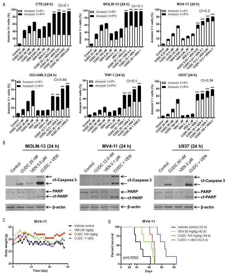
In order to determine if CUDC-907 has the capacity to enhance venetoclax-induced cell death, six AML cell lines were treated with venetoclax and CUDC-907, alone or combined, to determine the extent of the antileukemic activity of the combination (cell line characteristics are shown in the Online Supplementary Table S2). Co-treatment of AML cells with CUDC-907 and venetoclax increased the proportion of Annexin V positive cells (indicative of apoptosis) significantly compared to control and single treatment groups (Figure 1A). Combination index (CI) values demonstrate synergy between the two drugs in all AML cell lines tested. Western blots revealed increased cleavage of caspase 3 and PARP by combined drug treatments compared to single treatments (Figure 1B), demonstrating that the cells underwent apoptosis and further confirming the flow cytometry data. In order to test efficacy and tolerance of CUDC-907 and venetoclax co-treatment in vivo, immunocompromised NSGS mice were inoculated with MV4-11 cells and treated with vehicle control, 85 mg/kg/inj venetoclax, and/or 100 mg/kg/inj CUDC-907. The mice were treated for 8 days, given a 4- day break, and then treated for 6 additional days. The 4- day break was given out of an abundance of caution since a higher dosed group from a concurrent trial (150 mg/kg CUDC-907)11 experienced moderate body weight loss (3%). Co-treatment of venetoclax with CUDC-907 prolonged median survival from 33 days for control subjects to 52.5 days with minimal weight loss (Figure 1D-E). Taken together, CUDC-907 enhances venetoclax activity in AML cell lines both in vitro and in vivo.
Figure 1.
CUDC-907 and venetoclax synergistically induce apoptosis in acute myeloid leukemia cells in vitro and show in vivo efficacy in an acute myeloid leukemia cell line derived xenograft mouse model. (A) Acute myeloid leukemia (AML) cell lines were treated with vehicle control, venetoclax (VEN), CUDC-907 (CUDC), or in combination for 24 hours. Annexin V-FITC/PI staining was assessed by flow cytometry analysis. Mean percent Annexin V+ cells ± standard error of the mean are shown. Combination index (CI) values were calculated using CompuSyn software. ***P<0.001 compared to individual drug treatment. (B) AML cells were treated with CUDC-907 and venetoclax, alone or in combination, for 24 hours. Whole cell lysates were subjected to Western blotting and probed with anti-cleaved caspase 3 (cf-Caspase 3), -PARP (cf-PARP indicated cleaved PARP), or –b-actin antibody. (C) MV4-11 cells (1x10⁶ cells/mouse) were injected intravenously through the tail vein of immunocompromised NSGS mice. Three days post cell injection the mice were randomized (5 mice/group) and treated with vehicle control (3% 200 proof ethanol, 1% polyoxyethylene (20) sorbitan monooleate, and USP water), 85 mg/kg/inj venetoclax, 100 mg/kg/inj CUDC-907, or 85 mg/kg/inj venetoclax plus 100 mg/kg/inj CUDC-907 daily. The mice were treated for 8 consecutive days followed by 4 days off and then an additional 6 days. Body weight was measured on a daily basis and are graphed as mean ± standard error of the mean (panel C). Overall survival probability, estimated with the Kaplan-Meier method, is shown (panel D).
Figure 2.
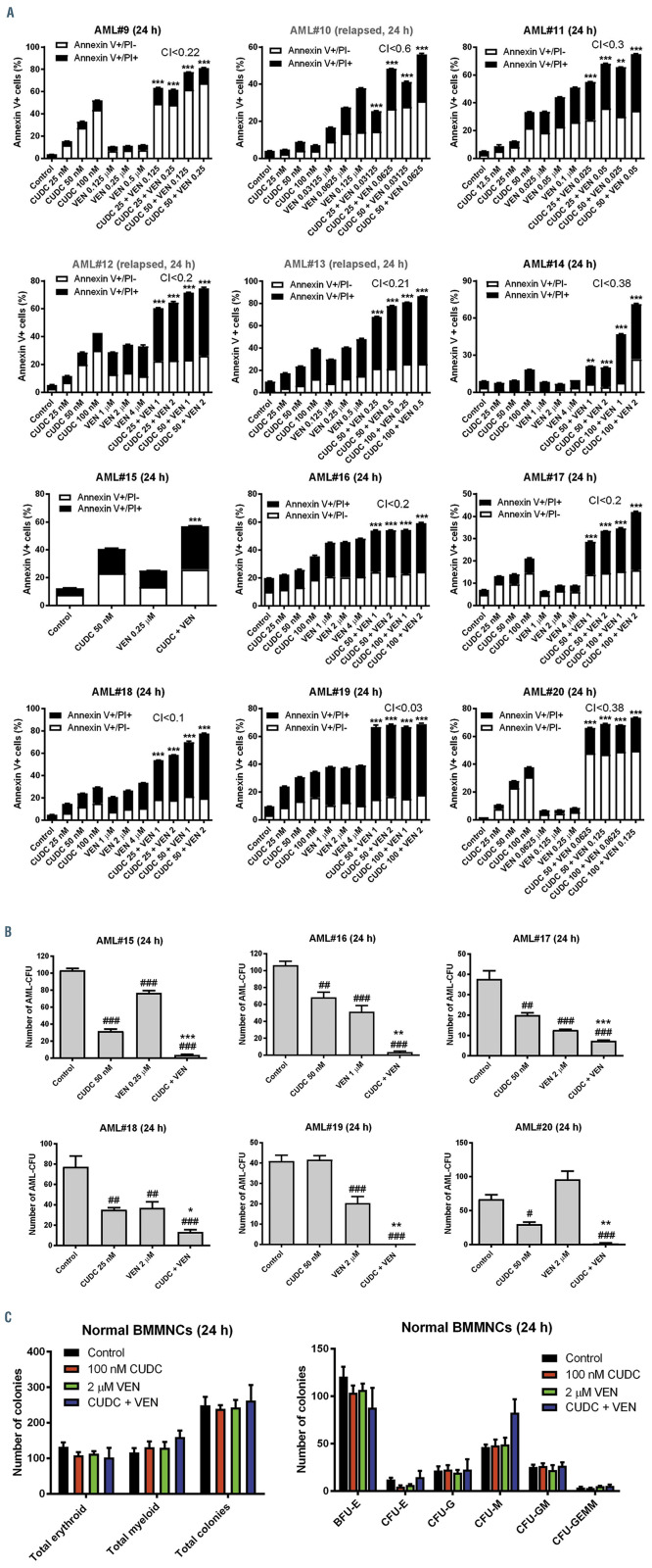
CUDC-907 and venetoclax synergize in primary acute myeloid leukemia cells and cooperatively prevent colony formation of acute myeloid leukemia progenitor cells while sparing normal hematopoietic progenitor cells ex vivo. (A) Primary acute myeloid leukemia (AML) patient samples were treated with vehicle control, venetoclax (VEN), CUDC-907 (CUDC), or in combination for 24 hours. Flow cytometry analysis of Annexin-V-FITC/PI staining was performed. Results are shown as mean percent Annexin V+ cells ± standard error of the mean (SEM). Combination index (CI) values were calculated using CompuSyn software. **P<0.01 and ***P<0.001 compared to single drug treatments. (B) Primary AML patient samples were cultured with vehicle control, venetoclax, CUDC-907, or in combination for 24 hours and then plated in methylcellulose. After incubation for 10-14 days, the number of surviving AML cells capable of generating leukemia colonies (AML-CFU) were enumerated. Data are presented as mean ± SEM. #P<0.05, ##P<0.01, and ###P<001 compared to control. *P<0.05, *P<0.01, and ***P<0.001 compared to single drug treatments. Technical triplicates were performed. (C) Normal human bone marrow mononuclear cells from a single donor were cultured with vehicle control, venetoclax, CUDC-907, or in combination for 24 hours, and then plated in methylcellulose. After incubation for 10-14 days, the number of surviving hematopoietic cells capable of generating colonies were enumerated. Total erythroid and myeloid colonies are presented as mean ± SEM (left panel). The number of BFU-E, CFU-E, CFU-G, CFU-M, CFU-GM, and CFU-GEMM colonies are presented as mean ± SEM (right panel).
CUDC-907 and venetoclax synergize in primary acute myeloid leukemia cells and co-operatively prevent colony formation of acute myeloid leukemia progenitor cells ex vivo
In order to enhance the clinical relevance of this study, primary AML patient samples were treated with CUDC- 907 and venetoclax at the indicated concentrations for 24 hours. Annexin V/PI staining and flow cytometry analysis results demonstrate synergy between the two drugs (CI <0.6) in all of the patient samples collected (with the exception of AML#15, which did not have enough sample to assess synergy), including AML relapse samples (AML#10, AML#12, AML#13, and AML#21; Figures 2A; Online Supplementary Figure S1). In order to determine the effect of the combination on leukemic progenitor cells, colony forming assays were performed using primary AML patient samples. CUDC-907 alone significantly reduced colony forming capacity in all primary AML cells, with the exception of AML#19 (Figure 2B). Venetoclax also significantly reduced colony formation in the primary samples, except AML#20. Combination therapy significantly reduced colony forming capacity in all of the patient samples tested compared to individual drug treatment (Figure 2B). Normal bone marrow mononuclear cells (BMMNC) were also tested to determine whether healthy hematopoietic progenitors would be targeted by CUDC-907 and venetoclax therapy. These treatments did not significantly affect colony forming capacity of the normal BMMNC population (Figure 2C), suggesting that the combination spares normal hematopoietic progenitors. In order to further determine the effect of the combination therapy on leukemia progenitor/ stem cells, primary AML cells were incubated with or without the human bone marrow stromal cell line HS-5. After allowing the cells to co-incubate for 24 hours, the cells were treated with CUDC-907 and venetoclax, alone or combined, for 24 hours and then measured the effects of these drug treatments on CD34+ AML progenitor/stem cells. CUDC-907 and venetoclax co-operated in inducing Annexin V positivity in these AML progenitor/stem cells in the absence of HS-5 cells (Online Supplementary Figure S2). Interestingly, the combination of CUDC-907 and venetoclax was still able to induce Annexin V positivity in these AML progenitor/stem cells even in the presence of HS-5 cells, though to a lesser extent.
Figure 3.
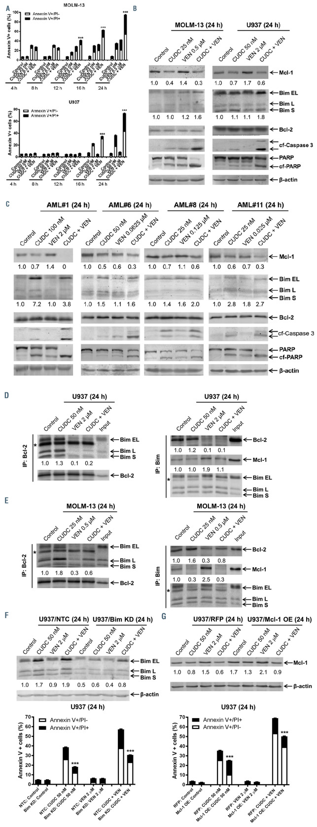
CUDC-907 upregulates Bim and downregulates Mcl-1 enhancing venetoclax activity against acute myeloid leukemia cells. (A) Acute myeloid leukemia (AML) cells were treated with vehicle control, venetoclax (VEN), CUDC-907 (CUDC), or in combination for up to 24 hours. Flow cytometry analysis of Annexin-V-FITC/PI staining was performed. Results are shown as mean percent Annexin V+ cells ± standard error of the mean (SEM). ***P<0.001 compared to single drug treatments. (B, C) AML cells were treated with vehicle control, venetoclax, CUDC-907, or in combination for 24 hours, and whole cell lysates were subjected to western blotting. Representative western blots are shown. The fold changes for the densitometry measurements, normalized to b-actin and then compared to no drug control, are indicated below the corresponding blot. Bim S, L, and EL indicate Bim short, long, and extra-long isoforms, respectively. (D, E) U937 and MOLM-13 cells were treated for 24 hours with vehicle control, venetoclax, CUDC-907, or in combination. Bcl-2 (left panel) or Bim (right panel) was immunoprecipitated from whole cell lysates. Western blots were probed with anti-Bim, -Bcl-2, or -Mcl-1 antibody. The fold changes for the densitometry measurements, normalized to b-actin and then compared to no drug control, are indicated below the corresponding blot. *Indicates the light chain of Bim or Bcl-2 antibody. (F) U937 cells were infected with NTC- (U937/NTC) or Bim-shRNA (U937/Bim) (panel F) or Precision LentiORF Mcl-1 (U937/Mcl-1) or RFP control (U937/RFP) (panel G) lentivirus particles overnight, then washed and incubated for 48 hours prior to adding puromycin or blasticidin, respectively, to the culture medium. The antibiotic-resistant cells were treated with vehicle control, venetoclax, CUDC-907, or in combination for 24 hours. Whole cell lysates were subjected to western blotting. Bim S, L, and EL indicate Bim short, long, and extralong isoforms, respectively. The fold changes for the Mcl-1 or Bim densitometry measurements, normalized to β-actin and then compared to no drug treatment control, are indicated (top panel). Annexin V/PI staining and flow cytometry analysis results are shown (bottom panel). ***P<0.001 compared to NTC or RFP.
Figure 4.
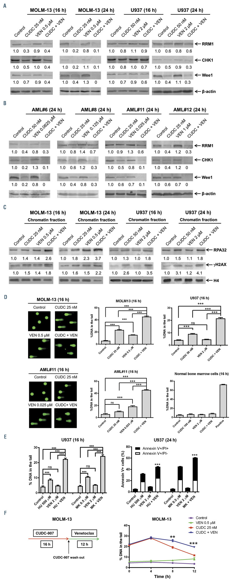
Venetoclax enhances CUDC-907-induced DNA damage in acute myeloid leukemia cells. (A, B) Acute myeloid leukemia (AML) cells were treated with vehicle control, venetoclax, CUDC-907, or in combination for 16 or 24 hours, and whole cell lysates were subjected to western blotting. Representative western blots are shown. The fold changes for the densitometry measurements, normalized to b-actin and then compared to no drug control, are indicated below the corresponding blot. (C) AML cells were treated as in panels A, B. The levels of RPA32 and γH2AX bound to chromatin were analyzed by western blotting. Densitometry measurements normalized to histone H4 and then compared to vehicle control are presented below the corresponding blot. (D) AML cells and normal bone marrow cells were treated for 16 hours with vehicle control, venetoclax, CUDC-907, venetoclax + CUDC-907, or a positive control (20 mM daunorubicin for 4 hours). Representative alkaline comet assay images for MOLM-13 and AML#11 are shown. Representative images for U937 and normal bone marrow cells are shown in the Online Supplementary Figure S4. Data are graphed as median percent DNA in the tail from three replicate gels ± standard error of the mean (SEM). ns indicates not significant and ***P<0.001. (E) U937 cells were treated with venetoclax in the presence or absence of hydroxyurea (HU) or MK-1775 (MK) for 16 hours and then subjected to the alkaline comet assay (left panel). Data are graphed as median percent DNA in the tail from three replicate gels ± SEM. ns indicates not significant and ***P<0.001. Representative images are shown in the Online Supplementary Figure S5. The treated U937 cells were also subjected to Annexin V/PI staining and flow cytometry analysis (right panel). Mean percent Annexin V+ cells ± SEM are shown. ***P<0.001 compared to single drug treatments. (F) MOLM- 13 cells were treated with or without CUDC-907 for 16 hours. The cells were washed with PBS three times and then split, half receiving fresh media and the other half receiving fresh media plus venetoclax. Cells were collected at 0, 4, 8, and 12 hours after addition of venetoclax. Alkaline comet assay results are shown as median percent DNA in tail from three replicate gels ± SEM. **P<0.01 and ***P<0.001 compared to CUDC-907 treatment. Representative images are shown in the Online Supplementary Figure S7.
Figure 5.
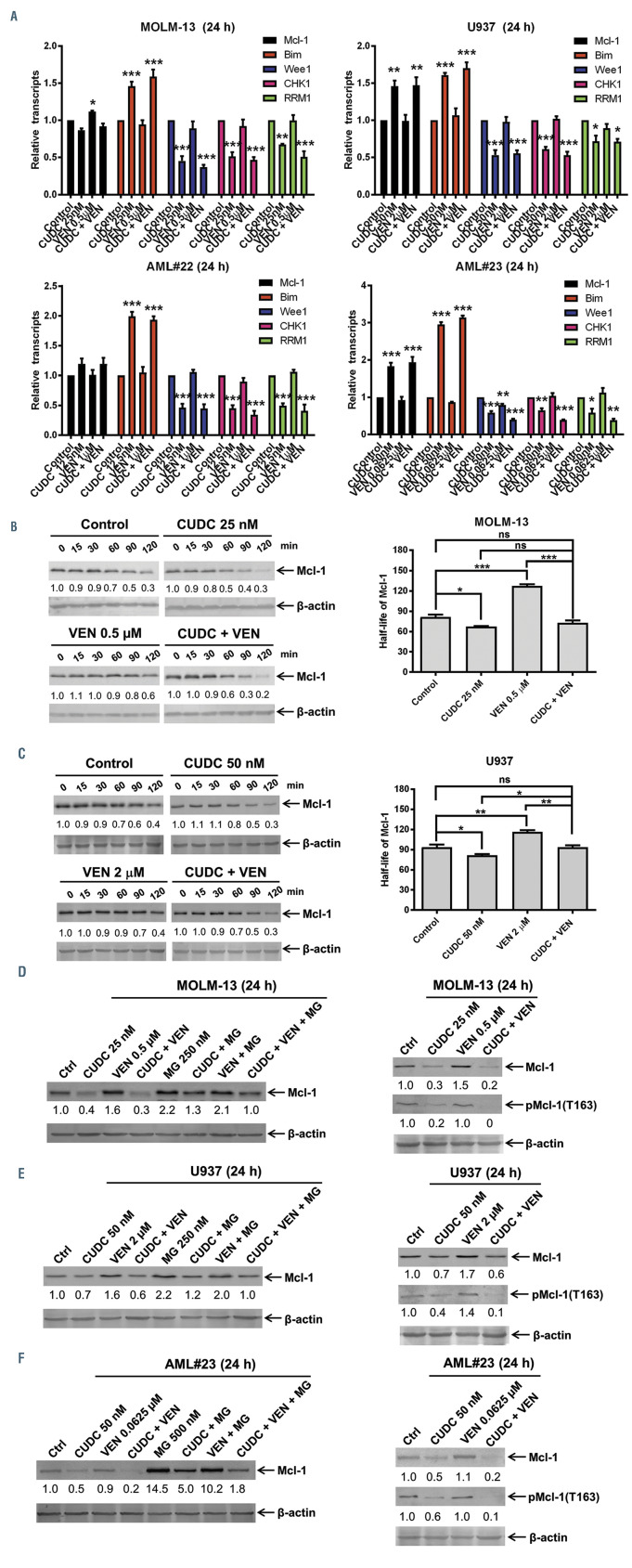
CUDC-907 synergizes with venetoclax by targeting Mcl-1 protein stability and transcriptionally regulating Bim, CHK1, Wee1, and RRM1. (A) Acute myeloid leukemia AML cells were treated for 24 h with vehicle control, venetoclax (VEN), CUDC-907 (CUDC), or in combination at the indicated concentrations. Total RNA was isolated and then Mcl-1, Bim, Wee1, CHK1, and RRM1 transcripts were determined by real-time RT-PCR. *P<0.05, **P<0.01, and ***P<0.001 compared to vehicle control. (B, C) MOLM-13 (panel B) and U937 (panel C) cells were treated with vehicle control, venetoclax, CUDC-907, or in combination for 12 hours, washed and then treated with 10 mg/mL cycloheximide for up to 2 hours. Western blots were probed with anti-Mcl-1 or -b-actin antibody. The fold changes for the densitometry measurements of Mcl-1 were normalized to β-actin and then compared to vehicle control. Representative western blots are shown on the left, while densitometry measurements are graphed and shown on the right. ns indicates not significant, *P<0.05, **P<0.01, and ***P<0.001. (D-F) MOLM-13 (panel D), U937 (panel E), and patient sample AML#23 (panel F) cells were treated with vehicle control, venetoclax, CUDC-907, or in combination with or without MG132, at the indicated concentrations, for 24 hours. Western blot analysis of whole cell lysates are shown. The fold changes for the densitometry measurements, normalized to b-actin and then compared to vehicle control, are shown.
CUDC-907 enhances venetoclax activity through upregulation of Bim and downregulation of Mcl-1
In order to begin to elucidate the mechanism of action of combined CUDC-907 and venetoclax, we treated AML cell lines for up to 24 hours to determine when enhancement of apoptosis occurs. Enhancement of venetoclaxinduced apoptosis by CUDC-907 was detected as early as 12 hours in MOLM-13 and MV4-11 cells and 16 hours in U937 cells (Figures 3A; Online Supplementary Figure S3A). Based on these results, AML cell lines MOLM-13 and U937 were treated with the indicated concentrations for 16 and 24 hours to determine the effects on Mcl-1, Bim, and Bcl-2 protein levels. Consistent with our previous report,1 venetoclax treatment resulted in an increase of Mcl-1 protein in both cell lines, which was completely abolished by the addition of CUDC-907, even when CUDC-907 treatment alone only moderately downregulated Mcl-1 (Figures 3B; Online Supplementary Figure S3B). We did not detect an obvious increase of Bim after CUDC-907 treatment, though we have previously demonstrated that at higher concentrations increased Bim protein levels could be detected.11 However, in the combination treated samples, Bim levels were substantially increased compared to CUDC-907 treatment alone. Consistent with the cell line data, CUDC-907 treatment reduced Mcl-1 protein, which was further reduced by combined drug treatment in four primary AML samples. As in the MOLM-13 and U937 AML cell lines, venetoclax treatment of AML#1 also resulted in an increase of Mcl-1 protein, again it was completely abolished by CUDC-907 (Figure 3C). In contrast to our cell line data, CUDC-907 alone and its combination with venetoclax caused marked increase in Bim protein. For both AML cell lines and primary patient samples, Bcl-2 protein levels were unaffected by treatment with CUDC-907 and venetoclax, alone or in combination. Further, combination treatment resulted in increased cleavage of caspase 3 and PARP in both AML cell lines and primary patient samples, confirming the flow cytometry results shown in Figure 2A; Figure 3A; Online Supplementary Figure S1; Online Supplementary Figure S3A. In order to determine if combined CUDC-907 and venetoclax treatment disrupts the binding of Bim to Mcl- 1 and Bcl-2, co-immunoprecipitation of Bcl-2 was performed using both U937 and MOLM-13 cells. CUDC-907 treatment caused increased binding of Bim to Bcl-2, while venetoclax treatment reduced Bim bound to Bcl-2 (Figure 3D-E). Co-immunoprecipitation of Bim revealed that venetoclax treatment increased binding of Bim to Mcl-1. CUDC-907 treatment reduced Bim bound to Mcl-1 in MOLM-13 cells, though not in U937 cells. However, combined treatment reduced both Bim bound to Bcl-2 and prevented increased Bim bound to Mcl-1 induced by venetoclax treatment. Knockdown of Bim (50%) significantly reduced apoptosis induced by CUDC-907 alone and in combination with venetoclax (Figure 3F). Mcl-1 overexpression also partially rescued cells from CUDC-907- induced apoptosis when treated alone and in combination with venetoclax (Figure 3G). Taken together, these results demonstrate that Bim and Mcl-1 play important roles in combined CUDC-907 and venetoclax treatment in AML cells. Further, CUDC-907 and venetoclax reciprocally overcome mechanisms of resistance to single drug treatment.
Figure 6.
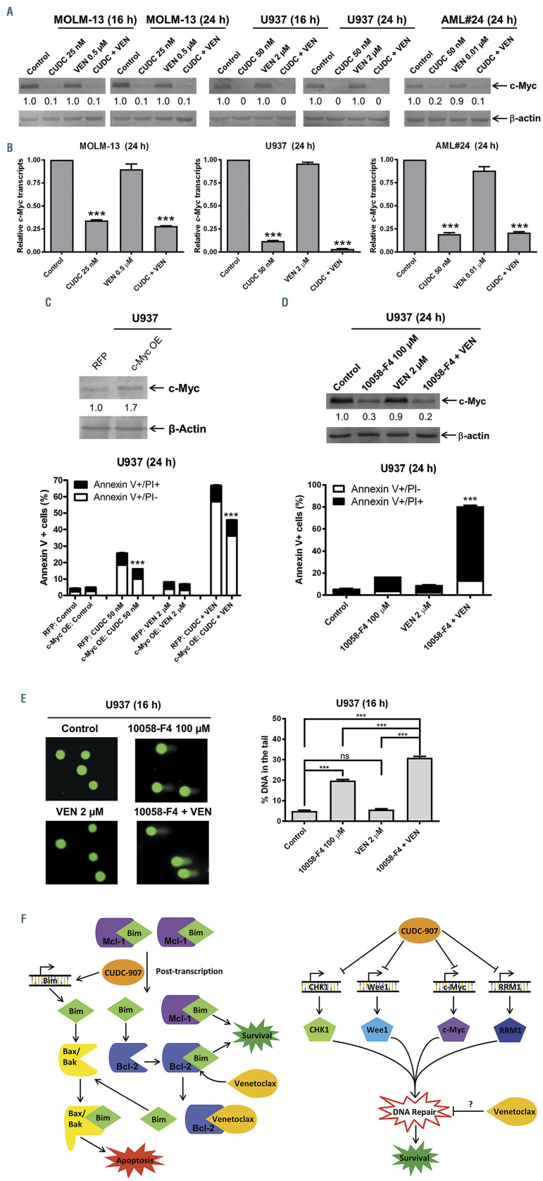
CUDC-907 downregulates c-Myc expression enhancing the antileukemic activity of venetoclax in acute myeloid leukemia cells. (A) Acute myeloid leukemia (AML) cells were treated, as indicated, for 16 or 24 hours and western blot analysis of whole cell lysates are shown together with the fold changes for the densitometry measurements, normalized to β-actin and then compared to vehicle control. (B) AML cells were treated as in panel A. Total RNA was isolated and c-Myc transcripts were measured by real-time RT-PCR. **P<0.001 compared to control. (C) U937 cells were infected with Precision LentiORF c-Myc or RFP control lentivirus particles overnight, then washed and incubated for 24 hours. Whole cell lysates from one aliquot of cells were subjected to western blotting (top panel). Densitometry measurements were normalized to b-actin; fold changes compared to RFP control are shown. The other aliquot of cells were treated with vehicle control, venetoclax, CUDC- 907, or in combination for 24 hours. Flow cytometry analysis results of Annexin V-FITC/PI staining are shown as mean Annexin V positive ± standard error of the mean (SEM) (bottom panel). ***P<0.001. (D) U937 cells were treated with vehicle control, 10058-F4, venetoclax, or in combination for 24 hours. Representative western blots of c-Myc and b-actin are shown (top panel). Densitometry measurements of c-Myc were normalized to β-actin; fold changes compared to vehicle control are shown. Flow cytometry analysis results of Annexin V-FITC/PI staining are shown as mean Annexin V positive ± SEM (bottom panel). ***P<0.001. (E) U937 cells were treated as in panel D and then subjected to alkaline comet assay. Representative images are shown (left panel). Results are shown as median percent DNA in tail from three replicate gels ± SEM (right panel). (F) Proposed mechanisms of action of venetoclax in combination with CUDC-907. CUDC-907 treatment increases Bim and decreases Mcl-1, but Bcl-2 sequesters Bim, which opposes apoptosis. Venetoclax treatment reduces Bim binding to Bcl-2, tipping the balance towards more “free” Bim, inducing apoptosis. CUDC-907 downregulates CHK1, Wee1, RRM1, and c-Myc, inducing DNA damage, while venetoclax prevents DNA repair, leading to accumulation of DNA damage and more cell death.
Venetoclax enhances CUDC-907-induced DNA damage in acute myeloid leukemia cells
Based on our previous study in which we show downregulation of key DNA repair proteins by CUDC-907 treatment,11 we sought to determine if these changes also occurred in the presence of venetoclax. Consistent with our published work, CUDC-907 downregulated RRM1, CHK1, and Wee1, which was maintained by the combined treatment in both AML cell lines and primary patient samples (Figure 4A-B). CUDC-907 treatment increased chromatin-bound RPA32 and γH2AX, reflecting increased DNA replication stress and DNA damage (Figure 4C). Venetoclax treatment increased chromatinbound RPA32 and γH2AX in MOLM-13 cells, though not in U937 cells. Combined CUDC-907 and venetoclax treatment resulted in more chromatin-bound RPA32 and γH2AX compared to single drug treatments. Alkaline comet assay results revealed that individual treatment with CUDC-907 induced DNA damage, while combined treatment significantly increased DNA damage compared to individual drug treatment for both AML cell lines and a primary AML patient sample (Figure 4D; Online Supplementary Figure S4). We previously reported that CHK1 plays a role in the mechanism of action of venetoclax in AML cells.6 In order to confirm the roles of Wee1 and RRM1 in venetoclax activity, U937 cells were treated with the selective Wee1 inhibitor MK-1775 or the ribonucleotide reductase inhibitor hydroxyurea (HU) alone or combined with venetoclax, and then subjected to comet assays. As shown in Figure 4E and Online Supplementary Figure S5, Wee1 inhibition and HU treatment both induced DNA damage, which was significantly increased by the addition of venetoclax. HU and MK-1775 treatment increased the percent of Annexin V positive cells, which was significantly enhanced by venetoclax. In order to further confirm the role of Wee1, Wee1 was overexpressed in U937 cells (Online Supplementary Figure S6A), which partially prevented apoptosis induced by CUDC- 907 alone and in combination with venetoclax (Online Supplementary Figure S6B). Further, overexpression of Wee1 decreased DNA damage induced by CUDC-907 treatment alone and in combination with venetoclax (Online Supplementary Figure S6C). These results show that Wee1 plays a role in apoptosis induced by CUDC-907 or combined CUDC-907 and venetoclax treatment in AML cells. In order to determine the effect of venetoclax on DNA damage, MOLM13 cells were treated with CUDC- 907 for 16 hours, washed, given fresh media with or without venetoclax for up to 12 hours. Following CUDC-907 treatment, DNA damage decreased, suggesting that repair progressed after removal of CUDC-907. However, addition of venetoclax significantly slowed down the decrease of the percent DNA in the comet tails, indicating inhibition of DNA repair (Figure 4F; Online Supplementary Figure S7). Taken together, these results suggest that venetoclax impairs repair of DNA damage induced by CUDC-907 in AML cells.
CUDC-907 decreases Mcl-1 protein stability and transcriptionally regulates Bim, CHK1, Wee1, and RRM1, enhancing venetoclax activity in acute myeloid leukemia cells
In order to begin to determine if CUDC-907 modulates Mcl-1, Bim, CHK1, Wee1, and RRM1 by altering transcription, transcripts were measured via real-time RT-PCR after drug treatment. In AML cell line U937 and primary patient sample AML#23, CUDC-907 treatment, both alone and in combination with venetoclax, significantly increased Mcl-1 transcripts, while in MOLM13 and AML#22 no significant change was detected (Figure 5A). CUDC-907 treatment alone and in combination with venetoclax significantly increased Bim and reduced CHK1, Wee1, and RRM1 transcripts in all samples tested. These results suggest that the upregulation of Bim and downregulation of CHK1, Wee1, and RRM1 by CUDC-907 treatment is likely through transcriptional mechanisms, while Mcl-1 is not. In order to determine if CUDC-907 and venetoclax have an impact on Mcl-1 protein stability, MOLM-13 and U937 cells were treated with CUDC-907 and venetoclax, alone or in combination, for 16 hours, washed, and then treated with cycloheximide (10 mg/mL) for up to 120 minutes. Western blots revealed that Mcl-1 levels decreased significantly faster in cells treated with CUDC-907 compared to control cells (MOLM13: 68 vs. 82 minutes, P=0.0189; U937: 82 vs. 94 minutes, P=0.0423). Venetoclax treatment significantly increased Mcl-1 halflife (MOLM13: 115 vs. 82 minutes, P=0.0005; U937: 115 vs. 94 minutes, P=0.0061), while combination treatment prevented venetoclax-induced Mcl-1 half-life increase (Figure 5B-C). Proteasome inhibition prevented downregulation of Mcl-1 by CUDC-907 both alone and in combination with venetoclax in AML cell lines and a primary AML patient sample (Figure 5D-F). Since phosphorylation of Mcl-1 at T163 has been shown to stabilize Mcl-1 by prolonging its half-life,25 we looked at phosphorylation of Mcl-1 post-drug treatment. CUDC-907 treatment alone and in combination with venetoclax reduced phosphorylation of Mcl-1 at T163 (Figure 5D-F). Taken together, these results suggest that CUDC-907 downregulates Mcl- 1 by decreasing its protein stability in AML cells.
CUDC-907 downregulates c-Myc expression enhancing the antileukemic activity of venetoclax in acute myeloid leukemia cells
Our previous work shows that CUDC-907 inhibits the expression of c-Myc,11 which is an oncoprotein that is frequently activated in AML cells and plays an important role in leukemogenesis.26,27 Western blot analyses revealed that CUDC-907 downregulation of c-Myc occurs in the presence or absence of venetoclax in AML cell lines and primary AML patient sample AML#24 (Figure 6A). c-Myc transcript levels were significantly reduced by CUDC-907 treatment alone and in combination with venetoclax (Figure 6B). In order to confirm the functional role of c- Myc, we transiently overexpressed c-Myc in U937 cells. Overexpression of c-Myc significantly reduced the effects of CUDC-907 alone and in combination with venetoclax (Figure 6C). Treatment with the c-Myc inhibitor 10058-F4 significantly enhanced venetoclax-induced apoptosis (Figure 6D). Further, c-Myc inhibition induced DNA damage that was further enhanced by combination with venetoclax (Figure 6E). These results indicate that c-Myc downregulation by CUDC-907 plays an important role in the enhancement of venetoclax-induced death of AML cells.
Discussion
We previously reported that Mcl-1 and Bim are important for the antileukemic activity of venetoclax1 and they are also important for CUDC-907 activity in AML cells.11 In this study, we found that CUDC-907 treatment decreases Mcl-1 protein, overcoming a mechanism of resistance to venetoclax. Our previous study showing that inhibition of Mcl-1 enhances the antileukemic activity of venetoclax,3 further supports that downregulation of Mcl- 1 by CUDC-907 plays an important role in the combined antileukemic activity of CUDC-907 and venetoclax. Venetoclax prevents Bcl-2 from sequestering Bim, overcoming a mechanism of resistance to CUDC-907. In agreement with our previous study,11 Bim transcripts were increased after treatment with CUDC-907 alone and in combination with venetoclax (Figure 5A). Further, CUDC- 907 treatment decreased Mcl-1 protein stability, which does not appear to be effected by venetoclax treatment. While Mcl-1 and Bim play important roles in CUDC-907 and venetoclax activity, knockdown of Bim and overexpression of Mcl-1 only partially prevented apoptosis induced by CUDC-907 alone and in combination with venetoclax, suggesting that other mechanisms exist.
In our previous study, we found that CHK1, Wee1, and RRM1 play important roles in the antileukemic activity of CUDC-907 against AML.11 We have also shown enhanced DNA damage in AML cells treated with combined venetoclax and DNA damaging agents1,6 and that CHK1 inhibition enhances the antileukemic activity of venetoclax.6 Pham and colleagues recently reported that venetoclax treatment activates the DNA damage response in lymphoma cells, though the mechanism is unknown.28 In agreement, we show that inhibition of Wee1 or RRM1 in combination with venetoclax co-operatively induce DNA damage in AML cells. Further, we found that venetoclax significantly prolonged persistence of CUDC-907-induced DNA damage, suggesting that Bcl-2 plays a role in the DNA repair.
There is preclinical evidence that CUDC-907 downregulates c-Myc in diffuse large B-cell lymphoma.9 Additionally, we previously reported that CUDC-907 downregulates c-Myc in vitro, in as little as 4 hours, and in vivo.11 This downregulation is maintained in the presence of venetoclax in vitro (Figure 6A). c-Myc overexpression partially rescues AML cells from both CUDC-907 monotherapy and combination therapy. Further, combination of venetoclax plus the c-Myc inhibitor 10058-F4 results in significant induction of apoptosis, supporting the assertion that c-Myc inhibition is a key mechanism underlying the combination’s synergy. Based on the roles PI3K/mTOR and c-Myc play in proliferation,29,30 this combination may inhibit cell proliferation in addition to inducing apoptosis. Similar to our results, Cinar et al. reported that the c-Myc inhibitor 10058-F4 enhances venetoclax activity in double-hit and triple-hit lymphoma.31 Though the mechanism remains to be fully understood, c-Myc inhibition and venetoclax cooperatively induce DNA damage in AML cells (Figure 6E), suggesting that Bcl-2 is involved in the DNA damage response.
Our data demonstrates that CUDC-907 can enhance venetoclax activity in vivo. The mice were given a 4-day break out of an abundance of caution since a higher dosed group in a concurrent trial (150 mg/kg CUDC-907, published in 11) experienced moderate body weight loss (3%). Additionally, treatment was stopped after 14 doses of CUDC-907, again due to the moderate body weight loss in the concurrent trial (150 mg/kg CUDC-907 group, which was completely reversible within 4 days). In hindsight, the body weight loss associated with combination treatment was minimal, suggesting that the 4-day drug holiday may not have been necessary or that we could have potentially given more than 14 doses. Nonetheless, our results show proof-of-concept; the combination of CUDC-907 and venetoclax increased the lifespan of the mice by 60%, while CUDC-907 and venetoclax alone increased the lifespan by 33% and 27%, respectively. Additionally, our unpublished data using the MV4-11- derived xenograft mouse model, shows that cytarabine in combination with venetoclax increased the lifespan of these mice by only 7.5% (unpublished data).
In summary, combined CUDC-907 and venetoclax shows great synergistic antileukemic activity against AML cells, at least partially mediated by Bim, Mcl-1, CHK1, Wee1, RRM1, and c-Myc (Figure 6F). Both CUDC-907 and venetoclax are orally available agents, which would simplify their administration in the clinical setting.32 More importantly, both agents are already in regular clinical use in experimental and non-experimental contexts. Venetoclax has been approved by the FDA for use in the context of AML, and CUDC-907 carries an FDA Fast Track designation for use in relapsed or refractory diffuse large B-cell lymphoma and a phase II trial is presently underway examining its efficacy in patients with MYCaltered relapsed/refractory diffuse large B-cell lymphoma. The results of this study support further development of this promising combination for the treatment of AML.
Supplementary Material
Funding Statement
FundingThis study was supported by Jilin University, Changchun, China, the Barbara Ann Karmanos Cancer Institute, Wayne State University School of Medicine, and by grants from Projects of Jilin Province Science and Technology Development Plan (20180101277JC), the National Natural Science Foundation of China (NSFC 31671438 and NSFC 31471295), National Key Research and Development Program of China (2017YFC1702103), Graduate Innovation Fund of Jilin University, Hyundai Hope on Wheels, LaFontaine Family/U Can-Cer Vive Foundation, Kids Without Cancer, Children’s Hospital of Michigan Foundation, Decerchio/Guisewite Family, Justin’s Gift, Elana Fund, Ginopolis/Karmanos Endowment and the Ring Screw Textron Endowed Chair for Pediatric Cancer Research. The Animal Model and Therapeutics Evaluation Core and Genomics Core are supported, in part, by NIH Center Grant P30 CA022453 to the Karmanos Cancer Institute at Wayne State University. The funders had no role in study design, data collection, analysis and interpretation of data, decision to publish, or preparation of the manuscript.
References
- 1.Niu X, Zhao J, Ma J, et al. Binding of released Bim to Mcl-1 is a mechanism of intrinsic resistance to ABT-199 which can be overcome by combination with daunorubicin or cytarabine in AML cells. Clin Cancer Res. 2016;22(17):4440-4451. [DOI] [PMC free article] [PubMed] [Google Scholar]
- 2.Knight T, Edwards H, Taub JW, Ge Y. Evaluating venetoclax and its potential in treatment-naive acute myeloid leukemia. Cancer Manag Res. 2019;11:3197-3213. [DOI] [PMC free article] [PubMed] [Google Scholar]
- 3.Luedtke DA, Niu X, Pan Y, et al. Inhibition of Mcl-1 enhances cell death induced by the Bcl-2-selective inhibitor ABT-199 in acute myeloid leukemia cells. Signal Transduct Target Ther. 2017;2:17012. [DOI] [PMC free article] [PubMed] [Google Scholar]
- 4.Xie C, Edwards H, Caldwell JT, Wang G, Taub JW, Ge Y. Obatoclax potentiates the cytotoxic effect of cytarabine on acute myeloid leukemia cells by enhancing DNA damage. Mol Oncol. 2015;9(2):409-421. [DOI] [PMC free article] [PubMed] [Google Scholar]
- 5.Knight T, Luedtke D, Edwards H, Taub JW, Ge Y. A delicate balance - the BCL-2 family and its role in apoptosis, oncogenesis, and cancer therapeutics. Biochem Pharmacol. 2019;162:250-261. [DOI] [PubMed] [Google Scholar]
- 6.Zhao J, Niu X, Li X, et al. Inhibition of CHK1 enhances cell death induced by the Bcl-2- selective inhibitor ABT-199 in acute myeloid leukemia cells. Oncotarget. 2016; 7(23):34785-34799. [DOI] [PMC free article] [PubMed] [Google Scholar]
- 7.Kotian S, Zhang L, Boufraqech M, et al. Dual Inhibition of HDAC and tyrosine kinase aignaling pathways with CUDC-907 inhibits thyroid cancer growth and metastases. Clin Cancer Res. 2017; 23(17):5044-5054. [DOI] [PMC free article] [PubMed] [Google Scholar]
- 8.Qian C, Lai CJ, Bao R, et al. Cancer network disruption by a single molecule inhibitor targeting both histone deacetylase activity and phosphatidylinositol 3-kinase signaling. Clin Cancer Res. 2012;18(15):4104-4113. [DOI] [PubMed] [Google Scholar]
- 9.Sun K, Atoyan R, Borek MA, et al. Dual HDAC and PI3K inhibitor CUDC-907 downregulates MYC and suppresses growth of MYC-dependent cancers. Mol Cancer Ther. 2017;16(2):285-299. [DOI] [PubMed] [Google Scholar]
- 10.Younes A, Berdeja JG, Patel MR, et al. Safety, tolerability, and preliminary activity of CUDC-907, a first-in-class, oral, dual inhibitor of HDAC and PI3K, in patients with relapsed or refractory lymphoma or multiple myeloma: an open-label, doseescalation, phase 1 trial. Lancet Oncol. 2016; 17(5):622-631. [DOI] [PMC free article] [PubMed] [Google Scholar]
- 11.Li X, Su Y, Madlambayan G, et al. Antileukemic activity and mechanism of action of the novel PI3K and histone deacetylase dual inhibitor CUDC-907 in acute myeloid leukemia. Haematologica. 2019;104(11):2225-2240. [DOI] [PMC free article] [PubMed] [Google Scholar]
- 12.Chen S, Dai Y, Pei XY, Grant S. Bim upregulation by histone deacetylase inhibitors mediates interactions with the Bcl-2 antagonist ABT-737: evidence for distinct roles for Bcl-2, Bcl-xL, and Mcl-1. Mol Cell Biol. 2009; 29(23):6149-6169. [DOI] [PMC free article] [PubMed] [Google Scholar]
- 13.Fiskus W, Sharma S, Saha S, et al. Pre-clinical efficacy of combined therapy with novel beta-catenin antagonist BC2059 and histone deacetylase inhibitor against AML cells. Leukemia. 2015;29(6):1267-1278. [DOI] [PMC free article] [PubMed] [Google Scholar]
- 14.Qi W, Zhang W, Edwards H, et al. Synergistic anti-leukemic interactions between panobinostat and MK-1775 in acute myeloid leukemia ex vivo. Cancer Biol Ther. 2015;16(12):1784-1793. [DOI] [PMC free article] [PubMed] [Google Scholar]
- 15.Aye Y, Li M, Long MJ, Weiss RS. Ribonucleotide reductase and cancer: biological mechanisms and targeted therapies. Oncogene. 2015;34(16):2011-2021. [DOI] [PubMed] [Google Scholar]
- 16.Rahmani M, Aust MM, Benson EC, Wallace L, Friedberg J, Grant S. PI3K/mTOR inhibition markedly potentiates HDAC inhibitor activity in NHL cells through BIM- and MCL- 1-dependent mechanisms in vitro and in vivo. Clin Cancer Res. 2014;20(18):4849-4860. [DOI] [PMC free article] [PubMed] [Google Scholar]
- 17.Salvatori B, Iosue I, Djodji Damas N, et al. Critical role of c-Myc in acute myeloid leukemia involving direct regulation of miR- 26a and histone methyltransferase EZH2. Genes Cancer. 2011;2(5):585-592. [DOI] [PMC free article] [PubMed] [Google Scholar]
- 18.Ohanian M, Rozovski U, Kanagal-Shamanna R, et al. MYC protein expression is an important prognostic factor in acute myeloid leukemia. Leuk Lymphoma. 2019; 60(1):37-48. [DOI] [PMC free article] [PubMed] [Google Scholar]
- 19.Niu X, Wang G, Wang Y, et al. Acute myeloid leukemia cells harboring MLL fusion genes or with the acute promyelocytic leukemia phenotype are sensitive to the Bcl-2-selective inhibitor ABT-199. Leukemia. 2014;28(7):1557-1560. [DOI] [PMC free article] [PubMed] [Google Scholar]
- 20.Ma J, Li X, Su Y, et al. Mechanisms responsible for the synergistic antileukemic interactions between ATR inhibition and cytarabine in acute myeloid leukemia cells. Sci Rep. 2017;7:41950. [DOI] [PMC free article] [PubMed] [Google Scholar]
- 21.Xie C, Edwards H, Xu X, et al. Mechanisms of synergistic antileukemic interactions between valproic acid and cytarabine in pediatric acute myeloid leukemia. Clin Cancer Res. 2010;16(22):5499-5510. [DOI] [PMC free article] [PubMed] [Google Scholar]
- 22.Edwards H, Xie C, LaFiura KM, et al. RUNX1 regulates phosphoinositide 3- kinase/AKT pathway: role in chemotherapy sensitivity in acute megakaryocytic leukemia. Blood. 2009;114(13):2744-2752. [DOI] [PMC free article] [PubMed] [Google Scholar]
- 23.Su Y, Li X, Ma J, et al. Targeting PI3K, mTOR, ERK, and Bcl-2 signaling network shows superior antileukemic activity against AML ex vivo. Biochem Pharmacol. 2018; 148:13-26. [DOI] [PMC free article] [PubMed] [Google Scholar]
- 24.Luedtke DA, Su Y, Liu S, et al. Inhibition of XPO1 enhances cell death induced by ABT- 199 in acute myeloid leukaemia via Mcl-1. J Cell Mol Med. 2018;22(12):6099-6111. [DOI] [PMC free article] [PubMed] [Google Scholar]
- 25.Domina AM, Vrana JA, Gregory MA, Hann SR, Craig RW. MCL1 is phosphorylated in the PEST region and stabilized upon ERK activation in viable cells, and at additional sites with cytotoxic okadaic acid or taxol. Oncogene. 2004;23(31):5301-5315. [DOI] [PubMed] [Google Scholar]
- 26.Hoffman B, Amanullah A, Shafarenko M, Liebermann DA. The proto-oncogene c-myc in hematopoietic development and leukemogenesis. Oncogene. 2002;21(21):3414-3421. [DOI] [PubMed] [Google Scholar]
- 27.Renneville A, Roumier C, Biggio V, et al. Cooperating gene mutations in acute myeloid leukemia: a review of the literature. Leukemia. 2008;22(5):915-931. [DOI] [PubMed] [Google Scholar]
- 28.Pham LV, Huang S, Zhang H, et al. Strategic therapeutic targeting to overcome venetoclax resistance in aggressive B-cell lymphomas. Clin Cancer Res. 2018; 24(16): 3967-3980. [DOI] [PubMed] [Google Scholar]
- 29.Gabay M, Li Y, Felsher DW. MYC activation is a hallmark of cancer initiation and maintenance. Cold Spring Harb Perspect Med. 2014;4(6). [DOI] [PMC free article] [PubMed] [Google Scholar]
- 30.Manning BD, Toker A. AKT/PKB signaling: navigating the network. Cell. 2017;169(3):381-405. [DOI] [PMC free article] [PubMed] [Google Scholar]
- 31.Cinar M, Rosenfelt F, Rokhsar S, et al. Concurrent inhibition of MYC and BCL2 is a potentially effective treatment strategy for double hit and triple hit B-cell lymphomas. Leuk Res. 2015;39(7):730-738. [DOI] [PubMed] [Google Scholar]
- 32.Eek D, Krohe M, Mazar I, et al. Patientreported preferences for oral versus intravenous administration for the treatment of cancer: a review of the literature. Patient Prefer Adherence. 2016;10:1609-1621. [DOI] [PMC free article] [PubMed] [Google Scholar]
Associated Data
This section collects any data citations, data availability statements, or supplementary materials included in this article.


