



Abstract
Purpose of review:
Despite significant progress in our understanding and clinical management of multisystem inflammatory syndrome in children (MIS-C), significant challenges remain. Here, we review recently published studies on the clinical diagnosis, risk stratification, and treatment of MIS-C, highlighting key gaps in research progress that are a microcosm for challenges in translational pediatrics research. We then discuss potential solutions in the realm of translational bioinformatics.
Recent findings:
Current case definitions are inconsistent and do not capture the underlying pathophysiology of MIS-C, which remains poorly understood. While overall mortality is low, some patients rapidly decompensate, and a test to identify those at risk for severe outcomes remains an unmet need. Treatment consists of various combinations of immunoglobulins, corticosteroids, and biologics, based on extrapolated data and expert opinion, while the benefits remain unclear as we await the completion of clinical trials.
Summary:
The small size and heterogeneity of the pediatric population contribute to unmet needs due to financial and logistical constraints of the current research infrastructure focused on eliminating most sources of heterogeneity, leading to ungeneralizable results. Data sharing and meta-analysis of gene expression shows promise to accelerate progress in the field of MIS-C as well as other childhood diseases beyond the current pandemic.
Keywords: translational medicine, pediatrics, bioinformatics, transcriptomics, MIS-C
Introduction
The rapid global spread of SARS-CoV-2 spawned an unprecedented public health crisis, triggering a colossal effort within the scientific community to unravel its pathogenesis, identify therapeutics, and develop vaccines. While our understanding of COVID-19 has increased dramatically since it was declared a pandemic in March 2020, it has been studied less in children. Compared to adults, reported case numbers and severity have been substantially lower in children [1] who have a U-shaped severity curve with chronic comorbidities (e.g., chronic lung disease, asthma, congenital heart disease, severe obesity, immunosuppression) [2] and age under 1 year being the two risk factors most consistently associated with severe acute COVID-19 [1,3–7]. As the pandemic evolved, clusters of case reports emerged in the spring of 2020, describing an unusual febrile illness temporally associated with SARS-CoV-2 infection in children. This alarming new entity, now defined as multisystem inflammatory syndrome in children (MIS-C), exhibited features of Kawasaki disease and toxic shock syndrome, and was associated with higher rates of critical illness, the need for extracorporeal support, and death [8–11]. Here, we review some of the major challenges remaining in our understanding and clinical management of MIS-C, a microcosm of challenges in pediatric translational research. We then present a computational framework for addressing these problems and designing future studies.
Diagnostic challenges
Subsequent to the initial European case reports, extraordinary efforts by the pediatric scientific community led to the publication of multiple case series and clinical guidelines [12–18], offering clinicians a rapid primer on the natural history and proposed management of this enigmatic disease. Therefore, current diagnosis and treatment of this immune-mediated, life-threatening disease with multi-organ manifestations relies heavily on evolving expert opinion, anecdotal experience, and extrapolation of data from different inflammatory conditions with similar clinical presentations.
Several factors exacerbate the complexity of managing MIS-C. First, while the pathophysiology is a topic of active investigation, it remains poorly understood [19, 20*, 21*,22,23]. Therefore, diagnosis is heavily reliant on syndromic case definitions without clear gold standard diagnostics linked to the underlying biology. There are multiple case definitions [14,15,17,24], which consist of clinical features and laboratory biomarkers that are nonspecific to MIS-C and overlap with other inflammatory, infectious, malignant, and rheumatic diseases. For example, Dufort et al. published the results of mandatory surveillance for MIS-C across 106 New York hospitals with 191 cases reported to the state health department as of May 10, 2020 [25]. Of the 161 cases meeting initial review criteria, 62 (38%) failed to meet the case definition but shared clinical and lab features of 99 who did. The case definition requirement for laboratory-confirmed SARS-CoV-2 infection is problematic, given that a smaller proportion of patients with MIS-C test positive by PCR (20-40%) than by serology (80-90%), which has variable sensitivity and is not universally available [9–12,25–27]. Moreover, there is no consensus on whether MIS-C is an acute or post-infectious disorder [25–29, 30*,31]. The predominance of gastrointestinal over respiratory symptoms supports the possibility of viral replication in the gastrointestinal tract, which would be missed on current testing [10,32*]. Finally, as the pandemic evolves and seropositivity rates for SARS-CoV-2 IgG continue to increase, the use of serologic testing to identify recent infection will become increasingly complicated.
Prognostic and Risk Stratification challenges
Perhaps one of the most concerning aspects of MIS-C is that some patients rapidly decompensate, and there are no definitive risk factors or biomarkers to identify high-risk patients [10,13]. While elevated procalcitonin, C-reactive protein (CRP), ferritin, and D-dimer correlate with severity, these nonspecific acute phase reactants have not demonstrated prognostic value as they fail to reliably distinguish between moderate and severe cases [3,12,27,33,34]. Some studies suggest that BNP/NT-proBNP may help identify MIS-C patients with left ventricular dysfunction. However, they are not truly predictive biomarkers as transient elevations occur without cardiac involvement, and significant elevations are typically seen in patients already presenting with clinical signs of [26,27,33,35–40]. Until truly predictive biomarkers are identified, providers on the front line need to retain a high level of suspicion and cast a wide net at the expense of unnecessary monitoring and interventions.
Treatment challenges
While the majority of children with MIS-C recover with supportive therapy tailored to their physiology (e.g., inotropes in cardiogenic shock), many institutions have published their own evolving guidelines [16,41–43] regarding the use of immunomodulatory drugs (corticosteroids, immunoglobulins, and biologics), whose potential benefits remain controversial in the absence of controlled trials [25,26,33,44]. Amidst substantial practice variability, a task force of experts in rheumatology, cardiology, infectious disease, and critical care recently published consensus guidelines based on available evidence to aid clinician decision-making while awaiting clinical trials [18]. The challenges facing low and middle-income countries with pediatric COVID-19 and MIS-C warrant special consideration, where some of the currently recommended biologic agents (e.g., Anakinra) are less attainable compared to corticosteroids, which may impose increased risk in populations where HIV or tuberculosis are prevalent [45–50]. An improved understanding of the underlying immune dynamics of pediatric patients with COVID-19 and MIS-C will be critical to establish which treatments are most effective at preventing or reversing life-threatening complications such as coronary aneurysms, refractory shock, and cytokine storm. The benefits of understanding the host immune response elements that contribute to severe disease could have broader benefits in the field of pediatrics, such as elucidating the etiology of similar conditions such as Kawasaki disease.
Applications of translational bioinformatics in Pediatrics
The challenges described in the diagnosis, risk stratification, and treatment of MIS-C are a microcosm of challenges to translational pediatrics research in general. The traditional clinical research paradigm involves a series of trials investigating 1-2 interventions in a single disease, which are particularly difficult to carry out in pediatrics due to small population size and patient heterogeneity. For example, the definitions of syndromes such as ARDS and sepsis encompass multiple sources of heterogeneity, including a wide range of severity, ages, comorbidities, and demographics [51,52]. The financial and logistical constraints of representing all these sources of heterogeneity translate into no single center having sufficient patient volume to produce generalizable knowledge. As a result, the current paradigm often fails to meet the needs of the pediatric population, and a significant proportion of the practice is based on evidence from small observational studies, expert opinion, or extrapolation of adult data, all of which are suboptimal and slow the advancement of the field, leaving urgent clinical questions unanswered. Translational bioinformatics can be a transformational tool to advance pediatric research and make it more efficient by addressing specific challenges unique to pediatric patient populations. There are many algorithms and approaches applicable across the research spectrum, from quality improvement to drug discovery, which are beyond the scope of this review. However, they are worth exploring here: [53,54]. Here, we will focus on how integrated meta-analysis of publicly available host transcriptome data can be leveraged to develop robust, independently validated biomarkers of immune-mediated disease across diverse populations while capturing clinically relevant biology, thus greatly shortening the paths between discovery, validation, and clinical implementation.
High-throughput “omics” technologies such as RNA-seq and microarray allow the simultaneous examination of thousands of genes and are thus an excellent way to study the host immune response. However, such datasets with more variables than samples can create results that are overfit and nonreproducible [55,56]. Integration using meta-analysis of gene expression is more robust as it produces results that validate in independent datasets [57]. However, this integration can be challenging due to inter-study biological, clinical, and technical heterogeneity. To address this, Khatri et al. developed a computational framework, called multi-cohort analysis, that accounts for this heterogeneity by following a set of guidelines described in Figure 1 [58]. The core benefit of this framework is the ability to translate ‘small data’ – datasets with tens of samples – into ‘Big Data’ – hundreds to thousands of samples, which collectively represent the heterogeneity observed in the real-world patient population. By capturing this heterogeneity, the results are significantly more robust and generalizable in independent datasets [59]. Haynes et al. created a user-friendly R package, called MetaIntegrator, which automates the majority of the multi-cohort analysis framework and guides the user from downloading the data, performing statistical analysis, and visualizing the results [59]. Several studies have repeatedly demonstrated that accounting for heterogeneity can lead to the identification of generalizable disease signatures that are reproducible and capable of accelerating translation to clinical practice [59–65*,66*].
Figure 1: Guidelines for integrating heterogeneous independent datasets.
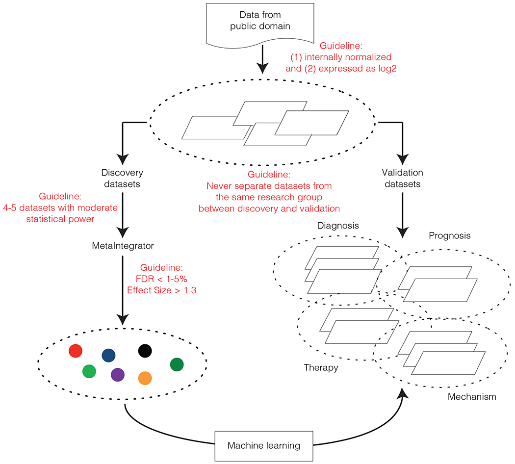
Datasets from public repositories such as the National Center for Biotechnology Information (NCBI) Gene Expression Omnibus (GEO) are divided into ‘discovery’ and ‘validation’ cohorts, where all datasets from one research group are either in the discovery or validation set. By ensuring that the data from the same group are not in both discovery and validation, we ensure that results from analysis of discovery datasets is always verified in completely independent datasets. Ideally, the discovery datasets should include 4-5 datasets that collectively represent the biological, clinical, and technical heterogeneity observed in the real-world patient population. These datasets do not need to have a large sample size, and should have moderate statistical power (e.g., 30-50 samples in each dataset). FDR = false discovery rate.
For example, in a landmark study, Sweeney et al. applied multi-cohort analysis in 663 samples across five independent cohorts and identified an 11-gene signature, called the Sepsis MetaScore (SMS), that distinguished sepsis from sterile inflammation in trauma cohorts [61]. The SMS distinguished time-matched septic from noninfected trauma patients across 4 independent validation cohorts of 218 samples with a mean AUROC 0.83 (range 0.73-0.89). In a follow-up study, Sweeney et al. validated the SMS in three independent cohorts of 213 neonates with sepsis versus controls [63]. The SMS had AUROC 0.92–0.93 in all three cohorts. Additionally, the SMS outperformed standard laboratory measurements such as C-reactive protein (CRP). The initial signature was not trained in any neonatal patients, further demonstrating the generalizability across heterogeneous patient populations. Similarly, a 3-gene whole blood-based diagnostic for acute tuberculosis is shown to work in both adult and pediatric patients with equal accuracy, irrespective of biological, clinical, and technical heterogeneity between datasets [62,67,68]. These results strongly suggest that integrating independent heterogeneous datasets can find biomarkers that generalize to both pediatric and adult populations.
This approach was recently expanded to a systems immunology integration of 4,780 blood transcriptome profiles from patients infected with one of 16 viruses (including SARS-CoV-2, Ebola, influenza, and chikungunya) across 34 independent cohorts from 18 countries, and single cell RNA-seq profiles of 702,970 immune cells from 289 samples across three independent cohorts [65*]. These cohorts spanned the age range of 0-90 years and severity from asymptomatic to fatal. This study revealed gene modules that distinguished patients with non-severe from severe viral infection with clinically useful accuracy and revealed important biological insights into the conserved host response to viral infections. These include a myeloid cell-dominated response with increased hematopoiesis, myelopoiesis, and myeloid-derived suppressor cells associated with increased severity. These findings are especially relevant to pediatrics as they illustrate how the conserved response across broad pathologies (e.g., viral infections) can be applicable beyond the current pandemic. It is possible that by the time clinical trials for MIS-C are completed pandemic may have passed. However, identification of the biological underpinnings of a conserved host response to hyperinflammatory conditions as a whole is possible with this approach, and may lead to important discoveries related to other diseases such Kawasaki Disease and Macrophage Activation Syndrome.
Conclusion
Despite an unprecedented amount of research, the etiology, pathogenesis, and causal relationship between MIS-C, SARS-CoV-2, and similar hyperinflammatory conditions such as Kawasaki disease remains unknown. An improved understanding of its underlying immunopathology is urgently needed to facilitate the development of better diagnostic and prognostic biomarkers to identify those at high risk of severe outcomes or inform treatment. The primary reason for these challenges is the sheer novelty of the disease, and the scientific community’s efforts to unravel its pathogenesis and identify potential therapies have been extraordinary. However, these challenges are also not unique to MIS-C; they represent similar challenges facing translational research in other pediatric diseases. Common barriers to quality research in pediatrics include the relatively low prevalence of diseases, as well as the heterogeneity of the patient populations and provider practices. Multi-center studies are often required to accrue sufficient numbers to achieve statistical power which take significant time and money to complete. Additionally, efforts to control for multiple sources of heterogeneity often lead to results that are statistically significant but clinically inapplicable because they fail to generalize. The approaches outlined in the latter half of this review have led to the identification of disease signatures that are diagnostic, prognostic, therapeutic, and mechanistic across a variety of immune-mediated diseases. We propose that a paradigm shift towards embracing integration of small, heterogeneous cohorts and leveraging the troves of public ‘omics’ data presents a transformative opportunity to tackle some of the most pressing challenges in pediatric translational research.
Key Points.
Challenges in the diagnosis, risk stratification, and treatment of MIS-C are a microcosm of greater challenges in pediatrics translational research
Small population size and heterogeneity are common barriers to progress in translational pediatrics research
Leveraging the growing publicly available gene expression data can drastically reduce the time and effort for biological hypothesis testing across numerous studies and diseases
Acknowledgements
Financial support and sponsorship.
PK is funded in part by the Bill and Melinda Gates Foundation (OPP1113682); the National Institute of Allergy and Infectious Diseases (NIAID) grants 1U19AI109662, U19AI057229, and 5R01AI125197; Department of Defense contracts W81XWH-18-1-0253 and W81XWH1910235; and the Ralph & Marian Falk Medical Research Trust.
Footnotes
Conflicts of interest.
PK is a shareholder and a consultant to Inflammatix, Inc.
References
- 1.Dong Y, Dong Y, Mo X, et al. Epidemiology of COVID-19 among children in China. Vol. 145, Pediatrics. 2020. [DOI] [PubMed] [Google Scholar]
- 2.Leeb RT, Price S, Sliwa S, et al. COVID-19 Trends Among School-Aged Children — United States, March 1–September 19, 2020. MMWR Morb Mortal Wkly Rep. 2020;69(39):1410–5. [DOI] [PMC free article] [PubMed] [Google Scholar]
- 3.Yasuhara J, Kuno T, Takagi H, Sumitomo N. Clinical characteristics of COVID-19 in children: A systematic review. Vol. 55, Pediatric Pulmonology. 2020. p. 2565–75. [DOI] [PubMed] [Google Scholar]
- 4.Götzinger F, Santiago-García B, Noguera-Julián A, et al. COVID-19 in children and adolescents in Europe: a multinational, multicentre cohort study. Lancet Child Adolesc Heal. 2020;4(9):653–61. [DOI] [PMC free article] [PubMed] [Google Scholar]
- 5.Wu Z, McGoogan JM. Characteristics of and Important Lessons from the Coronavirus Disease 2019 (COVID-19) Outbreak in China: Summary of a Report of 72314 Cases from the Chinese Center for Disease Control and Prevention. JAMA - J Am Med Assoc. 2020;323(13):1239–42. [DOI] [PubMed] [Google Scholar]
- 6.Liu Y, Yan LM, Wan L, et al. Viral dynamics in mild and severe cases of COVID-19. Vol. 20, The Lancet Infectious Diseases. 2020. p. 656–7. [DOI] [PMC free article] [PubMed] [Google Scholar]
- 7.Yang J, Zheng Y, Gou X, et al. Prevalence of comorbidities and its effects in coronavirus disease 2019 patients: A systematic review and meta-analysis. Int J Infect Dis. 2020;94:91–5. [DOI] [PMC free article] [PubMed] [Google Scholar]
- 8.Riphagen S, Gomez X, Gonzalez-Martinez C, et al. Hyperinflammatory shock in children during COVID-19 pandemic. Vol. 395, The Lancet. 2020. p. 1607–8. [DOI] [PMC free article] [PubMed] [Google Scholar]
- 9.Toubiana J, Poirault C, Corsia A, et al. Kawasaki-like multisystem inflammatory syndrome in children during the covid-19 pandemic in Paris, France: prospective observational study. BMJ. 2020;369:m2094. [DOI] [PMC free article] [PubMed] [Google Scholar]
- 10.Belhadjer Z, Méot M, Bajolle F, et al. Acute Heart Failure in Multisystem Inflammatory Syndrome in Children in the Context of Global SARS-CoV-2 Pandemic. Circulation. 2020;142(5):429–36. [DOI] [PubMed] [Google Scholar]
- 11.Verdoni L, Mazza A, Gervasoni A, et al. An outbreak of severe Kawasaki-like disease at the Italian epicentre of the SARS-CoV-2 epidemic: an observational cohort study. Lancet. 2020;395(10239):1771–8. [DOI] [PMC free article] [PubMed] [Google Scholar]
- 12.Cheung EW, Zachariah P, Gorelik M, et al. Multisystem Inflammatory Syndrome Related to COVID-19 in Previously Healthy Children and Adolescents in New York City. Vol. 324, JAMA - Journal of the American Medical Association. 2020. p. 294–6. [DOI] [PMC free article] [PubMed] [Google Scholar]
- 13.Chiotos K, Bassiri H, Behrens EM, et al. Multisystem Inflammatory Syndrome in Children During the Coronavirus 2019 Pandemic: A Case Series. J Pediatric Infect Dis Soc. 2020;9(3):393–8. [DOI] [PMC free article] [PubMed] [Google Scholar]
- 14.Multisystem inflammatory syndrome in children and adolescents with COVID-19 [Internet]. [cited 2021 Mar 1]. Available from: https://www.who.int/publications/i/item/multisystem-inflammatory-syndrome-in-children-and-adolescents-with-covid-19
- 15.Paediatric multisystem inflammatory syndrome temporally associated with COVID-19 (PIMS) - guidance for clinicians | RCPCH [Internet]. [cited 2021 Mar 1]. Available from: https://www.rcpch.ac.uk/resources/paediatric-multisystem-inflammatory-syndrome-temporally-associated-covid-19-pims-guidance
- 16.Seattle Children’s Hospital. Kazmier K, Albert J, de la Morena M, et al. 2020. December. COVID-19 Pathway [Internet]. [cited 2021 Mar 1]. Available from: https://www.seattlechildrens.org/pdf/covid-19-pathway.pdf
- 17.HAN Archive - 00432 | Health Alert Network (HAN) [Internet]. [cited 2021 Mar 1]. Available from: https://emergency.cdc.gov/han/2020/han00432.asp
- 18.Henderson LA, Canna SW, Friedman KG, et al. American College of Rheumatology Clinical Guidance for Pediatric Patients with Multisystem Inflammatory Syndrome in Children (MIS-C) Associated with SARS-CoV-2 and Hyperinflammation in COVID-19. Version 2. Arthritis Rheumatol. 2020; [DOI] [PMC free article] [PubMed] [Google Scholar]
- 19.Suratannon N, Dik WA, Chatchatee P, Martin van Hagen P. COVID-19 in children: Heterogeneity within the disease and hypothetical pathogenesis. Vol. 38, Asian Pacific Journal of Allergy and Immunology. 2020. p. 170–7. [DOI] [PubMed] [Google Scholar]
- *20.Consiglio CR, Cotugno N, Sardh F, et al. The Immunology of Multisystem Inflammatory Syndrome in Children with COVID-19. Cell. 2020;183(4):968–981.e7.This study applied systems-level analysis of blood immune cells, cytokines and autoantibodies to compare immune profiles of children with Kawasaki disease, SARS-CoV-2 infection, MIS-C, and healthy controls.
- *21.Carter MJ, Fish M, Jennings A, et al. Peripheral immunophenotypes in children with multisystem inflammatory syndrome associated with SARS-CoV-2 infection. Nat Med. 2020;26(11):1701–7.This study evaluated longitudinal changes in peripheral leukocyte phenotypes, expression profiles, and cytokine profiles between children with MIS-C and SARS-CoV-2.
- 22.Gruber CN, Patel RS, Trachtman R, et al. Mapping Systemic Inflammation and Antibody Responses in Multisystem Inflammatory Syndrome in Children (MIS-C). Cell. 2020;183(4):982–995.e14. [DOI] [PMC free article] [PubMed] [Google Scholar]
- 23.Diorio C, Henrickson SE, Vella LA, et al. Multisystem inflammatory syndrome in children and COVID-19 are distinct presentations of SARS–CoV-2. J Clin Invest. 2020;130(11):5967–75. [DOI] [PMC free article] [PubMed] [Google Scholar]
- 24.Health Advisory: Pediatric Multi-system Inflammatory Syndrome Temporally Associated with COVID-19 Interim Case Definition in New York State [Internet]. [cited 2021 Mar 1]. Available from: https://health.ny.gov/press/releases/2020/docs/2020-05-13_health_advisory.pdf
- 25.Dufort EM, Koumans EH, Chow EJ, et al. Multisystem Inflammatory Syndrome in Children in New York State. N Engl J Med. 2020;383(4):347–58. [DOI] [PMC free article] [PubMed] [Google Scholar]
- 26.Godfred-Cato S, Bryant B, Leung J, et al. COVID-19–Associated Multisystem Inflammatory Syndrome in Children — United States, March–July 2020. MMWR Morb Mortal Wkly Rep [Internet]. 2020. August 14 [cited 2021 Mar 1];69(32):1074–80. Available from: http://www.cdc.gov/mmwr/volumes/69/wr/mm6932e2.htm?s_cid=mm6932e2_w [DOI] [PMC free article] [PubMed] [Google Scholar]
- 27.Bautista-Rodriguez C, Sanchez-de-Toledo J, Clark BC, et al. Multisystem Inflammatory Syndrome in Children: An International Survey. Pediatrics. 2021;147(2):e2020024554. [DOI] [PubMed] [Google Scholar]
- 28.Rowley AH. Understanding SARS-CoV-2-related multisystem inflammatory syndrome in children. Nat Rev Immunol. 2020;20(8):453–4. [DOI] [PMC free article] [PubMed] [Google Scholar]
- 29.Blondiaux E, Parisot P, Redheuil A, et al. Cardiac MRI in children with multisystem inflammatory syndrome associated with COVID-19. Radiology. 2020;297(3):E283–8. [DOI] [PMC free article] [PubMed] [Google Scholar]
- *30.Cheng MH, Zhang S, Porritt RA, et al. Superantigenic character of an insert unique to SARS-CoV-2 spike supported by skewed TCR repertoire in patients with hyperinflammation. Proc Natl Acad Sci U S A. 2020;This study identified common sequence and structure motifs between SARS-CoV-2 spike protein and staphylococcal superantigens, suggesting a superantigenic effect as a potential contributor to MIS-C and adult COVID-19 cytokine storm.
- 31.Jiang L, Tang K, Levin M, et al. COVID-19 and multisystem inflammatory syndrome in children and adolescents. Vol. 20, The Lancet Infectious Diseases. 2020. p. e276–88. [DOI] [PMC free article] [PubMed] [Google Scholar]
- *32.Lamers MM, Beumer J, Vaart J Van Der, et al. SARS-CoV-2 productively infects human gut enterocytes. Science (80- ). 2020;369(6499):50–4.This study provides a model for coronavirus infection of enterocytes by identifying highly expressed SARS-CoV-2 receptor angiotensin-converting enzyme 2 (ACE2) on differentiated enterocytes from human small intestinal organoids and demonstrating infection by confocal and electron microscopy.
- 33.Feldstein LR, Rose EB, Horwitz SM, et al. Multisystem Inflammatory Syndrome in U.S. Children and Adolescents. N Engl J Med. 2020;383(4):334–46. [DOI] [PMC free article] [PubMed] [Google Scholar]
- 34.García-Salido A, de Carlos Vicente JC, Belda Hofheinz S, et al. Severe manifestations of SARS-CoV-2 in children and adolescents: from COVID-19 pneumonia to multisystem inflammatory syndrome: a multicentre study in pediatric intensive care units in Spain. Crit Care. 2020;24(1):666. [DOI] [PMC free article] [PubMed] [Google Scholar]
- 35.Melendez E, Whitney JE, Norton JS, et al. A pilot study of the association of amino-terminal pro-B-type natriuretic peptide and severity of illness in pediatric septic shock. Pediatr Crit Care Med. 2019;20(2):E55–60. [DOI] [PubMed] [Google Scholar]
- 36.Whittaker E, Bamford A, Kenny J, et al. Clinical Characteristics of 58 Children with a Pediatric Inflammatory Multisystem Syndrome Temporally Associated with SARS-CoV-2. JAMA - J Am Med Assoc. 2020;324(3):259–69. [DOI] [PMC free article] [PubMed] [Google Scholar]
- 37.Matsubara D, Kauffman HL, Wang Y, et al. Echocardiographic Findings in Pediatric Multisystem Inflammatory Syndrome Associated With COVID-19 in the United States. J Am Coll Cardiol. 2020;76(17):1947–61. [DOI] [PMC free article] [PubMed] [Google Scholar]
- 38.Theocharis P, Wong J, Pushparajah K, et al. Multimodality cardiac evaluation in children and young adults with multisystem inflammation associated with COVID-19. Eur Hear J - Cardiovasc Imaging. 2020; [DOI] [PMC free article] [PubMed] [Google Scholar]
- 39.Davies P, Evans C, Kanthimathinathan HK, et al. Intensive care admissions of children with paediatric inflammatory multisystem syndrome temporally associated with SARS-CoV-2 (PIMS-TS) in the UK: a multicentre observational study. Lancet Child Adolesc Heal. 2020;4(9):669–77. [DOI] [PMC free article] [PubMed] [Google Scholar]
- 40.Sperotto F, Friedman KG, Son MBF, et al. Cardiac manifestations in SARS-CoV-2-associated multisystem inflammatory syndrome in children: a comprehensive review and proposed clinical approach. Vol. 180, European Journal of Pediatrics. 2021. p. 307–22. [DOI] [PMC free article] [PubMed] [Google Scholar]
- 41.Multisystem Inflammatory Syndrome (MIS-C) Clinical Pathway — Emergency, ICU and Inpatient | Children’s Hospital of Philadelphia [Internet]. [cited 2021 Mar 1]. Available from: https://www.chop.edu/clinical-pathway/multisystem-inflammatory-syndrome-mis-c-clinical-pathway
- 42.Multisystem Inflammatory Syndrome in Children (MIS-C) Pathway [Internet]. [cited 2021 Mar 1]. Available from: https://med.stanford.edu/content/dam/sm/school/documents/rambam/Saraswati_MultisystemInflammatorySyndromeinChildren(MIS-C).pdf
- 43.Multisystem Inflammatory Syndrome in Children (MIS-C): Lucidchart [Internet]. [cited 2021 Mar 1]. Available from: https://lucid.app/documents/embeddedchart/e2fb545a-ac76-40b1-a11e-09bb6de8ec5b
- 44.Kaushik A, Gupta S, Sood M, et al. A systematic review of multisystem inflammatory syndrome in children associated with SARS-CoV-2 infection. Pediatric Infectious Disease Journal. 2020. p. E340–6. [DOI] [PubMed] [Google Scholar]
- 45.Balasubramanian S, Nagendran TM, Ramachandran B, Ramanan A V. Hyper-inflammatory Syndrome in a Child With COVID-19 Treated Successfully With Intravenous Immunoglobulin and Tocilizumab. Vol. 57, Indian Pediatrics. 2020. p. 681–3. [DOI] [PMC free article] [PubMed] [Google Scholar]
- 46.Rauf A, Vijayan A, John ST, et al. Multisystem Inflammatory Syndrome with Features of Atypical Kawasaki Disease during COVID-19 Pandemic. Indian J Pediatr. 2020;87(9):745–7. [DOI] [PMC free article] [PubMed] [Google Scholar]
- 47.Covid-19 [Internet]. [cited 2021 Mar 1]. Available from: https://covid19.gou.go.ug/
- 48.Gupta N, Praharaj I, Bhatnagar T, et al. Severe acute respiratory illness surveillance for coronavirus disease 2019, India, 2020. Vol. 151, Indian Journal of Medical Research. 2020. p. 236–40. [DOI] [PMC free article] [PubMed] [Google Scholar]
- 49.IEDCR [Internet]. [cited 2021 Mar 1]. Available from: https://iedcr.gov.bd/
- 50.Kache S, Chisti MJ, Gumbo F, et al. COVID-19 PICU guidelines: for high- and limited-resource settings. Pediatr Res. 2020;88(5):705–16. [DOI] [PMC free article] [PubMed] [Google Scholar]
- 51.Khemani RG, Smith L, Lopez-Fernandez YM, et al. Paediatric acute respiratory distress syndrome incidence and epidemiology (PARDIE): an international, observational study. Lancet Respir Med. 2019;7(2):115–28. [DOI] [PMC free article] [PubMed] [Google Scholar]
- 52.Goldstein B, Giroir B, Randolph A. International pediatric sepsis consensus conference: Definitions for sepsis and organ dysfunction in pediatrics. In: Pediatric Critical Care Medicine. 2005. [DOI] [PubMed] [Google Scholar]
- 53.Sanchez-Pinto LN, Luo Y, Churpek MM. Big Data and Data Science in Critical Care. Vol. 154, Chest. 2018. p. 1239–48. [DOI] [PMC free article] [PubMed] [Google Scholar]
- 54.Hutton J Pediatric Biomedical Informatics: Computer Applications in Pediatric Research. Second edition. Edited by Hutton J (editors). Singapore: Springer; 2016. [Google Scholar]
- 55.Shi L, Jones WD, Jensen RV, et al. The balance of reproducibility, sensitivity, and specificity of lists of differentially expressed genes in microarray studies. In: BMC Bioinformatics. 2008. [DOI] [PMC free article] [PubMed] [Google Scholar]
- 56.Ioannidis JPA, Ntzani EE, Trikalinos TA, Contopoulos-Ioannidis DG. Replication validity of genetic association studies. Nat Genet. 2001;29(3):306–9. [DOI] [PubMed] [Google Scholar]
- 57.Sweeney TE, Haynes WA, Vallania F, et al. Methods to increase reproducibility in differential gene expression via meta-analysis. Nucleic Acids Res. 2017;45(1). [DOI] [PMC free article] [PubMed] [Google Scholar]
- 58.Khatri P, Roedder S, Kimura N, et al. A common rejection module (CRM) for acute rejection across multiple organs identifies novel therapeutics for organ transplantation. J Exp Med. 2013;210(11):2205–21. [DOI] [PMC free article] [PubMed] [Google Scholar]
- 59.Haynes WA, Vallania F, Liu C, et al. Empowering multi-cohort gene expression analysis to increase reproducibility. In: Pacific Symposium on Biocomputing. 2017. p. 144–53. [DOI] [PMC free article] [PubMed] [Google Scholar]
- 60.Andres-Terre M, McGuire HM, Pouliot Y, et al. Integrated, Multi-cohort Analysis Identifies Conserved Transcriptional Signatures across Multiple Respiratory Viruses. Immunity. 2015;43(6):1199–211. [DOI] [PMC free article] [PubMed] [Google Scholar]
- 61.Sweeney TE, Shidham A, Wong HR, Khatri P. A comprehensive time-course-based multicohort analysis of sepsis and sterile inflammation reveals a robust diagnostic gene set. Sci Transl Med. 2015;7(287). [DOI] [PMC free article] [PubMed] [Google Scholar]
- 62.Warsinske HC, Rao AM, Moreira FMF, et al. Assessment of Validity of a Blood-Based 3-Gene Signature Score for Progression and Diagnosis of Tuberculosis, Disease Severity, and Treatment Response. JAMA Netw open. 2018;1(6):e183779. [DOI] [PMC free article] [PubMed] [Google Scholar]
- 63.Sweeney TE, Wynn JL, Cernada M, et al. Validation of the sepsis metascore for diagnosis of neonatal sepsis. J Pediatric Infect Dis Soc. 2018;7(2):129–35. [DOI] [PMC free article] [PubMed] [Google Scholar]
- 64.Mayhew MB, Buturovic L, Luethy R, et al. A generalizable 29-mRNA neural-network classifier for acute bacterial and viral infections. Nat Commun. 2020;11(1). [DOI] [PMC free article] [PubMed] [Google Scholar]
- *65.Zheng H, Rao AM, Dermadi D, et al. Multi-cohort analysis of host immune response identifies conserved protective and detrimental modules associated with severity irrespective of virus. medRxiv. 2020.This study integrated bulk blood transcriptome and scRNA-seq profiles across heterogeneous patient populations of all ages to identify genes associated with severe outcomes in viral infection across multiple viruses including SARS-CoV-2, and identified molecular signatures of severe disease related to myelopoiesis, hematopoiesis, myeloid-derived immune suppression, and a dysregulated interferon response.
- *66.Buturovic L, Zheng H, Tang B, et al. A 6-mRNA host response whole-blood classifier trained using patients with non-COVID-19 viral infections accurately predicts severity of COVID-19. medRxiv. 2020.This study presents a host-gene-expression-based classifier for severity of viral infections, validated across multiple viruses including COVID-19 which predicted 30-day mortality and discriminated between severe versus non-severe infection in multiple independent cohorts.
- 67.Sweeney TE, Braviak L, Tato CM, Khatri P. Genome-wide expression for diagnosis of pulmonary tuberculosis: A multicohort analysis. Lancet Respir Med. 2016;4(3):213–24. [DOI] [PMC free article] [PubMed] [Google Scholar]
- 68.Roy Chowdhury R, Vallania F, Yang Q, et al. A multi-cohort study of the immune factors associated with M. tuberculosis infection outcomes. Nature. 2018;560(7720):644–8. [DOI] [PMC free article] [PubMed] [Google Scholar]