



Abstract
Primary immunodeficiency disorders (PIDs), which are humoral, combined, and innate defects of the immune system, are relatively uncommon and may go undiagnosed in patients experiencing recurrent infections, resulting in increased morbidity and mortality. PIDs are clinically characterized by a broad spectrum of disorders, including repeated infections, autoimmune disorders, lymphoproliferative diseases, congenital anomalies, and increased risk of malignancy. Cardiothoracic imaging plays a crucial role in the diagnosis of PIDs owing to the high rates of repeated respiratory infections leading to bronchiectasis and other forms of chronic lung disease. Although PIDs as a group may seem similar in terms of radiologic features and clinical manifestations, there are specific entities that are pertinent to each PID on an individual level. For example, patients with common variable immunodeficiency may develop a unique granulomatous lymphocytic interstitial lung disease, and Good syndrome is associated with thymoma. Familiarity with the imaging characteristics of these disorders may expedite diagnosis and prognostication, and better direct therapy. Reviewing the thoracic manifestations of all PIDs is beyond the scope of this article; thus, the focus herein is on discussing the thoracic manifestations of the most common PIDs and their imaging features.
© RSNA, 2021
An earlier incorrect version appeared online. This article was corrected on March 25, 2021.
Summary
This update describes the thoracic imaging characteristics of the most frequent humoral, combined, and innate immunodeficiencies.
Essentials
■ Patients with primary immunodeficiency disorder (PID) develop multiple pulmonary disorders, autoimmune diseases, allergic manifestations, and lymphoproliferative disorders, and are at increased risk of malignancy.
■ Increased susceptibility in patients with PID predisposes to recurrent respiratory infections, often resulting in parenchymal lung destruction with subsequent chronic lung disease, frequently in the form of bronchiectasis.
■ Each individual PID has particular imaging features, and thoracic manifestations prevail over any other.
Introduction
The term primary immunodeficiency disorder (PID) encompasses a group of diseases caused by genetic defects in immune system development and/or function (1). While individually PIDs are rare, the cumulative incidence is about 10:100 000 live births worldwide annually, with increased prevalence among populations with autosomal recessive conditions (2).The International Union of Immunological Societies prefers the term inborn errors of immunity when referring to PID, and its expert committee on inborn errors of immunity categorizes PIDs into 10 major groups (3).
Alternatively, PIDs can be classified more broadly based on abnormalities of adaptive immunity (T-cell, B-cell, or combined immunodeficiencies [CIDs]) or innate immunity (eg, phagocyte and complement disorders) (1).
Patients with PIDs develop multiple pulmonary disorders, autoimmune diseases, and gastrointestinal complications and are at increased risk of lymphoproliferative disorders and solid malignant tumors. Pulmonary complications associated with PIDs are common and significantly contribute to morbidity and mortality (1). These pulmonary complications include both infectious and noninfectious complications, and imaging plays a key role in their identification and surveillance.
Reviewing the thoracic manifestations of all PIDs is beyond the scope of this article, so herein we focus on the thoracic manifestations of the most common PIDs and their imaging features (Table). These include mediastinal abnormalities, vascular and cardiac diseases, skeletal findings, airway diseases, and a myriad of findings in the pulmonary parenchyma. Although there is considerable overlap in the thoracic findings associated with PIDs, this article outlines the most common imaging features of each immunodeficiency state and describes its unique pattern of associated intrathoracic finding.
Thoracic Imaging Findings in Specific Types of Primary Immunodeficiency Disorder

Imaging Approach for Primary Immunodeficiencies
Radiologists play an important role in the evaluation and diagnosis of PIDs. A history of recurrent infections, particularly respiratory tract infections (RTIs), may be the first indication of impaired immunity. Initial radiologic evaluation usually starts with chest radiography, although chest CT is more sensitive and specific for detecting the infectious (eg, bronchiectasis) and noninfectious (eg, interstitial lung disease [ILD]) complications of PID. Familiarity with the imaging appearances of each PID is key, as identification of distinct patterns on chest CT by a radiologist may expedite diagnostic workup and treatment. The radiologist needs to have a high degree of suspicion to diagnose a PID. Typically, what will prompt the consideration of a PID will be a history of recurrent RTIs. The initial evaluation of a patient with RTIs should always be chest radiography followed by CT when the former is abnormal. CT has a high sensitivity for detecting general complications associated with RTIs (ie, bronchiectasis, ILD) and screening individual specific imaging features of PIDs.
B-Cell (Antibody-Deficiency) Immunodeficiencies
B-cell immunodeficiencies are the most common group of PID, accounting for approximately 50% of all cases (1). They are characterized by defective antibody production and generally present with recurrent bacterial infections, usually in the upper and lower respiratory tracts. Other clinical manifestations include autoimmune diseases, such as autoimmune hemolytic anemia and idiopathic thrombocytopenic purpura. Some B-cell immunodeficiencies are associated with increased risk of malignancy, particularly lymphoma and gastrointestinal malignancies (4).
Though a heterogeneous group of disorders, B-cell immunodeficiencies share common imaging features, including recurrent RTIs resulting in bronchiectasis, bronchial wall thickening, mucoid impaction, and air trapping (5). Bronchiectasis is most commonly seen in the lower lungs (4,6). The frequency of bronchial abnormalities in this group of patients is reported to be between 14% and 60%. ILD is reported to be 240 times more common among patients with B-cell PID than in the general population. Lymphocytic interstitial pneumonia and organizing pneumonia are the most commonly seen patterns of ILD in this population (6). Granulomatosis or sarcoidlike reaction is also common. Some findings are unique to individual B-cell PIDs; these are discussed in their corresponding sections (4).
Selective IgA Deficiency
Selective immunoglobulin (Ig) A deficiency is the most common PID, defined as decreased serum level of IgA in the presence of normal levels of other Ig isotypes. In the United States, the prevalence is up to 100:100 000 (7). Most individuals with IgA deficiency are asymptomatic and identified incidentally. Symptomatic patients may present with recurrent RTI. The most common pathogens include Haemophilus influenzae and Streptococcus pneumoniae. RTIs eventually lead to chronic airways disease in the form of obliterative bronchiolitis (8). Associated imaging findings include air trapping, emphysema, bronchial wall thickening, mucoid impaction, and atelectasis (4,8,9) (Fig 1). Patients with selective IgA deficiency are less likely to develop bronchiectasis than individuals with other PIDs owing to their functioning IgG antibodies (4). In a cohort of 330 patients with selective IgA deficiency, Domínguez et al (10) found bronchiectasis in only six (2%) of the patients. Autoimmune conditions, including thrombocytopenic purpura, hemolytic anemia, juvenile rheumatoid arthritis, and thyroiditis, are seen in up to 28% of these patients (8).
Figure 1:

Selective immunoglobulin A deficiency in a young patient. A, B, Transaxial CT images show basilar predominant bronchiectasis (white arrows), tree-in-bud nodularity (black arrow), and right middle lobe peribronchial consolidation (arrowhead).
X-Linked Agammaglobulinemia
X-linked agammaglobulinemia (XLA), also known as Bruton agammaglobulinemia, accounts for 6%–11% of PIDs and results from mutations in the Bruton tyrosine kinase (BTK) gene involved in early B-cell development. The mutation leads to a severe reduction (< 2%) in circulating B cells, severely reduced serum Ig levels of all classes, and lack of recall humoral response to antigens (11).
XLA is an X-linked recessive disease with a prevalence in the United States of one in 379 000 live births (11). The most common clinical manifestation is recurrent RTI, which affects the lower respiratory tract in up to 73% of patients. Chronic obstructive pulmonary disease may be seen as a result of bronchial inflammation and airway remodeling if intravenous Ig replacement is not initiated promptly. Other common extrathoracic manifestations include gastrointestinal tract disorders (eg, persistent diarrhea), as well as infections involving the central nervous system, skin, and ear. Noninfectious manifestations include arthritis, neutropenia, autoimmune disorders, and malignancies (11,12).
Repeated episodes of pneumonia often lead to bronchiectasis (9,12). Bronchiectasis occurs early in XLA, probably due to the precocious onset of pulmonary infections (13). Fekrvand et al (14) found bronchiectasis in 44.8% of patients at thin-section CT. Additional findings include mucoid impaction, bronchial wall thickening, and air trapping (6,9) (Fig 2).
Figure 2:

A–F, X-linked agammaglobulinemia (XLA) in multiple patients with acute and chronic lung findings. A–C, Acute pneumonia in three patients with XLA. A, Transaxial CT image in a young patient with XLA shows multifocal ground-glass opacities (arrows) in the right lung. B, Transaxial CT image in a young patient with XLA shows extensive centrilobular ground-glass nodularity (arrows) in the left lower lobe. C, Transaxial CT image in a 59-year-old man with history of XLA shows multiple foci consolidation and ground-glass opacity in the right upper lobe (arrows). D, E, XLA in a 17-year-old male patient. D, Axial CT image through the right lower lobe shows varicoid bronchiectasis in the right lower lobe with associated bronchial wall thickening (arrow). There is mucoid impaction in the form of tree-in-bud nodules (arrowhead). E, Coronal CT image of the lungs shows right lower lobe bronchiectasis, mosaic attenuation, and chronic left lower lobe atelectasis with bronchiectatic change (arrow). F, Mucoid impaction in XLA. Transaxial CT image through the lungs in a 34-year-old man with history of XLA shows left lower lobe bronchiectasis with extensive mucoid impaction (arrow) and resultant air trapping throughout the left lower lobe.
Common Variable Immunodeficiency
Common variable immunodeficiency (CVID) is a primary B-cell immunodeficiency disorder, characterized by hypogammaglobulinemia, and lacks a more specific genetic or phenotypic classification. CVID is the most common symptomatic PID, with an estimated prevalence of between 4:100 000 and 2:100 000 (15). Its heterogeneous clinical features include recurrent respiratory and gastrointestinal tract infections as well as noninfectious complications, including autoimmune and lymphoproliferative disorders (12,15).
At CT, findings include lower lung predominant bronchiectasis (in 25%–79% of patients) and can be cylindrical, varicoid, or cystic in morphology (4,12,15). Distal mucoid impaction may be recognized as centrilobular and tree-in-bud nodules (15). CT may show a mosaic attenuation pattern indicating areas of air trapping due to small and large airway impaction (12,16) (Figs 3, 4).
Figure 3:

A–D, Common variable immunodeficiency (CVID) in three patients with acute lung infections. A, B, Respiratory syncytial virus pneumonia in a patient with CVID. CT images demonstrate multifocal ground-glass opacities bilaterally (arrows). C, Transaxial CT image through the lower lungs shows bilateral multifocal areas of consolidation (arrows) in the setting of multifocal pneumonia. D, CT image of the right lung demonstrates cluster of tree-in-bud nodularity (arrow) in the superior segment of the right lower lobe compatible with an acute infectious process.
Figure 4:

A, Common variable immunodeficiency (CVID) in a 20-year-old man with bronchiectasis. CT image through the left lower lobes shows lower lung bronchiectasis (white arrow), mucus plugs, bronchial wall thickening, and chronic atelectasis of the left lower lobe (black arrow). A background of diffuse mosaicism is present. B, Unclassified interstitial lung disease (ILD) in a patient with CVID. CT image through the lower lobes shows basilar predominant reticular abnormality, ground-glass opacity, traction bronchiectasis, and interlobular septal thickening. C, Lymphocytic interstitial pneumonia in CVID. CT image through the lower lobes shows ill-defined centrilobular ground-glass nodules and interlobular septal thickening at the lung bases (arrows). D–F, A 45-year-old man with CVID and biopsy-proven granulomatous lymphocytic ILD. CT images in lung window through the upper and lower lobes, D, E, demonstrate foci of consolidation and ill-defined nodules (arrowheads), interlobular septal thickening (white arrows), and ground-glass opacities (black arrows). CT image in soft-tissue window through the lung bases, F, shows bilateral consolidated foci (arrows).
ILD may be seen in the setting of CVID, with both granulomatous and lymphocytic properties at histologic examination. Surgical biopsy shows findings of organizing pneumonia, lymphocytic interstitial pneumonia, and granulomatous disease. Patients with CVID can develop a unique disease known as granulomatous lymphocytic ILD (GLILD). This entity results in a mixed physiologic pattern including restriction and obstruction, along with low diffusion capacity. Imaging findings of GLILD include consolidation, nodularity, and interlobular septal thickening in a lower lung predominant distribution. Nodules may be solid or ground glass in attenuation (Fig 4). Additionally, patients with GLILD present almost invariably with lymphadenopathy and splenomegaly. Overall, patients with CVID who develop GLILD have lower survival than patients with CVID alone (15).
CVID has a well-documented association with lymphoproliferative disorders, especially non-Hodgkin lymphoma. In CVID, the risk of non-Hodgkin lymphoma has been estimated to be between 1.4% and 7%; these lymphomas are usually extranodal (17). Pulmonary lymphoma is characterized by nodules and masses, often in a perilymphatic distribution, which can coalesce into masslike consolidations on internal air bronchograms. Other common imaging findings in CVID include pulmonary consolidations and sarcoidosis-like lymphadenopathy (9).
Hyper-IgM Syndrome
Hyperimmunoglobulin M (hyper-IgM) syndrome is an X-linked PID caused by mutations in the CD40 ligand gene, leading to deficiencies in IgG, IgA, and IgE with preserved or elevated levels of IgM. The estimated prevalence in the United States is 0.1:100 000 (18,19). Patients with hyper-IgM syndrome usually develop symptoms in the 1st or 2nd year after birth. Affected patients experience recurrent bacterial and opportunistic infections (especially Cryptosporidium parvum and Pneumocystis jiroveci infections). More than 50% of patients have chronic or intermittent neutropenia, often associated with oral ulcers. Patients with hyper-IgM syndrome are prone to autoimmune manifestations, including hematologic abnormalities, arthritis, and inflammatory bowel disease. Furthermore, susceptibility to liver, pancreas, and biliary tract neoplasms has been reported (19).
Imaging findings include recurrent RTI leading to bronchiectasis and ILD (Fig 5). Other associated thoracic imaging findings include mediastinal lymphadenopathy, lymphoma, and pulmonary granulomas (9). P jiroveci pneumonia is the presenting infection in about 40% of patients with hyper-IgM syndrome (20). These patients present with bilateral ground-glass opacities in a perihilar predominant distribution. At chest CT, abnormal findings are present in 85.7% of patients. Bronchiectasis is seen in 14.5% and hilar lymphadenopathy in 16.1% of patients (21).
Figure 5:

Hyperimmunoglobulin M syndrome in a young patient with atypical pneumonia. Chest CT image shows multifocal ground-glass opacities bilaterally (arrows).
Good Syndrome
Good syndrome is a PID characterized by hypogammaglobulinemia, low-to-absent peripheral B cells, and impaired cell-mediated immunity (22). Its pathogenesis is not well understood. One hypothesis is that cytokines from the bone marrow or abnormal thymus affect production of B-cell precursors; another is that autoantibodies inhibit production of B cells (23). Good syndrome is an adult-onset PID, occurring in adults between the ages of 40 and 70 years (22). Pu et al (23) found that 6%–11% of patients with thymoma have Good syndrome.
An important feature of Good syndrome is the presence of a thymoma (23). These patients experience recurrent respiratory or systemic bacterial, viral, and fungal infections, and they are particularly susceptible to opportunistic organisms such as cytomegalovirus, herpes simplex virus, and P jiroveci. Autoimmune disorders in these patients include pure red cell aplasia and myasthenia gravis (22).
Similar to those of other humoral immunodeficiencies, imaging findings in Good syndrome include sequelae of recurrent RTI, predominantly bronchiectasis, which can involve upper and lower lung zones (24). Zaman et al (25) found that bronchiectasis occurs in up to 45% of patients. Imaging features associated with bronchiectasis, such as mucoid impaction, bronchial wall thickening, and air trapping, may also be seen (9).
Combined Immunodeficiencies
CIDs are a heterogeneous group of PIDs characterized by defective cellular and humoral immunity. The most frequent forms of CIDs are associated with genetic defects in development of T and/or B lymphocytes. Patients with CIDs display broad infectious susceptibility, especially to opportunistic infections, presenting very early in life with pneumonia, failure to thrive, candidiasis, and chronic diarrhea. Additionally, patients with CID are at increased risk of autoimmunity, lymphoproliferative disorders, and malignancies (26).
Intrathoracic imaging findings are heterogeneous in this group of diseases, depending on the underlying condition. These may include repeated RTI leading to bronchiectasis, air trapping, and ILD. Some CIDs have imaging findings due to their associated congenital malformations, and these are described below (4).
Severe Combined Immunodeficiency
Severe combined immunodeficiencies (SCIDs) are a group of inherited disorders responsible for severe dysfunction of the immune system resulting from impaired differentiation of both CD4+ and CD8+ T lymphocytes. This may or may not be associated with defects in B-cell and natural killer cell differentiation. The incidence of SCID is one in 40 000–100 000 live births in the United States (27). Patients with SCID display a broad infectious susceptibility, classically characterized by recurrent, severe, or opportunistic infections, including P. jiroveci pneumonia, cytomegalovirus, Mycobacterial infections, and recurrent oral thrush. Other clinical manifestations include chronic diarrhea and failure to thrive. Lymphoproliferative disorders have also been described in these patients (28). Patients with SCID may develop pulmonary alveolar proteinosis, which is caused by the aberrant accumulation of surfactant-derived components in the lungs (29). SCID is considered fatal without immune reconstitution by stem cell transplantation (27).
CT features of SCID include absence of thymic tissue, severe and recurrent bouts of pneumonia leading to bronchiectasis, mucus plugging, and air trapping. Interstitial thickening may also be present. Patients may also have intrathoracic lymphadenopathy (4,9) (Fig 6). Pulmonary alveolar proteinosis is seen in 43% of patients with adenosine deaminase deficient subtype of SCID (29). At CT, pulmonary alveolar proteinosis is characterized by scattered or diffuse ground-glass opacities with superimposed interlobular and intralobular interstitial thickening (30). Patients with adenosine deaminase deficient SCID present with skeletal abnormalities, including scapular spurring along with flaring and cupping of rib ends (4,9,31).
Figure 6:

Severe combined immunodeficiency (SCID) in multiple patients. A, B, A 20-year-old woman with SCID and pneumonia. Chest CT image shows a right upper lobe consolidation (black arrow) and significant mediastinal lymphadenopathy (white arrows). C, Young woman with SCID. Chest CT image through the lung bases shows bilateral lower lobe bronchiectasis and mucus plugging (arrows). D, Noncontrast chest CT image in a 21-year-old woman shows absence of thymic tissue (*).
22q11.2 Deletion Syndrome (DiGeorge Syndrome)
The 22q11.2 deletion syndrome (DiGeorge syndrome) is the most common chromosomal microdeletion disorder and results in defective development of the third and fourth pharyngeal pouches (4,32). The prevalence of this syndrome ranges from 16 to 33 in 100 000 live births (32). The classic presentation is a triad of thymic hypoplasia or aplasia, conotruncal vascular anomalies, and hypoplasia or absence of the parathyroid glands (4). Mild to moderate immunodeficiency may be present. Approximately 75%–80% of patients have low T-cell numbers, and 10% have thymic aplasia or hypoplasia (33). The syndrome is heterogeneous and may include multiple congenital anomalies and later-onset conditions, such as palatal, gastrointestinal, and renal abnormalities; autoimmune disease; cognitive delays; and psychiatric illness (32).
Thoracic imaging features include thymic aplasia or hypoplasia and conotruncal vascular anomalies (4) (Fig 7). Forty-nine percent to 83% of patients with 22q11.2 deletion syndrome have congenital heart disease. Most common anomalies include tetralogy of Fallot, interrupted aortic arch, and truncus arteriosus (4,34). The 22q.11.2 deletions have also been described in up to 25% of patients with aortic arch abnormalities without intracardiac defects, including double aortic arch, right aortic arch with mirror branching, and left aortic arch with aberrant right subclavian artery (34). Tracheomegaly has also been described in these patients (35).
Figure 7:

A–C, The 22q11.2 deletion syndrome in two patients with congenital heart disease. A, Volume-rendered CT image of the heart shows changes of repaired interruption of the aortic arch (arrow). B, C, Contrast-enhanced cardiac CT images show repaired tetralogy of Fallot with pulmonary artery stents, ventricular septal defect patch (black arrow), and transannular patch repair (white arrow).
Hyper-IgE Syndrome
Hyperimmunoglobulin E (hyper-IgE) syndromes comprise a group of rare PID caused mostly by autosomal dominant signal transducer and activator of transcription 3 (STAT3) gene mutation, termed Job syndrome (4). Autosomal dominant hyper-IgE syndrome is a rare disorder with an estimated annual incidence of 0.1:100 000 (36). The STAT3 mutation leads to impaired signal transduction of many cytokines, including interleukin 6, 10, and 17. It is clinically characterized by a classic triad of eczema, recurrent skin and pulmonary infections (mostly bacterial and fungal organisms), and elevated IgE levels (37). Other clinical manifestations include skeletal abnormalities, coarse facial features, retained primary teeth, susceptibility to fractures, and aneurysms (4,36).
Patients with hyper-IgE syndrome are susceptible to recurrent infections by Staphylococcus and Candida species. Staphylococcal lung infections often lead to pneumatocele formation. Pneumatoceles may be multiple, large, and bilateral and appear at CT as large cystic or cavitary lesions most commonly involving the lower lobes, middle lobe, and lingula (35) (Figs 8, 9). Pneumatoceles are seen in up to 27% of patients with hyper-IgE syndrome. Other findings include bronchiectasis (67% of patients), empyema, and bronchopleural fistula. Fractures of varying age are often seen (4,9,38).
Figure 8:

A–D, Hyperimmunoglobulin (hyper-IgE) syndrome (Job syndrome) in four patients. A, A 19-year-old woman with Job syndrome and right upper lobe pneumonia. Chest CT image shows a right upper lobe consolidation (arrow). B, Staphylococcal empyema in a 45-year-old woman with Job syndrome. Contrast-enhanced chest CT image shows a mildly loculated right pleural effusion with associated compressive atelectasis. C, Chest CT image shows a large, thick-walled abscess in the right upper lobe (black arrow) and a pneumatocele in the left upper lobe (white arrow). D, Chest CT image in a young patient with hyper-IgE syndrome and multiple cavitary and cystic lesions in the right lung (white arrow). One of these ruptured into the anterior mediastinum (black arrow).
Figure 9:

A–C, Hyperimmunoglobulin E syndrome in a 19-year-old woman with cavitary change. Initial, A, axial and, B, coronal chest CT images show a consolidation in the right lower lobe with cavitary changes (arrows). C, Axial chest CT image at 2-year follow-up shows a large pneumatocele in the region of the prior consolidation.
Ataxia Telangiectasia
Ataxia telangiectasia is an autosomal recessive disorder caused by mutations in the ATM (ataxia telangiectasia mutated) gene, which encodes a protein of the same name that coordinates cellular signaling pathways in response to DNA double-strand breaks, oxidative stress, and other genotoxic stress. The estimated prevalence is 1:100000 children (39). Immunodeficiency is the result of low circulating levels of T and B lymphocytes, as well as impaired lymphocyte function. Clinically, ataxia telangiectasia is characterized by cerebellar degeneration, oculocutaneous telangiectasia, immunodeficiency with RTIs, cancer susceptibility, and radiation sensitivity (Fig 10). Additional manifestations may include neurologic, immunologic, vascular, pulmonary, metabolic, and dermatologic complications (39).
Figure 10:

A–B, Ataxia telangiectasia in a 7-month-old infant with neuroblastoma. A, Contrast-enhanced chest CT image demonstrates widespread heterogeneously enhancing paraspinal masses displacing mediastinal structures anteriorly and encasing the descending thoracic aorta. B, Iodine 123 metaiodobenzylguanidine (MIBG) SPECT demonstrates diffuse MIBG uptake throughout these paraspinal masses, which is consistent with neuroblastoma.
Recurrent RTIs by common bacterial pathogens (H influenzae, S pneumoniae, Pseudomonas aeruginosa, and Staphylococcus aureus) lead to bronchiectasis, typically by the end of the 1st decade. Bronchiectasis is seen in 10%–47% of patients (39). Pneumothorax is common, particularly in patients who develop fibrosis. In a study by Schroeder et al (40), ILD was present in 26% of patients with ataxia telangiectasia and either chronic respiratory symptoms or pulmonary disease listed as cause of death. CT shows basilar predominant reticular abnormality, interlobular septal thickening, and pleural thickening, without honeycombing or emphysema (9,39).
Repeated radiography in ataxia telangiectasia should be avoided, as this patient population has an amplified radiation sensitivity. In fact, exposure to diagnostic radiography early in life is associated with increased risk of childhood leukemia. Similarly, if CT is clinically justifiable a low-dose examination is preferred (39).
Wiskott-Aldrich Syndrome
Wiskott-Aldrich syndrome (WAS) is a rare X-linked recessive disorder caused in part by defects in the number and function of anti-inflammatory myeloid and lymphoid lineage cells (41). The incidence of WAS is between two in 100 000 and 0.4 in 100 000 live births (41). WAS has a wide clinical spectrum ranging from mild, isolated thrombocytopenia to life-threatening hemorrhage, immunodeficiency with severe infections, eczema, autoimmunity, and malignancy (4,41). Severe infections with opportunistic pathogens, such as P jiroveci pneumonia, herpes simplex virus, and Aspergillus, also occur (4). Malignancy is frequently seen patients with WAS, with lymphoproliferative disorders and myelodysplasia being the most common (42). The incidence of lymphoproliferative disorders is approximately 2% at 10 years of age and 10% at 30 years of age. Extranodal disease is common, predominantly involving the central nervous system and lungs. Moreover, Kaposi sarcoma is seen in 15% of patients with WAS and lymphoproliferative disorders (43).
Imaging findings in WAS include recurrent pneumonia (including P jiroveci pneumonia) (Fig 11), hemorrhage related to thrombocytopenia, or lymphoma with nodal involvement. In these patients, pulmonary involvement with lymphoma is not uncommon (9,43). Some unique findings of WAS in contrast with other PIDs are necrotizing vasculitis and aneurysmal arterial dilatation. There have been a few reported cases of aortic aneurysm in patients with WAS (44).
Figure 11:
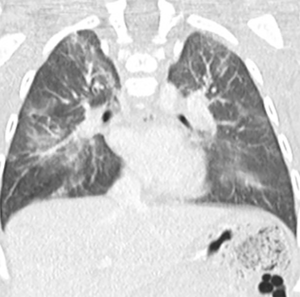
Wiskott-Aldrich syndrome in a 6-month-old infant with multifocal pneumonia. Coronal reformatted chest CT image shows multifocal ground-glass opacities bilaterally, a finding compatible with infection.
Innate Immunodeficiencies
Innate immunodeficiencies form a group of PIDs affecting a set of cells and mechanisms involved in the host defense by a nonspecific and fast response. These include immunodeficiencies due to phagocytic defects and complement deficiencies (1). Depending on the genetic defect involved, most patients with innate immunity defects have a predisposition to one isolated type of infection (bacterial, viral, or fungal) (45). Patients affected by phagocytic disorders experience severe pyogenic bacterial and fungal infections of the skin, respiratory tract, and viscera, as well as painful sores around the mouth. In contrast, individuals affected by complement deficiencies present with systemic autoimmune disease that resembles lupus erythematosus, or with severe or recurrent infections with encapsulated organisms (1).
There is a wide spectrum of imaging findings associated with innate immunodeficiencies; these findings vary according to the specific disorder. Common radiologic manifestations include recurrent and chronic pneumonia, lung abscess, nonspecific hilar and mediastinal lymphadenopathy, hepatosplenomegaly, suppurative lymphadenitis, subcutaneous and chest wall abscesses, and osteomyelitis (4). Sherwani et al (46) showed that the most common thoracic imaging findings in patients with phagocytic disorders are consolidation and nodules as well as absence of bronchiectasis.
Chronic Granulomatous Disease
Chronic granulomatous disease (CGD) is a PID caused by defects in any of the five subunits of the nicotinamide adenine dinucleotide phosphate oxidase complex responsible for the respiratory burst in phagocytic leukocytes (4,47). As a result of this defect, the phagocytic cells’ reactive oxygen species, which are crucial to killing microorganisms, are not produced. The prevalence of CGD is approximately 0.5:100 000 individuals in North America and Europe, but it varies significantly worldwide (47).
CGD is characterized by recurrent, life-threatening bacterial and fungal infections of the lungs, skin, lymph nodes, liver, brain, and bones. The lungs are the most commonly involved organ. Frequent pathogens include S aureus, Aspergillus, Nocardia, Serratia, and Salmonella species (47). Invasive aspergillosis constitutes the major cause of mortality (48).
Thoracic imaging findings are nonspecific, including consolidation, ground-glass or tree-in-bud opacities, or scattered nodules (49), which can be present in bacterial or fungal infections and may be complicated by abscess formation (50). Attention should be paid to potential chest wall invasion as this is seen in up to one in four patients with CGD and fungal pneumonia, including soft-tissue abscesses or osteomyelitis (48).
Chronic and recurrent lung infections can lead to scarring or destruction of the pulmonary parenchyma (35,50). CT following treatment of infection may show local fibrosis, septal thickening, and bronchiectasis (48), in addition to architectural distortion, emphysema, and nodular calcifications (35) (Fig 12). Lymphadenitis is found in 60% of patients with CGD and manifests with enlarged, centrally necrotic lymph nodes (50). Esophagitis and esophageal strictures have also been described in this population (4,35).
Figure 12:

A–D, Chronic granulomatous disease (CGD) in three patients. A, Chest CT image in a young patient with CGD shows a cavitary pneumonia (arrow) in the right upper lobe. B, Chest CT image in a young patient with CGD shows a focus of consolidation (arrow) in the left lower lobe. C, Chest CT image in a child with CGD demonstrates extensive consolidative and cavitary changes throughout the lower lobes (arrows). D, Autopsy specimen of the same patient shows the extensive cavitary/necrotizing foci in the lungs.
Conclusions
PIDs are relatively uncommon disorders that frequently affect the lungs and other intrathoracic organs, such as the lymph nodes, thymus, and heart. Although there is considerable overlap in the radiologic findings (Fig 13) associated with PIDs, there are unique features and patterns associated with each entity that can help distinguish between different diseases. The radiologist has an important role in the care of these patients, including raising the possibility of PIDs as a de novo diagnosis and, importantly, in recognizing potential complications on follow-up imaging studies resulting from repeated infection or other diseases such as ILD and related malignancy.
Figure 13:

Comparative Venn diagram depicts imaging features of the broad groups of primary immunodeficiency disorders. Not every disease presents with these findings. ILD = interstitial lung disease, * = recurrent respiratory tract infections (RTIs) lead to bronchiectasis.
Disclosures of Conflicts of Interest: J.A.R. disclosed no relevant relationships. T.J.B. disclosed no relevant relationships. C.S.R. disclosed no relevant relationships. D.B.G. disclosed no relevant relationships. L.P.B. disclosed no relevant relationships. D.V. disclosed no relevant relationships.
Abbreviations:
- CGD
- chronic granulomatous disease
- CID
- combined immunodeficiency
- CVID
- common variable immunodeficiency
- GLILD
- granulomatous lymphocytic ILD
- hyper-IgE
- hyperimmunoglobulin E
- hyper-IgM
- hyperimmunoglobulin M
- Ig
- immunoglobulin
- ILD
- interstitial lung disease
- PID
- primary immunodeficiency disorder
- RTI
- respiratory tract infection
- SCID
- severe CID
- STAT3
- signal transducer and activator of transcription 3
- WAS
- Wiskott-Aldrich syndrome
- XLA
- X-linked agammaglobulinemia
References
- 1.McCusker C, Upton J, Warrington R. Primary immunodeficiency. Allergy Asthma Clin Immunol 2018;14(Suppl 2):61. [DOI] [PMC free article] [PubMed] [Google Scholar]
- 2.Booth C, Romano R, Roncarolo MG, Thrasher AJ. Gene therapy for primary immunodeficiency. Hum Mol Genet 2019;28(R1):R15–R23. [DOI] [PubMed] [Google Scholar]
- 3.Bousfiha A, Jeddane L, Picard C, et al. Human Inborn Errors of Immunity: 2019 Update of the IUIS Phenotypical Classification. J Clin Immunol 2020;40(1):66–81. [DOI] [PMC free article] [PubMed] [Google Scholar]
- 4.Wu EY, Ehrlich L, Handly B, Frush DP, Buckley RH. Clinical and imaging considerations in primary immunodeficiency disorders: an update. Pediatr Radiol 2016;46(12):1630–1644. [DOI] [PMC free article] [PubMed] [Google Scholar]
- 5.D’Angelo P, Cereser L, Bednarova S, Carli MD, Pellegrin AD, Zuiani C. Chest HRCT findings in patients with humoral Primary Immunodeficiencies and recurrent respiratory infections: review of literature and personal experience. European Society of Radiology, 2017. [Google Scholar]
- 6.Bierry G, Boileau J, Barnig C, et al. Thoracic manifestations of primary humoral immunodeficiency: a comprehensive review. RadioGraphics 2009;29(7):1909–1920. [DOI] [PubMed] [Google Scholar]
- 7.Wang N, Shen N, Vyse TJ, et al. Selective IgA deficiency in autoimmune diseases. Mol Med 2011;17(11-12):1383–1396. [DOI] [PMC free article] [PubMed] [Google Scholar]
- 8.Yel L. Selective IgA deficiency. J Clin Immunol 2010;30(1):10–16. [DOI] [PMC free article] [PubMed] [Google Scholar]
- 9.Patel P, Karafotias I, Tran SD, Mulholland N, Yusuf G, Stefanidis K. Thoracic imaging findings in patients with primary humoral immunodeficiency. ESTI 2019 conference, 2019. [Google Scholar]
- 10.Domínguez O, Giner MT, Alsina L, Martín MA, Lozano J, Plaza AM. Clinical phenotypes associated with selective IgA deficiency: a review of 330 cases and a proposed follow-up protocol [in Spanish]. An Pediatr (Barc) 2012;76(5):261–267. [DOI] [PubMed] [Google Scholar]
- 11.Chen XF, Wang WF, Zhang YD, Zhao W, Wu J, Chen TX. Clinical characteristics and genetic profiles of 174 patients with X-linked agammaglobulinemia: Report from Shanghai, China (2000-2015). Medicine (Baltimore) 2016;95(32):e4544. [DOI] [PMC free article] [PubMed] [Google Scholar]
- 12.Milliron B, Henry TS, Veeraraghavan S, Little BP. Bronchiectasis: Mechanisms and Imaging Clues of Associated Common and Uncommon Diseases. RadioGraphics 2015;35(4):1011–1030. [DOI] [PubMed] [Google Scholar]
- 13.Botnaru V, Munteanu O, Cemîrtan S, Rusu D, Slevestru R. A rare genetic cause of bronchiectasis. Pneumologia 2016;65(4):216–221. [PubMed] [Google Scholar]
- 14.Fekrvand S, Yazdani R, Olbrich P, et al. Evaluation of respiratory complications in patients with X-linked and autosomal recessive agammaglobulinemia. Pediatr Allergy Immunol 2020;31(4):405–417. [DOI] [PubMed] [Google Scholar]
- 15.Bang TJ, Richards JC, Olson AL, Groshong SD, Gelfand EW, Lynch DA. Pulmonary Manifestations of Common Variable Immunodeficiency. J Thorac Imaging 2018;33(6):377–383. [DOI] [PubMed] [Google Scholar]
- 16.Boloursaz MR, Khalilzadeh S, Nezhad PR, et al. Chest CT Manifestations in Children with CVID: A 10-Year Report. Tanaffos 2012;11(4):56–59. [PMC free article] [PubMed] [Google Scholar]
- 17.Cunningham-Rundles C, Cooper DL, Duffy TP, Strauchen J. Lymphomas of mucosal-associated lymphoid tissue in common variable immunodeficiency. Am J Hematol 2002;69(3):171–178. [DOI] [PubMed] [Google Scholar]
- 18.de la Morena MT, Leonard D, Torgerson TR, et al. Long-term outcomes of 176 patients with X-linked hyper-IgM syndrome treated with or without hematopoietic cell transplantation. J Allergy Clin Immunol 2017;139(4):1282–1292. [DOI] [PMC free article] [PubMed] [Google Scholar]
- 19.Wang LL, Zhou W, Zhao W, et al. Clinical features and genetic analysis of 20 Chinese patients with X-linked hyper-IgM syndrome. J Immunol Res 2014;2014:683160. [DOI] [PMC free article] [PubMed] [Google Scholar]
- 20.Nandan D, Nag VK, Trivedi N, Singh S. X-linked Hyper-IgM Syndrome with Bronchiectasis. J Lab Physicians 2014;6(2):114–116. [DOI] [PMC free article] [PubMed] [Google Scholar]
- 21.Moazzami B, Yazdani R, Azizi G, et al. Respiratory Complications in Patients with Hyper IgM Syndrome. J Clin Immunol 2019;39(6):557–568. [DOI] [PubMed] [Google Scholar]
- 22.Multani A, Gomez CA, Montoya JG. Prevention of infectious diseases in patients with Good syndrome. Curr Opin Infect Dis 2018;31(4):267–277. [DOI] [PubMed] [Google Scholar]
- 23.Pu C, Sukhal S, Fakhran S. Humoral Immunity in Bronchiectasis: Finding Good’s Syndrome. Case Rep Pulmonol 2015;2015:531731. [DOI] [PMC free article] [PubMed] [Google Scholar]
- 24.Popper ST, Lanks CW. Bronchiectasis and Thymoma: Have You Got the Good’s? Proceedings of UCLA Health 2018;22. https://www.proceedings.med.ucla.edu/wp-content/uploads/2018/09/Lanks-A180817CL-Harbor-BLM-edited.pdf. Accessed May 16, 2020. [Google Scholar]
- 25.Zaman M, Huissoon A, Buckland M, et al. Clinical and laboratory features of seventy-eight UK patients with Good’s syndrome (thymoma and hypogammaglobulinaemia). Clin Exp Immunol 2019;195(1):132–138. [DOI] [PMC free article] [PubMed] [Google Scholar]
- 26.Al-Herz W, Notarangelo LD, Sadek A, Buckley R; USIDNET Consortium . Combined immunodeficiency in the United States and Kuwait: Comparison of patients’ characteristics and molecular diagnosis. Clin Immunol 2015;161(2):170–173. [DOI] [PMC free article] [PubMed] [Google Scholar]
- 27.Liao CY, Yu HW, Cheng CN, et al. A novel pathogenic mutation on Interleukin-7 receptor leading to severe combined immunodeficiency identified with newborn screening and whole exome sequencing. J Microbiol Immunol Infect 2020;53(1):99–105. [DOI] [PubMed] [Google Scholar]
- 28.Akar HH, Patiroglu T, Hershfield M, van der Burg M. Combined immunodeficiencies: twenty years experience from a single center in Turkey. Cent Eur J Immunol 2016;41(1):107–115. [DOI] [PMC free article] [PubMed] [Google Scholar]
- 29.Grunebaum E, Cutz E, Roifman CM. Pulmonary alveolar proteinosis in patients with adenosine deaminase deficiency. J Allergy Clin Immunol 2012;129(6):1588–1593. [DOI] [PubMed] [Google Scholar]
- 30.Choi HK, Park CM, Goo JM, Lee HJ. Pulmonary alveolar proteinosis versus exogenous lipoid pneumonia showing crazy-paving pattern: Comparison of their clinical features and high-resolution CT findings. Acta Radiol 2010;51(4):407–412. [DOI] [PubMed] [Google Scholar]
- 31.Verhagen MV, Trevisan V, Adu J, Owens CM, Booth C, Calder A. Chest Radiographs for Distinguishing ADA-SCID from Other Forms of SCID. J Clin Immunol 2020;40(2):259–266. [DOI] [PubMed] [Google Scholar]
- 32.McDonald-McGinn DM, Sullivan KE, Marino B, et al. 22q11.2 deletion syndrome. Nat Rev Dis Primers 2015;1(1):15071. [DOI] [PMC free article] [PubMed] [Google Scholar]
- 33.Morsheimer M, Brown Whitehorn TF, Heimall J, Sullivan KE. The immune deficiency of chromosome 22q11.2 deletion syndrome. Am J Med Genet A 2017;173(9):2366–2372. [DOI] [PubMed] [Google Scholar]
- 34.McElhinney DB, Clark BJ 3rd, Weinberg PM, et al. Association of chromosome 22q11 deletion with isolated anomalies of aortic arch laterality and branching. J Am Coll Cardiol 2001;37(8):2114–2119. [DOI] [PubMed] [Google Scholar]
- 35.García-Peña P, Boixadera H, Barber I, Toran N, Lucaya J, Enríquez G. Thoracic findings of systemic diseases at high-resolution CT in children. RadioGraphics 2011;31(2):465–482. [DOI] [PubMed] [Google Scholar]
- 36.Gernez Y, Freeman AF, Holland SM, et al. Autosomal Dominant Hyper-IgE Syndrome in the USIDNET Registry. J Allergy Clin Immunol Pract 2018;6(3):996–1001. [DOI] [PMC free article] [PubMed] [Google Scholar]
- 37.Freeman AF, Olivier KN. Hyper-IgE Syndromes and the Lung. Clin Chest Med 2016;37(3):557–567. [DOI] [PMC free article] [PubMed] [Google Scholar]
- 38.Bogdanovski K, Urban AK, Darnell D, Peterson J, Welch P, Freeman AF. Characterization of Lung Disease in Autosomal Dominant Hyper IgE Syndrome. Am J Respir Crit Care Med 2018;197:A4797. [Google Scholar]
- 39.Bhatt JM, Bush A, van Gerven M, et al. ERS statement on the multidisciplinary respiratory management of ataxia telangiectasia. Eur Respir Rev 2015;24(138):565–581. [DOI] [PMC free article] [PubMed] [Google Scholar]
- 40.Schroeder SA, Swift M, Sandoval C, Langston C. Interstitial lung disease in patients with ataxia-telangiectasia. Pediatr Pulmonol 2005;39(6):537–543. [DOI] [PubMed] [Google Scholar]
- 41.Rivers E, Worth A, Thrasher AJ, Burns SO. How I manage patients with Wiskott Aldrich syndrome. Br J Haematol 2019;185(4):647–655. [DOI] [PMC free article] [PubMed] [Google Scholar]
- 42.Imai K, Morio T, Zhu Y, et al. Clinical course of patients with WASP gene mutations. Blood 2004;103(2):456–464. [DOI] [PubMed] [Google Scholar]
- 43.Cheminant M, Mahlaoui N, Desconclois C, et al. Lymphoproliferative disease in patients with Wiskott-Aldrich syndrome: Analysis of the French Registry of Primary Immunodeficiencies. J Allergy Clin Immunol 2019;143(6):2311–2315.e7. [DOI] [PubMed] [Google Scholar]
- 44.Pellier I, Dupuis Girod S, Loisel D, et al. Occurrence of aortic aneurysms in 5 cases of Wiskott-Aldrich syndrome. Pediatrics 2011;127(2):e498–e504. [DOI] [PubMed] [Google Scholar]
- 45.Duchamp M, Miot C, Bustamante JC, Picard C. Innate immunity primary immunodeficiencies and infections [in French]. Arch Pediatr 2016;23(7):760–768. [DOI] [PubMed] [Google Scholar]
- 46.Sherwani P, Bhalla AS, Jana M, et al. Thoracic Manifestations of Primary Immunodeficiency Disorders. Indian J Pediatr 2020;87(10):846–849. [DOI] [PubMed] [Google Scholar]
- 47.Arnold DE, Heimall JR. A Review of Chronic Granulomatous Disease. Adv Ther 2017;34(12):2543–2557. [DOI] [PMC free article] [PubMed] [Google Scholar]
- 48.Salvator H, Mahlaoui N, Catherinot E, et al. Pulmonary manifestations in adult patients with chronic granulomatous disease. Eur Respir J 2015;45(6):1613–1623. [DOI] [PubMed] [Google Scholar]
- 49.Henrickson SE, Jongco AM, Thomsen KF, Garabedian EK, Thomsen IP. Noninfectious Manifestations and Complications of Chronic Granulomatous Disease. J Pediatric Infect Dis Soc 2018;7(suppl_1):S18–S24. [DOI] [PMC free article] [PubMed] [Google Scholar]
- 50.Lee M, Lee MS, Lee JS, Ko SY, Jeong SY. Spectrum of imaging findings of chronic granulomatous disease: a single center experience. Diagn Interv Radiol 2017;23(6):472–477 [Published correction appears in Diagn Interv Radiol 2018;24(1):62.]. [DOI] [PMC free article] [PubMed] [Google Scholar]