When light-matter polaritons travel in periodic media, they acquire properties akin to Bloch quasi-particles in crystals.
Abstract
Efficient control of photons is enabled by hybridizing light with matter. The resulting light-matter quasi-particles can be readily programmed by manipulating either their photonic or matter constituents. Here, we hybridized infrared photons with graphene Dirac electrons to form surface plasmon polaritons (SPPs) and uncovered a previously unexplored means to control SPPs in structures with periodically modulated carrier density. In these periodic structures, common SPPs with continuous dispersion are transformed into Bloch polaritons with attendant discrete bands separated by bandgaps. We explored directional Bloch polaritons and steered their propagation by dialing the proper gate voltage. Fourier analysis of the near-field images corroborates that this on-demand nano-optics functionality is rooted in the polaritonic band structure. Our programmable polaritonic platform paves the way for the much-sought benefits of on-the-chip photonic circuits.
INTRODUCTION
Photons provide an unparalleled resource for quantum technology (1–3), including quantum communications (4, 5), sensing (6), and computation (7, 8). However, free-space photons do not directly interact with electric or magnetic fields, making it difficult to efficiently program their properties. Hybridizing photons with matter gives rise to composite light-matter quasi-particles known as polaritons and creates new pathways for programmable optical phenomena (9–11). For example, the hybridization of photons and excitons in semiconductors results in exciton-polaritons that are amenable to both electronic and optical means of manipulation (12–20). Another prominent example of coupled light-matter quasi-particles are surface plasmon polaritons (SPPs), which are hybrids of photons and free electrons in conducting surfaces (21). Sophisticated designs of plasmonic structures have uncovered numerous ways to program SPPs (22–24). In complete analogy to Bloch electrons in solids or photons in photonic crystals, the dispersion of SPPs in periodic media reveals discrete bands separated by bandgaps (25–28). Thus, polaritonic crystals enable new means for programming the directional motion of SPPs by electrostatic control in planar gated structures.
Graphene SPPs are deeply subdiffractional optical modes (29, 30) with high quality factor. Systematic efforts uncovered the inherent tunability of graphene SPP wavelengths by gate voltage (31–38)—a feat that is difficult to achieve using alternative plasmonic media. We devised and demonstrated an as-yet-unexplored method for steering the direction of SPP travel in a pristine graphene layer. Specifically, we created a graphene polaritonic crystal (28) by introducing a periodic spatial modulation of the Fermi energy (39). In these designer crystals, graphene SPPs with continuous dispersion are transformed into Bloch polaritons residing in discrete bands (26, 28, 40–42). The propagation direction of Bloch polaritons is governed by the polaritonic bands in which they reside and can be readily controlled by gate voltage. Prior demonstrations of directional SPPs all relied on manipulating the photonic constituents of the polaritonic quasi-particles by selecting the handedness of the exciting laser beam (22, 43–50). We programmed Bloch polaritons by controlling their matter constituents using gate voltages.
RESULTS AND DISCUSSION
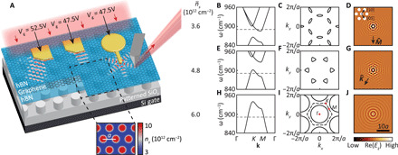
A schematic of our graphene polaritonic crystal is shown in Fig. 1A. A graphene device fully encapsulated between thin layers of hexagonal boron nitride (hBN) resides on top of a Si substrate with a thermally grown SiO2 layer. The SiO2 layer is patterned into a hexagonal array of pillars with lattice periodicity a = 85 nm (28, 39). An infrared laser beam at energy ω = 890 cm−1 illuminates the entire device. Patterned gold launchers on top of the device excite Bloch polaritons propagating along the polaritonic crystal (Fig. 1A) (51). Our patterning techniques largely preserve the quality of the graphene devices (39), and we achieved a polariton damping rate of γ = 12 cm−1 at low temperature.
Fig. 1. A platform for programmable Bloch polaritons.

(A) Schematic of a back-gated graphene polaritonic crystal. Gold launchers excite Bloch polaritons propagating along designated directions controlled by the gate voltage Vg. Inset shows the carrier density distribution in the graphene layer with average carrier density . (B) Simulated polaritonic band structure at . Dashed line corresponds to ω = 890 cm−1. (C) Simulated equi-energy contours at and ω = 890 cm−1, showing the polaritonic pockets around M points. (D) Simulated electric field Re(Ez) of polaritons excited by a point source at and ω = 890 cm−1. Bloch polaritons propagate predominantly along directions. Inset shows an enlarged real-space lattice pattern. (E) to (G), (H) to (J), same as (B) to (D) at and , respectively. At , Bloch polaritons reside in the lower band (E), emerge in the K pockets (F), and propagate along directions (G). At , Bloch polaritons reside entirely in the lower band (H), exhibit circular equi-energy contours (I) and propagate isotropically in all directions (J). Inset of (I) shows the first BZ marked with symmetry points.
Optical phenomena in our patterned polaritonic platform can be categorized under the notion of a polaritonic crystal (25). Specifically, the patterned substrate introduces a nanoscale periodic variation of the dielectric constants in the immediate proximity of the graphene layer. Under the application of a back gate, the variations in the capacitive coupling is imprinted as carrier density modulation in graphene (Fig. 1A, inset). Propagating polaritons scatter and interfere in regions with varying carrier densities, leading to the formation of the polaritonic band structure and Brillouin zones (BZ) (Fig. 1, B, E, and H) (26). At crystal momentum away from the BZ boundary, polariton dispersion is minimally affected by the periodic modulation and largely follows the dispersion in unpatterned devices as , where EF is the graphene Fermi energy and k is the SPP momentum. Near the boundary of the BZ, polariton dispersion is strongly modified, which leads to the formation of a complete bandgap for laser energies between 870 and 890 cm−1 (Fig. 1B). At a probe laser energy of ω = 890 cm−1, we excite polaritons residing in the upper polaritonic band. By tuning the graphene carrier density from to , the polaritonic band structure shifts upward in energy and the polaritonic bandgap emerges at 950 cm−1 (Fig. 1H and movie S1). In the latter setting, the incident laser beam excites polaritons in the lower polaritonic band. By tuning the gate voltage, we gain access to both the upper and the lower polaritonic bands (Fig. 1, B, E, and H) at a given laser energy ω.
To gain insight into the properties of Bloch polaritons, we explored the equi-energy contours (Fig. 1, C, F, and I) composed of all modes supported by the polaritonic band structure at a given laser energy ω. When ω intersects with the upper polaritonic band (, Fig. 1B) just above the bandgap, equi-energy contours emerge around M points in the BZ (Fig. 1C). The propagation direction of Bloch polaritons is governed by their group velocity, which is largely aligned with the crystal momentum in BZ. This latter conjecture is supported by real-space polariton propagation simulations where we plotted the electric field Re(Ez) of Bloch polaritons launched by a point source in the polaritonic crystal (Fig. 1D). Simulations confirm that Bloch polaritons only propagate along directions. Provided that the laser energy falls in the polaritonic bandgap, polariton propagation is completely inhibited (not shown). Next, when the lower polaritonic band is raised further toward ω (, Fig. 1E), equi-energy contours reemerge in the K/K′ valleys (Fig. 1F). Bloch polaritons switch their propagation direction from to (Fig. 1G). Last, when the carrier density is increased to , the equi-energy contours evolve to near-perfect circles around the center of the BZ (Fig. 1I), and Bloch polaritons propagate isotropically in all directions (Fig. 1J), similar to their counterparts in unpatterned graphene devices. The above modeling uncovers the utility of graphene polaritonic crystals for programmable steering of Bloch polaritons.
We used low-temperature near-field optical microscopy to image Bloch polariton propagation in real space and to extract the equi-energy contours in momentum space. In these experiments, the entire device is illuminated by an infrared light from a CO2 laser. The metallic tip of an atomic force microscope (AFM) acts as an optical antenna that outcouples Bloch polaritons into free-space photons, enabling real-space polaritonic imaging deeply below the diffraction limit (31, 32). The tip-scattered light is registered by a detector, and the amplitude s(r) and phase ϕ(r) of the corresponding near-field signal are extracted by a proper demodulation scheme (Materials and Methods). We mainly analyzed the near-field amplitude images s(r). All the near-field measurements were performed at T = 60 K to reduce SPP losses due to phonon scattering (34) and to minimize broadening of the polaritonic band structure.
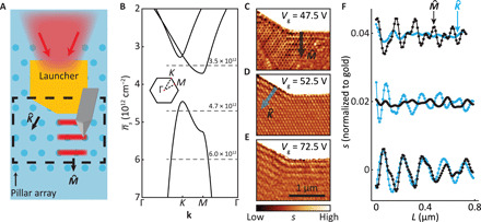
Near-field images close to a gold launcher show directional propagation of Bloch polaritons (Fig. 2). The leading edges of the launcher form a 30° angle, thus exciting Bloch polaritons propagating along and directions (arrows in Fig. 2A). In the course of our measurements, we kept the laser energy at ω = 890 cm−1 while tuning the gate voltage. It is instructive to plot the gate-voltage dependence of the band structure at ω = 890 cm−1 (Fig. 2B and fig. S1). This new representation contains the same physics as Fig. 1 (B, E, and H) but facilitates a more direct connection to our imaging data. Specifically, Fig. 2B reveals that polaritons reside near M points at lower carrier densities, whereas their K point counterparts are activated at higher carrier densities. At gate voltage Vg = 47.5 V (Fig. 2C), Bloch polaritons only propagate along the direction. This phenomenon is particularly apparent when comparing the line profiles along (black) and (cyan) directions (Fig. 2F). A combination of Vg = 47.5 V and ω = 890 cm−1 produces polaritonic fringes only in the direction, which also indicates that polaritons reside in the upper polaritonic band (Fig. 2B, top dashed line). At a slightly higher gate voltage Vg = 52.5 V, Bloch polariton propagation is inhibited in the direction, and the launcher only excites SPPs traveling diagonally along the direction (Fig. 2D). Line profiles in Fig. 2F confirm that Bloch polaritons propagate predominantly in the direction and that they can only be excited in the lower polaritonic band (Fig. 2B, middle dashed line). At even higher gate voltage Vg = 72.5 V, the laser energy is away from the polaritonic bandgap (Fig. 2B, bottom dashed line) and Bloch polaritons propagate isotropically in all directions (Fig. 2, E and F). Near-field images also reveal periodic dark spots, which are attributed to variation in the local polaritonic density of states (28) that is peripheral to our focus on directional Bloch polaritons. We have investigated multiple devices at a variety of gate voltages across the bandgap (fig. S2). All devices show consistent control of polariton propagation by gate voltage. The agreement between our experimental observations and band structure calculations demonstrates the ability to steer Bloch polaritons along the specified trajectories in our planar structure using the electric field effect.
Fig. 2. Directional launching of Bloch polaritons.

(A) Schematic of the device with the scanned region marked in the dashed box. The shades of blue colors represent different carrier densities in graphene. (B) Dependence of the polariton momentum k on average carrier density at ω = 890 cm−1. Dashed lines correspond to the experimental conditions in (C) to (E). Inset: BZ of the polaritonic crystal marked with symmetry points. (C) Near-field image acquired at T = 60 K and Vg = 47.5 V. Bloch polaritons propagate in direction, as marked by the black arrow. (D) and (E) same as (C) at Vg = 52.5 V and Vg = 72.5 V, respectively. (D) shows that Bloch polaritons propagate predominantly in direction, as marked by the cyan arrow. (E) reveals isotropic polariton propagation in all directions. (F) Averaged line profiles along and directions acquired from (C) to (E). Line profiles are normalized to gold and shifted vertically by multiples of 0.02 units for clarity.
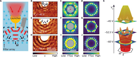
Fourier analysis of the polaritonic patterns confirms that the directional launching is governed by the polaritonic band structure. Experimental inputs for this analysis are acquired by imaging Bloch polaritons emanating from the gold launcher in Fig. 3A, which resembles a point source for polaritonic waves (fig. S3). A gate voltage of Vg = −45 V implies that Bloch polaritons reside above the polaritonic bandgap and propagate along the direction (Fig. 3B). The symmetrized Fourier transform (Materials and Methods) of the near-field image (Fig. 3C) yields prominent features around M points in the BZ, forming a hexagonal motif. A similar pattern is also observed in modeling results in Fig. 3D. The agreement between gross features in the data and the modeling corroborates that the directional Bloch polariton propagation is governed by the band structure. Propagating Bloch polaritons vanish for gate voltages within the bandgap at Vg = −50 V (fig. S4). At even lower gate voltages such as Vg = −52.5 V, polariton propagation direction is switched to (Fig. 3E). The Fourier transform of the near-field image reveals the formation of polaritonic pockets near K points (Fig. 3, F and G, the complete field of view of the Fourier image is shown in fig. S5). Last, at Vg = −60 V, equi-energy contours appear as near-perfect circles within the first BZ (Fig. 3, I and J). The three selected panels of Fig. 3 (C, F, and I) are displayed again in Fig. 3K and are seen to be in close agreement with the simulated polaritonic band structure.
Fig. 3. Fourier analysis of polaritonic images.

(A) Schematic of the device with the scanned region marked in the dashed box. (B) Near-field image obtained at Vg = −45 V, showing Bloch polaritons traveling predominantly in directions. (C) Symmetrized Fourier transform (FT) of the image in (B). The six bright features represent Bloch polaritonic modes around M points. Polaritonic features close to the Γ point are extraneous to our study and thus are shaded in the gray circle. Inset: First BZ marked with symmetry points. (D) Simulated Fourier transform at . (E) to (G) same as (B) to (D) at Vg = −52.5 V and . Bloch polaritons propagate predominantly along directions (E). The Fourier transform (F) shows six polaritonic pockets near K points in accord with the simulation (G). (H) to (J) same as (B) to (D) at Vg = −60 V and . The near-field image shows that Bloch polaritons propagate isotropically in all directions (H) and the Fourier transform (I) shows circular equi-energy contours, which agrees with the simulation (J). (K) Stacked equi-energy contours for selected gate voltages and overlaid with the theoretical polaritonic band structure.
Outlook
We demonstrated programmable Bloch polaritons in a graphene polaritonic crystal platform. Bloch polaritons propagate along designated directions set by the applied gate voltage. The same general approach is well suited to the manipulation of other polaritonic systems, such as exciton polaritons and magnon polaritons. Relatively straightforward improvements of our platform aimed at reducing plasmonic damping and working gate voltages will enable operation at ambient conditions. Furthermore, our back-gated platform is compatible with ubiquitous metal oxide semiconductor field effect transistor technology and sets the stage for integrated photonic circuits (52, 53).
MATERIALS AND METHODS
Cryogenic near-field imaging techniques
Cryogenic near-field imaging measurements were performed using a home-built scattering-type scanning near-field optical microscope (s-SNOM) (54). The s-SNOM apparatus is based on a taping mode AFM, coupled to a continuous-wave CO2 laser (Access Laser), operating in ultrahigh vacuum (UHV) and cryogenic temperatures. The incoming laser beam is focused on the sample using a high-NA off-axis parabolic mirror inside the UHV chamber. The metallic AFM tip is tapped above the sample surface at a frequency of ~250 kHz. The tip-scattered light is demodulated using a pseudo-heterodyne detection scheme to extract both the near-field amplitude s(r, ω) and the phase ϕ(r, ω). The images shown in the main text are the near-field amplitude s(r, ω) normalized to the gold launcher. Gold deposited using thermal evaporation serves as a good reference for the demodulated signal. To properly suppress background contributions to the near-field signal, we demodulate at the third harmonic of the tip tapping frequency (28).
Sample fabrication
The patterned dielectric superlattices (PDSLs) (28, 39) consisting of a hexagonal array of pillars were fabricated by plasma etching the SiO2 using a thin polymethyl methacrylate (PMMA) mask. Si substrates with thermally grown SiO2 of thickness 285 nm were spin-coated with a layer of 495 A2 PMMA of thickness 50 nm. The hexagonal pattern was written by e-beam lithography in a Nanobeam nB4 system at a current of 300 to 400 pA. SiO2 pillars were etched in an Oxford Plasmalab 80 Plus system using a mixture of CHF3 gas (40 sccm) and Ar gas (5 sccm) to a depth of ~50 nm. The PMMA mask was removed through an O2 plasma etching, and the PDSL was cleaned by piranha chemical etching. The resulting superlattice is an array of pillars with 46- to 48-nm diameter and 58-nm height that form a hexagonal lattice, with lattice periodicity 85 nm in the device shown in Fig. 2 and 80 nm in the device shown in Fig. 3.
A mechanically exfoliated graphene and hBN heterostructure was placed onto the PDSL by mechanical transfer (55). The heterostructure consists of a top h11BN layer of ~4 nm, a layer of graphene, and a bottom h11BN layer of ~4 nm and was assembled using a polypropylene carbonate (PPC) transfer slide. Given the challenges involved in picking up an hBN layer as thin as 4 nm, such as the topmost layer, using PPC, we dug a circular hole (diameter 7 to 11 μm) out of a thick (30 to 60 nm) hBN flake using e-beam lithography. This thick hBN flake could be easily picked up by the PPC, and the thin h11BN and graphene flakes were positioned so that they covered the circular hole from below and could be picked up by the van der Waals interaction between them and the thick top hBN flake. Therefore, inside the circular hole (not shown in experimental images) we had a thin h11BN-graphene-h11BN heterostructure. Metal contacts and launchers were deposited using standard e-beam lithography processes. The graphene device is a single crystal within our field of view, as confirmed by near-field imaging results. All the reported back gate voltages are measured from the charge neutrality point of the devices. The residual charge density in the absence of gate voltage is estimated to be ns = 1.75 × 1011 cm−2.
The monoisotopic h11BN single crystals were synthesized by the metal flux method (56). High-purity elemental 11B (99.41%) was first mixed with Ni and Cr powders in a weight ratio of 1:12:12. These materials were then loaded into an alumina crucible and heated to a molten state with a flowing mixture of nitrogen (95%) and hydrogen (5%) at 1550°C. The h11BN crystals were precipitated on the flux surface by slowly cooling (1°C/hour) to 1500°C. After the growth process, the mixture temperature was quickly quenched to room temperature. The crystal flakes were obtained from the solidified flux with tape.
Symmetrization of Fourier space images
Near-field images acquired near the apex of the gold launcher were cropped to remove the gold launcher. The cropped near-field images were subsequently Fourier-transformed, using a Hann window, to get the raw image in reciprocal space. To enhance the signal-to-noise ratio and better reveal the excited polaritonic modes, we applied all the symmetry operations of the hexagonal lattice to the raw Fourier image and averaged the resulting images. More specifically, we consecutively rotated the raw Fourier image in 60° intervals, leading to six transformed images. We then applied the mirror operation to these six transformed images, resulting in six additional images. The average of all 12 transformed images results in the Fourier images shown in Fig. 3. The Fourier transform method is commonly used to analyze polaritonic equi-energy contours (57–60).
Acknowledgments
Funding: Experimental research on Bloch polaritons is solely supported by the Energy Frontier Research Center on Programmable Quantum Materials funded by the US Department of Energy (DOE), Office of Science, Basic Energy Sciences (BES), under award DE-SC0019443. The development of infrared near-field imaging capabilities at Columbia was supported through the Vannevar Bush Faculty Fellow ONR-VB: N00014-19-1-2630 (D.N.B). D.N.B is a Moore Investigator in Quantum Materials EPIQS #9455. The monoisotopic hBN crystal growth (J.H.E) was supported by the National Science Foundation (NSF) award CMMI #1538127. Theory and modeling results obtained by G.S. and M.J. were supported by the Office of Naval Research (ONR) under grant N00014-21-1-2056, and by NSF under grants No. DMR-1741788 and DMR-1719875. M.J. also acknowledges support by the fellowship from Kwanjeong Educational Foundation. Theory and modeling results obtained by M.F. were supported by the ONR grant ONR-N000014-18-1-2722. Author contributions: L.X., A.S.M., and Y.D. performed the nanoscale infrared measurements and characterizations. Y.L., C.F., S.Z., S.L., K.W., T.T., J.H.E., and C.R.D. designed and created the device structures. L.X., M.J., F.L.R., M.M.F., and G.S. provided theoretical calculations. D.N.B. supervised the project. L.X. and C.L. analyzed the data. L.X., Y.L., M.J., and D.N.B. cowrote the manuscript with input from all coauthors. Competing interests: The authors declare that they have no competing interests. Data and materials availability: All data needed to evaluate the conclusions in the paper are present in the paper and/or the Supplementary Materials. Raw experimental data can be found in the Dryad repository (61).
SUPPLEMENTARY MATERIALS
Supplementary material for this article is available at http://advances.sciencemag.org/cgi/content/full/7/19/eabe8087/DC1
REFERENCES AND NOTES
- 1.Wang J., Sciarrino F., Laing A., Thompson M. G., Integrated photonic quantum technologies. Nat. Photonics 14, 273–284 (2020). [Google Scholar]
- 2.Elshaari A. W., Pernice W., Srinivasan K., Benson O., Zwiller V., Hybrid integrated quantum photonic circuits. Nat. Photonics 14, 285–298 (2020). [DOI] [PMC free article] [PubMed] [Google Scholar]
- 3.Chang D. E., Vuletić V., Lukin M. D., Quantum nonlinear optics - Photon by photon. Nat. Photonics 8, 685–694 (2014). [Google Scholar]
- 4.Slussarenko S., Pryde G. J., Photonic quantum information processing: A concise review. Appl. Phys. Rev. 6, 041303 (2019). [Google Scholar]
- 5.Flamini F., Spagnolo N., Sciarrino F., Photonic quantum information processing: A review. Rep. Prog. Phys. 82, 016001 (2018). [DOI] [PubMed] [Google Scholar]
- 6.Pirandola S., Bardhan B. R., Gehring T., Weedbrook C., Lloyd S., Advances in photonic quantum sensing. Nat. Photonics 12, 724–733 (2018). [Google Scholar]
- 7.Carolan J., Harrold C., Sparrow C., Martín-López E., Russell N. J., Silverstone J. W., Shadbolt P. J., Matsuda N., Oguma M., Itoh M., Marshall G. D., Thompson M. G., Matthews J. C. F., Hashimoto T., O’Brien J. L., Laing A., Universal linear optics. Science 349, 711–716 (2015). [DOI] [PubMed] [Google Scholar]
- 8.Qiang X., Zhou X., Wang J., Wilkes C. M., Loke T., O’Gara S., Kling L., Marshall G. D., Santagati R., Ralph T. C., Wang J. B., O’Brien J. L., Thompson M. G., Matthews J. C. F., Large-scale silicon quantum photonics implementing arbitrary two-qubit processing. Nat. Photonics 12, 534–539 (2018). [Google Scholar]
- 9.Basov D. N., Fogler M. M., García De Abajo F. J., Polaritons in van der Waals materials. Science 354, aag1992 (2016). [DOI] [PubMed] [Google Scholar]
- 10.Basov D. N., Asenjo-garcia A., Schuck P. J., Zhu X., Rubio A., Polariton panorama. Nanophotonics 10, 549–577 (2020). [Google Scholar]
- 11.Ballarini D., De Liberato S., Polaritonics: From microcavities to sub-wavelength confinement. Nanophotonics 8, 641–654 (2019). [Google Scholar]
- 12.Chervy T., Knüppel P., Abbaspour H., Lupatini M., Fält S., Wegscheider W., Kroner M., Imamolu A., Accelerating polaritons with external electric and magnetic fields. Phys. Rev. X. 10, 011040 (2020). [Google Scholar]
- 13.Latini S., Ronca E., De Giovannini U., Hübener H., Rubio A., Cavity control of excitons in two-dimensional materials. Nano Lett. 19, 3473–3479 (2019). [DOI] [PMC free article] [PubMed] [Google Scholar]
- 14.Loginov D. K., Belov P. A., Davydov V. G., Gerlovin I. Y., Ignatiev I. V., Kavokin A. V., Masumoto Y., Exciton-polariton interference controlled by electric field. Phys. Rev. Res. 2, 033510 (2020). [Google Scholar]
- 15.Suárez-Forero D. G., Riminucci F., Ardizzone V., de Giorgi M., Dominici L., Todisco F., Lerario G., Pfeiffer L. N., Gigli G., Ballarini D., Sanvitto D., Electrically controlled waveguide polariton laser. Optica 7, 1579–1586 (2020). [Google Scholar]
- 16.Byrnes T., Kim N. Y., Yamamoto Y., Exciton-polariton condensates. Nat. Phys. 10, 803–813 (2014). [Google Scholar]
- 17.Su R., Ghosh S., Wang J., Liu S., Diederichs C., Liew T. C. H., Xiong Q., Observation of exciton polariton condensation in a perovskite lattice at room temperature. Nat. Phys. 16, 301–306 (2020). [Google Scholar]
- 18.Klembt S., Harder T. H., Egorov O. A., Winkler K., Ge R., Bandres M. A., Emmerling M., Worschech L., Liew T. C. H., Segev M., Schneider C., Höfling S., Exciton-polariton topological insulator. Nature 562, 552–556 (2018). [DOI] [PubMed] [Google Scholar]
- 19.Liu W., Ji Z., Wang Y., Modi G., Hwang M., Zheng B., Sorger V. J., Pan A., Agarwal R., Generation of helical topological exciton-polaritons. Science 370, 600–604 (2020). [DOI] [PubMed] [Google Scholar]
- 20.Solnyshkov D. D., Malpuech G., St-Jean P., Ravets S., Bloch J., Amo A., Microcavity polaritons for topological photonics. Opt. Mater. Exp. 11, 1119–1142 (2021). [Google Scholar]
- 21.Zayats A. V., Smolyaninov I. I., Maradudin A. A., Nano-optics of surface plasmon polaritons. Phys. Rep. 408, 131–314 (2005). [Google Scholar]
- 22.Lin J., Mueller J. P. B., Wang Q., Yuan G., Antoniou N., Yuan X. C., Capasso F., Polarization-controlled tunable directional coupling of surface plasmon polaritons. Science 340, 331–334 (2013). [DOI] [PubMed] [Google Scholar]
- 23.Song J. C. W., Rudner M. S., Chiral plasmons without magnetic field. Proc. Natl. Acad. Sci. U.S.A. 113, 4658–4663 (2016). [DOI] [PMC free article] [PubMed] [Google Scholar]
- 24.Jin D., Christensen T., Soljačić M., Fang N. X., Lu L., Zhang X., Infrared topological plasmons in graphene. Phys. Rev. Lett. 118, 245301 (2017). [DOI] [PubMed] [Google Scholar]
- 25.J. J. D. Joannopoulos, S. Johnson, J. N. J. Winn, R. R. D. Meade, Photonic Crystals: Molding the Flow of Light (Princeton Univ. Press, 2008). [Google Scholar]
- 26.Jung M., Fan Z., Shvets G., Midinfrared plasmonic valleytronics in metagate-tuned graphene. Phys. Rev. Lett. 121, 086807 (2018). [DOI] [PubMed] [Google Scholar]
- 27.Sunku S. S., Ni G. X., Jiang B. Y., Yoo H., Sternbach A., McLeod A. S., Stauber T., Xiong L., Taniguchi T., Watanabe K., Kim P., Fogler M. M., Basov D. N., Photonic crystals for nano-light in moiré graphene superlattices. Science 362, 1153–1156 (2018). [DOI] [PubMed] [Google Scholar]
- 28.Xiong L., Forsythe C., Jung M., McLeod A. S., Sunku S. S., Shao Y. M., Ni G. X., Sternbach A. J., Liu S., Edgar J. H., Mele E. J., Fogler M. M., Shvets G., Dean C. R., Basov D. N., Photonic crystal for graphene plasmons. Nat. Commun. 10, 4780 (2019). [DOI] [PMC free article] [PubMed] [Google Scholar]
- 29.Iranzo D. A., Nanot S., Dias E. J. C., Epstein I., Peng C., Efetov D. K., Lundeberg M. B., Parret R., Osmond J., Hong J. Y., Kong J., Englund D. R., Peres N. M. R., Koppens F. H. L., Probing the ultimate plasmon confinement limits with a van der waals heterostructure. Science 295, 291–295 (2018). [DOI] [PubMed] [Google Scholar]
- 30.Epstein I., Alcaraz D., Huang Z., Pusapati V. V., Hugonin J. P., Kumar A., Deputy X. M., Khodkov T., Rappoport T. G., Peres N. M. R., Smith D. R., Koppens F. H. L., Far-field excitation of single graphene plasmon cavities with ultracompressed mode volumes. Science 1223, 1219–1223 (2020). [DOI] [PubMed] [Google Scholar]
- 31.Chen J., Badioli M., Alonso-González P., Thongrattanasiri S., Huth F., Osmond J., Spasenovic M., Centeno A., Pesquera A., Godignon P., Zurutuza Elorza A., Camara N., García De Abajo F. J., Hillenbrand R., Koppens F. H. L., Optical nano-imaging of gate-tunable graphene plasmons. Nature 487, 77–81 (2012). [DOI] [PubMed] [Google Scholar]
- 32.Fei Z., Rodin A. S., Andreev G. O., Bao W., Mcleod A. S., Wagner M., Zhang L. M., Zhao Z., Thiemens M., Dominguez G., Fogler M. M., Castro Neto A. H., Lau C. N., Keilmann F., Basov D. N., Gate-tuning of graphene plasmons revealed by infrared nano-imaging. Nature 487, 82–85 (2012). [DOI] [PubMed] [Google Scholar]
- 33.Woessner A., Lundeberg M. B., Gao Y., Principi A., Alonso-González P., Carrega M., Watanabe K., Taniguchi T., Vignale G., Polini M., Hone J., Hillenbrand R., Koppens F. H. L., Highly confined low-loss plasmons in graphene-boron nitride heterostructures. Nat. Mater. 14, 421–425 (2015). [DOI] [PubMed] [Google Scholar]
- 34.Ni G. X., McLeod A. S., Sun Z., Wang L., Xiong L., Post K. W., Sunku S. S., Jiang B. Y., Hone J., Dean C. R., Fogler M. M., Basov D. N., Fundamental limits to graphene plasmonics. Nature 557, 530–533 (2018). [DOI] [PubMed] [Google Scholar]
- 35.Woessner A., Gao Y., Torre I., Lundeberg M. B., Tan C., Watanabe K., Taniguchi T., Hillenbrand R., Hone J., Polini M., Koppens F. H. L., Electrical 2π phase control of infrared light in a 350-nm footprint using graphene plasmons. Nat. Photonics 11, 421–424 (2017). [Google Scholar]
- 36.Gerber J. A., Berweger S., O’Callahan B. T., Raschke M. B., Phase-resolved surface plasmon interferometry of graphene. Phys. Rev. Lett. 113, 055502 (2014). [DOI] [PubMed] [Google Scholar]
- 37.Maia F. C. B., O’Callahan B. T., Cadore A. R., Barcelos I. D., Campos L. C., Watanabe K., Taniguchi T., Deneke C., Belyanin A., Raschke M. B., Freitas R. O., Anisotropic flow control and gate modulation of hybrid phonon-polaritons. Nano Lett. 19, 708–715 (2019). [DOI] [PubMed] [Google Scholar]
- 38.Lundeberg M. B., Gao Y., Asgari R., Tan C., Van Duppen B., Autore M., Alonso-González P., Woessner A., Watanabe K., Taniguchi T., Hillenbrand R., Hone J., Polini M., Koppens F. H. L., Tuning quantum nonlocal effects in graphene plasmonics. Science 357, 187–191 (2017). [DOI] [PubMed] [Google Scholar]
- 39.Forsythe C., Zhou X., Watanabe K., Taniguchi T., Pasupathy A., Moon P., Koshino M., Kim P., Dean C. R., Band structure engineering of 2D materials using patterned dielectric superlattices. Nat. Nanotechnol. 13, 566–571 (2018). [DOI] [PubMed] [Google Scholar]
- 40.Silveiro I., Manjavacas A., Thongrattanasiri S., García De Abajo F. J., Plasmonic energy transfer in periodically doped graphene. New J. Phys. 15, 033042 (2013). [Google Scholar]
- 41.Qiu P., Liang R., Qiu W., Chen H., Ren J., Lin Z., Wang J.-X., Kan Q., Pan J.-Q., Topologically protected edge states in graphene plasmonic crystals. Opt. Express 25, 22587–22594 (2017). [DOI] [PubMed] [Google Scholar]
- 42.Jung M., Gladstone R. G., Shvets G., Nanopolaritonic second-order topological insulator based on graphene plasmons. Adv. Photonics 2, 046003 (2020). [Google Scholar]
- 43.López-Tejeira F., Rodrigo S. G., Martín-Moreno L., García-Vidal F. J., Devaux E., Ebbesen T. W., Krenn J. R., Radko I. P., Bozhevolnyi S. I., González M. U., Weeber J. C., Dereux A., Efficient unidirectional nanoslit couplers for surface plasmons. Nat. Phys. 3, 324–328 (2007). [Google Scholar]
- 44.Liu Y., Palomba S., Park Y., Zentgraf T., Yin X., Zhang X., Compact magnetic antennas for directional excitation of surface plasmons. Nano Lett. 12, 4853–4858 (2012). [DOI] [PubMed] [Google Scholar]
- 45.Radko I. P., Bozhevolnyi S. I., Brucoli G., Martin-Moreno L., Garcia-Vidal F. J., Boltasseva A., Efficient unidirectional ridge excitation of surface plasmons. Opt. Express 17, 7228–7232 (2009). [DOI] [PubMed] [Google Scholar]
- 46.Rodríguez-Fortũno F. J., Marino G., Ginzburg P., O’Connor D., Martínez A., Wurtz G. A., Zayats A. V., Near-field interference for the unidirectional excitation of electromagnetic guided modes. Science 340, 328–330 (2013). [DOI] [PubMed] [Google Scholar]
- 47.Shitrit N., Yulevich I., Maguid E., Ozeri D., Veksler D., Kleiner V., Hasman E., Spin-optical metamaterial route to spin-controlled photonics. Science 340, 724–726 (2013). [DOI] [PubMed] [Google Scholar]
- 48.Huang L., Chen X., Bai B., Tan Q., Jin G., Zentgraf T., Zhang S., Helicity dependent directional surface plasmon polariton excitation using a metasurface with interfacial phase discontinuity. Light Sci. Appl. 2, e70 (2013). [Google Scholar]
- 49.Petersen J., Volz J., Rauschenbeutel A., Nanophotonics. Chiral nanophotonic waveguide interface based on spin-orbit interaction of light. Science 346, 67–71 (2014). [DOI] [PubMed] [Google Scholar]
- 50.Wang S., Zhao C., Li X., Dynamical manipulation of surface plasmon polaritons. Appl. Sci. 9, 3297 (2019). [Google Scholar]
- 51.Alonso-González P., Nikitin A. Y., Golmar F., Centeno A., Pesquera A., Vélez S., Chen J., Navickaite G., Koppens F., Zurutuza A., Casanova F., Hueso L. E., Hillenbrand R., Controlling graphene plasmons with resonant metal antennas and spatial conductivity patterns. Science 344, 1369–1373 (2014). [DOI] [PubMed] [Google Scholar]
- 52.Davis T. J., Gómez D. E., Roberts A., Plasmonic circuits for manipulating optical information. Nanophotonics 6, 543–559 (2017). [Google Scholar]
- 53.Bogaerts W., Pérez D., Capmany J., Miller D. A. B., Poon J., Englund D., Morichetti F., Melloni A., Programmable photonic circuits. Nature 586, 207–216 (2020). [DOI] [PubMed] [Google Scholar]
- 54.McLeod A. S., Van Heumen E., Ramirez J. G., Wang S., Saerbeck T., Guenon S., Goldflam M., Anderegg L., Kelly P., Mueller A., Liu M. K., Schuller I. K., Basov D. N., Nanotextured phase coexistence in the correlated insulator V2O3. Nat. Phys. 13, 80–86 (2017). [Google Scholar]
- 55.Dean C. R., Young A. F., Meric I., Lee C., Wang L., Sorgenfrei S., Watanabe K., Taniguchi T., Kim P., Shepard K. L., Hone J., Boron nitride substrates for high-quality graphene electronics. Nat. Nanotechnol. 5, 722–726 (2010). [DOI] [PubMed] [Google Scholar]
- 56.Liu S., He R., Xue L., Li J., Liu B., Edgar J. H., Single crystal growth of millimeter-sized monoisotopic hexagonal boron nitride. Chem. Mater. 30, 6222–6225 (2018). [Google Scholar]
- 57.Li P., Dolado I., Alfaro-Mozaz F. J., Casanova F., Hueso L. E., Liu S., Edgar J. H., Nikitin A. Y., Vélez S., Hillenbrand R., Infrared hyperbolic metasurface based on nanostructured van der Waals materials. Science 359, 892–896 (2018). [DOI] [PubMed] [Google Scholar]
- 58.Ma W., Alonso-González P., Li S., Nikitin A. Y., Yuan J., Martín-Sánchez J., Taboada-Gutiérrez J., Amenabar I., Li P., Vélez S., Tollan C., Dai Z., Zhang Y., Sriram S., Kalantar-Zadeh K., Lee S. T., Hillenbrand R., Bao Q., In-plane anisotropic and ultra-low-loss polaritons in a natural van der Waals crystal. Nature 562, 557–562 (2018). [DOI] [PubMed] [Google Scholar]
- 59.Zheng Z., Xu N., Oscurato S. L., Tamagnone M., Sun F., Jiang Y., Ke Y., Chen J., Huang W., Wilson W. L., Ambrosio A., Deng S., Chen H., A mid-infrared biaxial hyperbolic van der Waals crystal. Sci. Adv. 5, eaav8690 (2019). [DOI] [PMC free article] [PubMed] [Google Scholar]
- 60.Li P., Hu G., Dolado I., Tymchenko M., Qiu C.-W., Alfaro-Mozaz F. J., Casanova F., Hueso L. E., Liu S., Edgar J. H., Vélez S., Alu A., Hillenbrand R., Collective near-field coupling in infrared-phononic metasurfaces for nano-light canalization. Nat. Commun. 11, 3663 (2020). [DOI] [PMC free article] [PubMed] [Google Scholar]
- 61.L. Xiong, Programmable Bloch Polaritons in Graphene (Dryad Dataset, 2021); 10.5061/dryad.5mkkwh74r. [DOI] [PMC free article] [PubMed]
Associated Data
This section collects any data citations, data availability statements, or supplementary materials included in this article.
Supplementary Materials
Supplementary material for this article is available at http://advances.sciencemag.org/cgi/content/full/7/19/eabe8087/DC1


