Abstract
Background
Endoscopic therapy has been widely applied to prevent variceal rebleeding, but data addressing the effect of endoscopic variceal eradication (VE) are lacking. We aimed to clarify the clinical impact of VE and reveal the long-term incidence and mortality of gastrointestinal rebleeding.
Methods
This prospective study included 228 cirrhotic patients who underwent secondary prophylaxis for variceal bleeding and achieved VE through a systematic procedure we proposed as endoscopic sequential therapy (EST). Rebleeding rates before and after VE were compared and cumulative incidence of rebleeding and mortality were calculated using the Kaplan-Meier method. A logistic regression model and P for trend were used to investigate the optimal time limit for VE.
Results
During a median (interquartile range) follow-up duration of 33.0 (23.0–48.75) months, rebleeding was identified in 28 patients (12.3%) after VE and in 27 patients (11.8%) during endoscopic sessions. The cumulative incidence of rebleeding before and after VE was 8.4% and 1.8% at 6 months, and 14.9% and 4.0% at 1 year respectively (P<0.001). The long-term incidence of all-cause/variceal rebleeding following VE was 10.4%/9.1%, and 31.5%/23.5% at 2 and 5 years respectively. Eleven patients (4.8%) died and the 5-year mortality was 9.3%. VE achieved within 6 months was associated with fewer rebleeding events compared to VE achieved after 6 months (5.5% vs. 20.0%, P=0.002), while logistic regression revealed an overall increasing trend in the odds ratio of rebleeding (vs. patients with VE time ≤6 months) for patients with 6< VE time ≤12 months and VE time >12 months (P for trend <0.001).
Conclusions
VE further reduces rebleeding based on routine endoscopic prophylaxis and improves long-term prognosis. VE within 6 months seems to be the optimal timing and should therefore be advocated.
Keywords: Variceal hemorrhage, variceal eradication (VE), endoscopy, sequential therapy, liver cirrhosis
Introduction
Esophagogastric variceal bleeding (EGVB) is a serious decompensating event in liver cirrhosis with poor long-term prognosis, accounting for 20% of 5-year mortality as an isolated complication and over 80% of 5-year mortality when associated with other complications (1). If untreated with proper subsequent secondary prophylaxis after hemostasis, recurrent EGVB occurs in 60% of patients, mostly within 1–2 years following the first episode of bleeding (2). In past decades, endoscopic therapies, including endoscopic band ligation (EBL), endoscopic cyanoacrylate injection (ECI) and endoscopic injection sclerotherapy (EIS), have been developed and widely applied as first-line treatment options for EGVB, leading to a substantial reduction in rebleeding and subsequent deaths (1,3,4). However, despite the therapeutic improvements, long-term rebleeding rates are still unsatisfactory and are reportedly as high as 30% at 1 year and 50% at 5 years (5-7).
Variceal eradication (VE), manifested as invisible or small residual esophagogastric varices (EGV), has been recommended as the therapeutic goal of endoscopic treatment and can be achieved by repeated sessions of operational endoscopies (1,3). Limited data show that VE reduces the rebleeding rate by more than 50% compared to the absence of endoscopic prophylaxis (8,9). However, the actual impact of VE on prognosis has seldom been evaluated in cohorts of patients receiving endoscopic prophylaxis. Moreover, the additional long-term benefit following VE achievement has not been studied. Meanwhile, a standard endoscopic protocol integrating treatment for both esophageal varices (EV) and gastric varices (GV) has not been established (5,10). Therefore, our study aimed to clarify the impact of endoscopic VE on rebleeding and mortality in cirrhotic patients undergoing secondary prophylaxis and proposes a systematic procedure, endoscopic sequential therapy (EST), highlighting complete eradication of EV and GV. We present the following article in accordance with the STROBE reporting checklist (available at http://dx.doi.org/10.21037/atm-20-3401).
Methods
Study design and participants
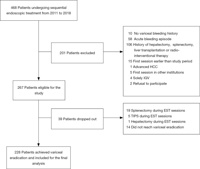
This prospective cohort study was conducted at a university-affiliated referral hospital. Consecutive patients with liver cirrhosis who underwent EBL, ECI or EIS as endoscopic treatment for EGV between October 2011 and August 2018 were screened for the study. Patients undergoing the first session of operational endoscopy for secondary prophylaxis of variceal bleeding in our endoscopy center were included. Patients were excluded if they met any of the following criteria: absence of variceal bleeding history; endoscopic treatment during the acute bleeding phase; history of prior hepatectomy, splenectomy, radio-intervention therapy or liver transplantation; coexistence of advanced hepatocellular carcinoma (HCC); sole presence of isolated GV; or refusal to participate in the study. The patient selection flow chart is shown in Figure 1. This study was approved by the institutional review board of The Third Affiliated Hospital of Sun Yat-sen University (No. 2-79) and conducted in accordance with the Declaration of Helsinki (as revised in 2013). Written informed consent was obtained from all study participants.
Figure 1.
Study population selection. A total of 228 patients were finally included out of 468 patients undergoing endoscopic treatment for esophagogastric varices. HCC, hepatocellular carcinoma; EST, endoscopic sequential therapy; IGV, isolated gastric varices; TIPS, transjugular intrahepatic portosystemic shunt.
Data collection and study follow-up
Baseline demographic, clinical and laboratory characteristics, along with endoscopic features, were collected. Endoscopic features were assessed based on the general rules of recording endoscopic findings for EGV (11). After baseline assessment, all recruited patients underwent an initial endoscopic treatment session for EGV, and the sessions were repeated every 4–6 weeks thereafter until eradication of the varices. EV eradication was defined as the absence of visible continuous varices upon air insufflation or any residual varices with red signs that could be ligated or injected. GV eradication was defined as the time when the entire GV became solidified by cyanoacrylate injection or as the absence of any size of varices with red signs (11,12). All patients included in the final analysis successfully achieved VE, which had to be agreed upon by two experienced endoscopists. Patients were followed up every 3–6 months with clinical assessment including endoscopic examinations, laboratory tests, concomitant non-endoscopic treatments, and development of gastrointestinal bleeding or death.
Study outcomes
The primary outcome was recurrent upper gastrointestinal bleeding or death at 1, 2 or 5 years after VE achievement, while variceal rebleeding was considered a subtype of all-cause gastrointestinal rebleeding. The secondary outcome was 6-month and 1-year rebleeding during endoscopic sessions before VE achievement. Outcome data were obtained primarily from study follow-up records. Patients who did not visit the clinic regularly were followed up through telephone inquiry. Rebleeding was defined as overt gastrointestinal bleeding (e.g., hematemesis, melena) associated with unstable vital signs (systolic blood pressure ≤90 mmHg or pulse ≥110 bpm), a significant drop in hemoglobin (at least 20 g/L), the need for blood transfusion with hemoglobin less than 70 g/L, and/or endoscopic verified bleeding (5). In addition, cases achieving VE within 6 months were categorized into the fast-VE group, while cases achieving VE after more than 6 months were categorized into the slow-VE group.
Performance of EST
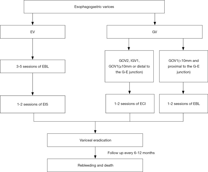
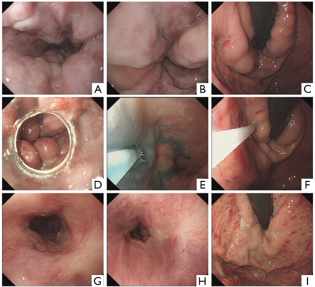
EST is a combination strategy developed in our department defined as ECI (when necessary) sequenced by repeated sessions of EBL and EIS (when necessary). In general, GV ≥10 mm in diameter and/or with high-risk bleeding stigmata were treated with ECI at the first visit, followed by repeated EBL for EV or proper GV during the same session or in subsequent sessions every 4–6 weeks. When EV became discontinuous or were surrounded by scars, under which condition the EBL was inapplicable, EIS was performed for the remnant EV to achieve VE. ECI was performed using a GIF-H260 (Olympus Corp., Tokyo, Japan) or EG-L590 (Fujinon Inc., Saitama, Japan) endoscope and a 23-gauge disposable injection needle catheter (1,800 mm in length). A mixture of N-butyl-2-cyanoacrylate (Compont Medical Corp., Beijing, China) and lipiodol (Guerbet, Villepinte, France) at a 1:1 ratio was injected with the total dosage decided by the operator depending on the type and size of the GV. EBL was performed using multiband devices (Wilson-Cook Medical Inc., Winston-Salem, NC, or SpeedBand, Boston Scientific Corp., Natick, MA). The procedure started at or just below the gastroesophageal junction and each varix was ligated, but no more than 7 bands were applied per session. EIS was performed using 1% lauromacrogol injection (Tianyu Chang’an Corp., Xi’an, China). No more than 5 mL sclerosant was injected into each site, and the total amount did not exceed 20 mL per session. The schematic workflow and endoscopic views of EST are shown in Figures 2 and 3, respectively. The performance principle was in accordance with European and Chinese guidelines while considering clinical experiences and local expertise (3,4,9,13-16). All procedures were performed by the same medical team, and the specific option for each endoscopic session was selected at the endoscopist’s discretion.
Figure 2.
Workflow of endoscopic sequential therapy and subsequent follow up. EV, esophageal varices; GV, gastric varices; EBL, endoscopic band ligation; EIS, endoscopic injection sclerotherapy; ECI, endoscopic cyanoacrylate injection; GOV, gastroesophageal varices; IGV, isolated gastric varices.
Figure 3.
Endoscopic views in different phases of endoscopic sequential therapy. (A,B) Severe EV in the middle and lower section of esophagus before EBL; (C) severe GV in the stomach along the lesser curvature before ECI; (D) performance of EBL; (E) performance of EIS on remnant EV with tracer of methylene blue solution after repeated EBL; (F) performance of ECI; (G,H) complete eradication of EV in the middle and lower section of esophagus after two sessions of EBL and one session of EIS; (I) complete eradication/consolidation of GV after one session of ECI. EV, esophageal varices; GV, gastric varices; EBL, endoscopic band ligation; EIS, endoscopic injection sclerotherapy; ECI, endoscopic cyanoacrylate injection.
Statistical analysis
Categorical variables were compared using Chi-square or Fisher’s exact test when appropriate, and quantitative variables were compared using Student’s t-test or Mann-Whitney U test, as applicable. The Kaplan-Meier method was used to estimate the cumulative incidence of rebleeding before and after VE. To illustrate the impact of VE on rebleeding, the log-rank test was used to compare the incidence of rebleeding before and after VE, as shown in different survival curves. Meanwhile, variceal rebleeding was considered a subtype of all-cause rebleeding, and the log-rank test was also performed to compare the incidence of variceal rebleeding before and after VE. In the rebleeding analysis, the endpoint was rebleeding, and data were censored at the time of last clinical visit, death or transfer to other non-endoscopic therapies, except locoregional therapies for small HCC. In the mortality analysis, the endpoint was death or liver transplantation, and data were censored at the time of last clinical visit or when transferred to other therapies as above. Moreover, rebleeding and mortality rates were compared between the fast-VE and slow-VE groups using the Chi-square test, and logistic regression was used to investigate the optimal timing for VE. A two-tailed P value less than 0.05 was considered statistically significant. All data were analysed using SPSS version 21.0 software (IBM Corp., Armonk, NY) and R statistics version 3.4.1 (R Core Team, Vienna, Austria).
Results
Patient characteristics and endoscopic treatment strategies
During the study period, 468 consecutive patients underwent sequential endoscopic treatment. Among them, 201 patients were excluded based on the exclusion criteria, and another 39 patients were excluded during the EST sessions. As a result, 228 patients (85.4%) achieved VE and were included in the study cohort (Figure 1) with a median (interquartile range, IQR) follow-up time of 33.0 (23.0–48.75) months. The cohort consisted of 178 (78.1%) men and 50 (21.9%) women with a mean age of 49.3±10.5 years. One hundred thirty-three (58.3%) patients had Child-Pugh A cirrhosis, 95 (41.7%) patients had Child-Pugh B or C cirrhosis, and 9 (3.9%) patients had Model for End-Stage Liver Disease (MELD) scores of 20 or above. Hepatitis B infection was the major underlying etiology (62.7%), followed by alcohol consumption (11.8%). Eighty-five (37.3%) patients were receiving non-selective beta-blockers (NSBBs) on admission. Two patients were diagnosed with small HCC without radiographic evidence of macrovascular invasion, and 14 patients had portal vein thrombosis at enrollment. Clinical and endoscopic characteristics of the included patients are shown in Table 1. On average, 3.6 sessions of EST were performed to achieve VE for each patient, and the mean time duration required was 8.5 months. EBL was the mainstay treatment option and was performed in nearly all patients (97.8%). The total number of bands applied per patient was 18 (Table S1).
Table 1. Baseline characteristics of patients on secondary prophylaxis.
| Characteristic | Total cohort (n=228) |
|---|---|
| Age (years) | 49.3±10.5 |
| Sex (male, %) | 178 (78.1) |
| Etiology of liver cirrhosis, n (%) | |
| HBV | 143 (62.7) |
| HCV | 14 (6.1) |
| Alcohol | 27 (11.8) |
| Alcohol + viral | 18 (7.9) |
| Others | 26 (11.4) |
| Hypertension, n (%) | 16 (7.0) |
| Diabetes mellitus, n (%) | 37 (16.2) |
| Ascites, n (%) | 95 (41.7) |
| Hepatocellular carcinoma, n (%) | 2 (0.9) |
| Portal vein thrombosis, n (%) | 14 (6.1) |
| Use of β-blocker, n (%) | 85 (37.3) |
| Laboratory test | |
| Hemoglobin level (g/L) | 90.1±22.2 |
| Platelet count (×109/L) | 87.0±43.3 |
| ALT level (U/L) | 39.5±42.7 |
| Total bilirubin level (µmol/L) | 26.2±27.3 |
| Prothrombin INR | 1.37±0.4 |
| Child-Pugh classification, n (%) | |
| A | 133 (58.3) |
| B | 79 (34.6) |
| C | 16 (7.0) |
| MELD score, n (%) | |
| 0–9 | 84 (36.8) |
| 10–19 | 135 (59.2) |
| 20–30 | 9 (3.9) |
| Endoscopic findings at initial therapy, n (%) | |
| Type, EV + GV/EV | 185/43 (81.1/18.9) |
| Location, Li + Lm or Li/Li + Lm + Ls | 214/14 (93.9/6.1) |
| EV size, F1 + F2/F3 | 17/211 (7.5/92.5) |
| GV size, F1 + F2/F3 | 88/99 (38.6/43.4) |
| EV, red sign, n (%) | 214 (93.9) |
| GV, red sign, n (%) | 110 (48.2) |
Data presented as mean ± SD or number of patients (percentage) where appropriate. MELD, Model for End-Stage Liver Disease; INR, international normalized ratio; EV, esophageal varices; GV, gastric varices.
Clinical outcome of rebleeding over time
A total of 28 patients (12.3%) experienced rebleeding after VE. Among the rebleeding episodes, 17 (60.7%) involved bleeding from varices (7 EV, 7 GV, 3 EV and GV), and 11 involved other causes (Table 2). The median (IQR) time duration from eradication to rebleeding was 16.5 (9.25–27.5) months. The cumulative incidence of rebleeding at 1, 2 and 5 years was 4.0%, 10.4%, and 31.5%, respectively. Regarding variceal rebleeding specifically, the incidence at the above corresponding years was 2.6%, 9.1%, and 23.5%, respectively (Figure 4). In contrast, 27 patients (11.8%) presented with rebleeding during the endoscopic sessions before VE, and most of the bleeding cases were variceal-related (23 cases, 85.2%). The majority of patients achieved VE within 12 months (182 cases, 79.8%). The cumulative incidence of rebleeding before VE at 6 months and 1 year was 8.4% and 14.9%, respectively, which was significantly higher than rebleeding after VE (log-rank P<0.001) (Figure 5A). When considering variceal rebleeding cases alone, short-term rebleeding before VE was also higher than after VE (log-rank P<0.001) (Figure 5B). With respect to treatment selection, patients receiving endoscopy plus NSBBs combination therapy showed similar rebleeding rates compared to endoscopic therapy alone, whether measured as all-cause rebleeding or variceal rebleeding during the follow-up period after VE (log-rank P=0.05 and 0.06, respectively) (Figures S1,S2).
Table 2. Clinical outcomes of patients on secondary prophylaxis.
| Outcomes | Total cohort (n=228) |
|---|---|
| Overall follow-up duration, (months, median, IQR) | 33.0 (23.0–48.75) |
| Follow-up duration after eradication, (months, median, IQR) | 24.0 (17.0–35.0) |
| Duration to rebleeding1 (months, median, IQR) | 16.5 (9.25–27.5) |
| Rebleeding source, n (%) | 28 (12.3) |
| EV | 7 (3.1) |
| GV | 7 (3.1) |
| EV + GV | 3 (1.3) |
| Procedure-related | 1 (0.4) |
| Peptic ulcer | 3 (1.3) |
| Others | 3 (1.3) |
| Unknown | 4 (1.8) |
| Rebleeding treatment, n (%) | |
| Medical | 13 (5.7) |
| Endoscopic | 14 (6.1) |
| Surgical or radio-interventional | 1 (0.4) |
| Duration to interval rebleeding2 (months, median, IQR) | 4 (0–11) |
| Interval rebleeding source, n (%) | 27 (11.8) |
| EV | 10 (4.4) |
| GV | 9 (3.9) |
| EV + GV | 4 (1.8) |
| Procedure-related | 2 (0.7) |
| Unknown | 2 (0.7) |
| Transfer to other therapies, n (%) | 24 (10.5) |
| Surgical therapy | 20 (8.8) |
| Radio-interventional therapy | 3 (1.3) |
| Liver transplantation | 1 (0.4) |
| Duration to death3 (months, median, IQR) | 21.0 (12.0–29.0) |
| Cause of death, n (%) | 11 (4.8) |
| Variceal bleeding | 1 (0.4) |
| Non-variceal bleeding | 0 (0) |
| Hepatocellular carcinoma | 4 (1.8) |
| Liver failure | 3 (1.3) |
| Others | 3 (1.3) |
1, for rebleeding cases after variceal eradication; 2, for rebleeding cases during endoscopic sessions; 3, for dead cases. Data presented as median and IQR or number of patients (percentage) where appropriate. IQR, interquartile range; EV, esophageal varices; GV, gastric varices; EBL, Endoscopic banding ligation; ECI, endoscopic cyanoacrylate injection; EIS, endoscopic injection sclerotherapy.
Figure 4.
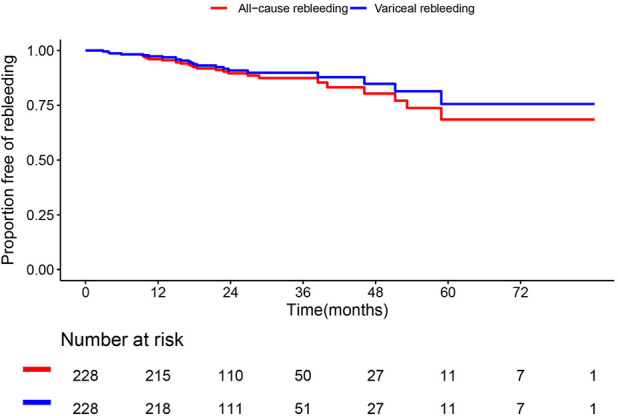
Kaplan-Meier analysis of rebleeding after variceal eradication. Survival curves of patients showing cumulative incidence of all-cause rebleeding (red) and variceal rebleeding (blue).
Figure 5.
Kaplan-Meier analysis of rebleeding before and after variceal eradication. (A) Survival curves of patients showed that the cumulative incidences of all-cause rebleeding before VE at 6 months and 1 year were significantly higher than those of rebleeding after VE. (B) When considering variceal rebleeding cases alone, rebleeding incidences before VE at 6 months and 1 year were also significantly higher than those of rebleeding after VE. VE, variceal eradication.
Clinical outcome of death over time and patients transferred to non-endoscopic therapies
Eleven patients (4.8%) died during the entire follow-up period. Causes of death included HCC (4 cases), liver failure (3 cases), systemic organ failure (2 cases) and cerebral hemorrhage (1 case). Only one case died of variceal bleeding (Table 2). Accordingly, the cumulative incidence of all-cause mortality at 1, 2 and 5 years was 0.9%, 4.4% and 9.3%, respectively. No deaths occurred during EST sessions before VE. In contrast, 24 patients (10.5%) were transferred to surgeries, interventional portosystemic shunt therapies and liver transplantations. Meanwhile, on-site endoscopy failure, which was defined as bleeding within 5 days of initial endoscopy, did not occur in our study. During the entire follow-up, no serious adverse events in the forms of perforation, strictures, chest empyema or pericardial effusion were observed, whereas transient dysphagia, chest pain and epigastric pain were occasional but did not require specific treatment.
Comparison of rebleeding and mortality for the fast-VE group and slow-VE group
One hundred twenty-eight patients (56.1%) achieved VE within 6 months, categorized as the fast-VE group, while the remaining 100 patients were categorized as the slow-VE group. Seven cases (5.5%) in the fast-VE group and 20 cases (20%) in the slow-VE group experienced rebleeding events during endoscopic sessions (Chi-square P=0.002) (Figure 6). Logistic regression showed that prolonged eradication time significantly increased the risk of rebleeding compared to restricted VE time within 6 months (OR 2.88, 95% CI: 0.95–8.70 for the group of 6< VE time ≤12 months; OR 5.91, 95% CI: 2.20–15.89 for the group of VE time >12 months) with P for trend <0.001 (Table 3). However, regarding rebleeding and mortality after VE, the two groups showed no significant differences (Chi-square P=0.603 for rebleeding; Chi-square P=0.660 for mortality). Furthermore, no significant differences in baseline clinical parameters were observed between the two groups (Table S2).
Figure 6.
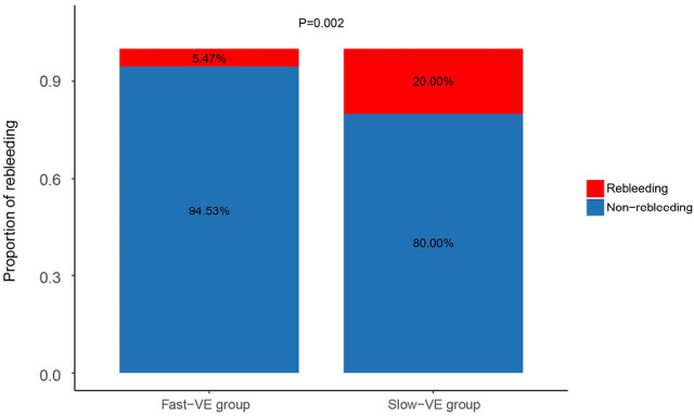
Comparison of rebleeding risk between fast-VE group and slow-VE group. Fast-VE group (VE achieved within 6 months) presented with less rebleeding events than slow-VE group (VE achieved after more than 6 months). VE, variceal eradication.
Table 3. Rebleeding risk stratified by different time periods used for variceal eradication.
| Time for VE | Rebleeding risk before VE, OR (95%CI) |
|---|---|
| ≤6 months | 1 |
| 6< time ≤12 months | 2.88 (0.95–8.70) |
| >12 months | 5.91 (2.20–15.89) |
| P for trend | <0.001 |
VE, variceal eradication; OR, odds ratio; CI, confidence interval.
Discussion
This study clarified the impact of VE on reducing recurrent bleeding in an endoscopic prophylaxis cohort. The natural history of cirrhotic patients in terms of rebleeding and mortality following VE was also delineated. To the best of our knowledge, real world prognostic data with clearly defined endoscopic eradication of both EV and GV have never been reported. Moreover, an eradication time limit of 6 months was found to be optimal, resulting in less interval rebleeding during endoscopic sessions.
According to prior studies, endoscopic therapy for secondary prophylaxis is still associated with a 20% to 30% rate of 1-year rebleeding, and procedure-related deaths might even occur (5-7,17,18). Therefore, the therapeutic goal of VE was proposed adhering to clinical guidelines. Recent data showed that the rebleeding proportion before VE was 24% to 32% (8,19), which was much higher than in our study (12%). In contrast, the rebleeding proportion after VE was reported to be 11.3% to 25% (8,20) and our data were within this range (12.3%). However, these studies only treated EV and did not provide rebleeding probability in a time-dependent manner, making the impact of a complete VE still unclear. Although the overall percentages before and after eradication were similar in our study, the beneficial impact became marked when taking the follow-up period into account, and an additional 10% decrease in the one-year rebleeding incidence was observed. On the other hand, most previous studies analysed data on EV alone, while few studies evaluated patients undergoing endoscopic treatment for both EV and coexisting junctional or fundal GV (5,10). Considering that GV occurs in 50% of cirrhotic patients and contributes to 10–20% of variceal bleeding (1,4,21,22), a complete occlusion of both EV and GV achieved by EST in our study was a sound and more ideal endoscopic endpoint than EV eradication alone. Since the mortality is still as high as 20% at 6 weeks after a separate bleeding episode, preventing bleeding during endoscopic intervals is of great importance and aggressive VE in our study should be considered favorable for waning rebleeding and mortality (23,24).
There was no standard time limit or number of sessions needed for VE, but most studies have reported a mean number of 3–6 sessions, and the recommended interval for band ligation is 2–4 weeks (1,3,4,8,19,25). In our study, we believe that 6 months is an optimal VE time limit because a large group of our patients underwent concurrent cyanoacrylate injection for GV, which may require 6–8 weeks for better reassessment on endoscopy. When taking time into account, the rebleeding rate before VE is still unsatisfactory, and we found that most bleeding events occurred in the slow-VE group. This might be due to poor compliance of this group of patients, having a greatly prolonged endoscopic interval and extended time duration to the eradication, which increased the bleeding risk of remaining high-risk varices awaiting treatment (26-28). Another reason for the high proportion of variceal-related rebleeding before VE was presumed to be the observed low prevalence of the use of non-selective β-blockers with less than 40% in our cohort, while the status of hemodynamic response and long-term drug adherence of these patients were both unknown. In practice, compromised patient compliance is a major threat to clinicians but is very common in chronic liver disease (29-32). However, the improved post-eradication prognosis revealed by our data reflects the advantage of VE, which may even benefit poor compliance patients who do not regularly follow up. In addition, during the endoscopic intervals before VE, we noted that rebleeding events were usually not fatal and could achieve hemostasis with timely endoscopic interventions. Nevertheless, our data strengthened the concept of fast eradication complying with clinical guidelines, and reinforced health education and optimized follow-up management are highly needed.
There are several limitations of our study. First, as a single-center study performed in an academic referral hospital, patient selection bias is inevitable, which may weaken the generalizability of our data. Second, all patients underwent the same endoscopic management strategy and there was no traditional mono EBL therapy group or mono medical treatment group for comparison. Therefore, we failed to compare the efficacy of EST to EBL or medication directly. However, in our study group of over 80% patients with concomitant high-risk EV and GV, monotherapy was of ethical concern and may hardly be applied in practice. Third, eradication time was longer than previously reported due to poor compliance of some patients, which may impair the efficacy of endoscopy therapy and increase interval bleeding events. Therefore, improved patient education is imperative. Fourth, although we screened all cases undergoing EST within the study period, the sample size was still insufficient and may attenuate the statistical power. Lastly, since the included cases were mostly mild to moderate liver disease patients, our findings may not be applicable to populations with more severe liver disease.
Conclusions
VE further reduces rebleeding based on routine endoscopic prophylaxis and improves long-term prognosis. Rapid VE within 6 months seems to be an optimal time duration for the entire endoscopy period and should therefore be advocated. EST is an effective way to achieve complete VE in real-world practice. Multicenter studies comprising larger population sizes remain highly warranted to further validate the beneficial effect of VE and the efficacy of EST compared to other strategies in the future.
Supplementary
The article’s supplementary files as
Acknowledgments
The authors would like to thank every physician in our department for the helpful discussions and AJE for its linguistic assistance during the preparation of this manuscript.
Funding: This study was supported by the National Natural Science Foundation of China (U1501224), the Natural Science Foundation of Guangdong Province Team Project (2018B030312009), the Science and Technology Developmental Foundation of Guangdong Province (2017B020226003), and the Science and Technology Program of Guangzhou City (201604020118). The funders had no role in the design, implementation or interpretation of results of this study.
Ethical Statement: The authors are accountable for all aspects of the work in ensuring that questions related to the accuracy of integrity of any part of the work are appropriately investigated and resolved. This study was approved by the institutional review board of The Third Affiliated Hospital of Sun Yat-sen University (No. 2-79) and conducted in accordance with the Declaration of Helsinki (as revised in 2013). Written informed consent was obtained from all study participants.
Footnotes
Reporting Checklist: The authors have completed the STROBE reporting checklist. Available at http://dx.doi.org/10.21037/atm-20-3401
Data Sharing Statement: Available at http://dx.doi.org/10.21037/atm-20-3401
Conflicts of Interest: All authors have completed the ICMJE uniform disclosure form (available at http://dx.doi.org/10.21037/atm-20-3401). The authors have no conflicts of interest to declare.
References
- 1.Garcia-Tsao G, Abraldes JG, Berzigotti A, et al. Portal hypertensive bleeding in cirrhosis: Risk stratification, diagnosis, and management: 2016 practice guidance by the American Association for the study of liver diseases. Hepatology 2017;65:310-35. 10.1002/hep.28906 [DOI] [PubMed] [Google Scholar]
- 2.Bosch J, Garcia-Pagan JC. Prevention of variceal rebleeding. Lancet 2003;361:952-4. 10.1016/S0140-6736(03)12778-X [DOI] [PubMed] [Google Scholar]
- 3.de Franchis R, Baveno VI. Faculty. Expanding consensus in portal hypertension: Report of the Baveno VI Consensus Workshop: Stratifying risk and individualizing care for portal hypertension. J Hepatol 2015;63:743-52. 10.1016/j.jhep.2015.05.022 [DOI] [PubMed] [Google Scholar]
- 4.Tripathi D, Stanley AJ, Hayes PC, et al. U.K. guidelines on the management of variceal haemorrhage in cirrhotic patients. Gut 2015;64:1680-704. 10.1136/gutjnl-2015-309262 [DOI] [PMC free article] [PubMed] [Google Scholar]
- 5.Cho H, Nagata N, Shimbo T, et al. Recurrence and prognosis of patients emergently hospitalized for acute esophageal variceal bleeding: A long-term cohort study. Hepatol Res 2016;46:1338-46. 10.1111/hepr.12692 [DOI] [PubMed] [Google Scholar]
- 6.Chalasani N, Kahi C, Francois F, et al. Improved patient survival after acute variceal bleeding: a multicenter, cohort study. Am J Gastroenterol 2003;98:653-9. 10.1111/j.1572-0241.2003.07294.x [DOI] [PubMed] [Google Scholar]
- 7.Pfisterer N, Dexheimer C, Fuchs EM, et al. Betablockers do not increase efficacy of band ligation in primary prophylaxis but they improve survival in secondary prophylaxis of variceal bleeding. Aliment Pharmacol Ther 2018;47:966-79. 10.1111/apt.14485 [DOI] [PubMed] [Google Scholar]
- 8.Hou MC, Lin HC, Lee FY, et al. Recurrence of esophageal varices following endoscopic treatment and its impact on rebleeding: comparison of sclerotherapy and ligation. J Hepatol 2000;32:202-8. 10.1016/S0168-8278(00)80064-1 [DOI] [PubMed] [Google Scholar]
- 9.Wang X, Wu B. Critical issues in the diagnosis and treatment of liver cirrhosis. Gastroenterol Rep (Oxf) 2019;7:227-30. 10.1093/gastro/goz024 [DOI] [PMC free article] [PubMed] [Google Scholar]
- 10.Branch-Elliman W, Perumalswami P, Factor SH, et al. Rates of recurrent variceal bleeding are low with modern esophageal banding strategies: a retrospective cohort study. Scand J Gastroenterol 2015;50:1059-67. 10.3109/00365521.2015.1027263 [DOI] [PubMed] [Google Scholar]
- 11.Tajiri T, Yoshida H, Obara K, et al. General rules for recording endoscopic findings of esophagogastric varices (2nd edition). Dig Endosc 2010;22:1-9. [DOI] [PubMed] [Google Scholar]
- 12.Mishra SR, Chander SB, Kumar A, et al. Endoscopic cyanoacrylate injection versus beta-blocker for secondary prophylaxis of gastric variceal bleed: a randomised controlled trial. Gut 2010;59:729-35. 10.1136/gut.2009.192039 [DOI] [PubMed] [Google Scholar]
- 13.Cheng YS, Pan S, Lien GS, et al. Adjuvant sclerotherapy after ligation for the treatment of esophageal varices: a prospective, randomized long-term study. Gastrointest Endosc 2001;53:566-71. 10.1067/mge.2001.114061 [DOI] [PubMed] [Google Scholar]
- 14.Jang WS, Shin HP, Lee JI, et al. Proton pump inhibitor administration delays rebleeding after endoscopic gastric variceal obturation. World J Gastroenterol 2014;20:17127-31. 10.3748/wjg.v20.i45.17127 [DOI] [PMC free article] [PubMed] [Google Scholar]
- 15.Guo YW, Miao HB, Wen ZF, et al. Procedure-related complications in gastric variceal obturation with tissue glue. World J Gastroenterol 2017;23:7746-55. 10.3748/wjg.v23.i43.7746 [DOI] [PMC free article] [PubMed] [Google Scholar]
- 16.Tao J, Li J, Chen X, et al. Endoscopic variceal sequential ligation does not increase risk of gastroesophageal reflux disease in cirrhosis patients. Dig Dis Sci 2020;65:329-35. 10.1007/s10620-019-05740-1 [DOI] [PMC free article] [PubMed] [Google Scholar]
- 17.Liu C, Liu Y, Shao R, et al. The predictive value of baseline hepatic venous pressure gradient for variceal rebleeding in cirrhotic patients receiving secondary prevention. Ann Transl Med 2020;8:91. 10.21037/atm.2019.12.143 [DOI] [PMC free article] [PubMed] [Google Scholar]
- 18.Lv Y, Zuo L, Zhu X, et al. Identifying optimal candidates for early TIPS among patients with cirrhosis and acute variceal bleeding: a multicentre observational study. Gut 2019;68:1297-310. 10.1136/gutjnl-2018-317057 [DOI] [PubMed] [Google Scholar]
- 19.dos Santos JM, Ferreira AR, Fagundes ED, et al. Endoscopic and pharmacological secondary prophylaxis in children and adolescents with esophageal varices. J Pediatr Gastroenterol Nutr 2013;56:93-8. 10.1097/MPG.0b013e318267c334 [DOI] [PubMed] [Google Scholar]
- 20.de la Peña J, Brullet E, Sanchez-Hernandez E, et al. Variceal ligation plus nadolol compared with ligation for prophylaxis of variceal rebleeding: a multicenter trial. Hepatology 2005;41:572-8. 10.1002/hep.20584 [DOI] [PubMed] [Google Scholar]
- 21.Park SW, Seo YS, Lee HA, et al. Changes in Cardiac Varices and Their Clinical Significance after Eradication of Esophageal Varices by Band Ligation. Can J Gastroenterol Hepatol 2016;2016:2198163. 10.1155/2016/2198163 [DOI] [PMC free article] [PubMed] [Google Scholar]
- 22.Xiaoqing Z, Na L, Lili M, et al. Endoscopic Cyanoacrylate Injection with Lauromacrogol for Gastric Varices: Long-Term Outcomes and Predictors in a Retrospective Cohort Study. J Laparoendosc Adv Surg Tech A 2019;29:1135-43. 10.1089/lap.2019.0360 [DOI] [PubMed] [Google Scholar]
- 23.Garcia-Tsao G, Bosch J. Management of varices and variceal hemorrhage in cirrhosis. N Engl J Med 2010;362:823-32. Erratum in: N Engl J Med 2011;364:490. 10.1056/NEJMra0901512 [DOI] [PubMed] [Google Scholar]
- 24.Lo GH. Endoscopic treatments for portal hypertension. Hepatol Int 2018;12:91-101. 10.1007/s12072-017-9828-8 [DOI] [PubMed] [Google Scholar]
- 25.Kumar A, Jha SK, Sharma P, et al. Addition of propranolol and isosorbide mononitrate to endoscopic variceal ligation does not reduce variceal rebleeding incidence. Gastroenterology 2009;137:892-901. 10.1053/j.gastro.2009.05.049 [DOI] [PubMed] [Google Scholar]
- 26.Yoshida H, Mamada Y, Taniai N, et al. A randomized control trial of bi-monthly versus bi-weekly endoscopic variceal ligation of esophageal varices. Am J Gastroenterol 2005;100:2005-9. 10.1111/j.1572-0241.2005.41864.x [DOI] [PubMed] [Google Scholar]
- 27.Lo GH. The role of endoscopy in secondary prophylaxis of esophageal varices. Clin Liver Dis 2010;14:307-23. 10.1016/j.cld.2010.03.009 [DOI] [PubMed] [Google Scholar]
- 28.Hwang JH, Shergill AK, Acosta RD, et al. The role of endoscopy in the management of variceal hemorrhage. Gastrointest Endosc 2014;80:221-7. 10.1016/j.gie.2013.07.023 [DOI] [PubMed] [Google Scholar]
- 29.Kones R, Rumana U, Morales-Salinas A. Confronting the most challenging risk factor: non-adherence. Lancet 2019;393:105-6. 10.1016/S0140-6736(18)33079-4 [DOI] [PubMed] [Google Scholar]
- 30.Shukla R, Kramer J, Cao Y, et al. Risk and predictors of variceal bleeding in cirrhosis patients receiving primary prophylaxis with non-selective beta-blockers. Am J Gastroenterol 2016;111:1778-87. 10.1038/ajg.2016.440 [DOI] [PubMed] [Google Scholar]
- 31.Polis S, Zang L, Mainali B, et al. Factors associated with medication adherence in patients living with cirrhosis. J Clin Nurs 2016;25:204-12. 10.1111/jocn.13083 [DOI] [PubMed] [Google Scholar]
- 32.Zhao C, Jin M, Le RH, et al. Poor adherence to hepatocellular carcinoma surveillance: A systematic review and meta-analysis of a complex issue. Liver Int 2018;38:503-14. 10.1111/liv.13555 [DOI] [PubMed] [Google Scholar]
Associated Data
This section collects any data citations, data availability statements, or supplementary materials included in this article.
Supplementary Materials
The article’s supplementary files as


