
Figure 1. Processing Steps to Generate 3-mm-Thick Ring From CSF to Adjacent White Matter.
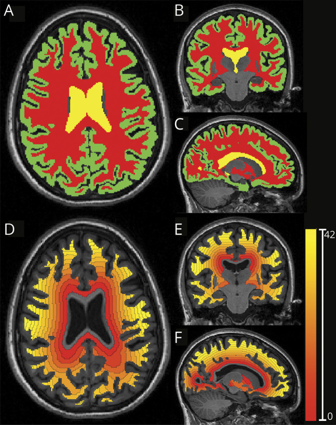
(A–C) Axial, coronal, and sagittal views of a T1-weighted images segmented with a multiatlas segmentation approach generating masks for the white matter, cortex, and ventricles (red, green, and yellow, respectively). (D–F) Axial, coronal, and sagittal views of the corresponding distance map from the ventricular surfaces toward the cortex, calculated in the white matter mask and divided into 3-mm-thick rings.