Abstract
Background
RAS/BRAFV600E mutations are the most remarkable oncogenic driver mutations in colorectal cancer (CRC) and play an important role in treatment selection. No data are available regarding the clinical and prognostic features of patients with detailed RAS/BRAFV600E-mutant metastatic CRC (mCRC) in Japan.
Methods
A total of 152 chemotherapy-naïve patients with mCRC were included in this study between August 2018 and July 2019. Tumor samples were collected, and RAS/BRAFV600E status was investigated. RAS/BRAFV600E status was examined using a MEBGEN RASKET-B kit and polymerase chain reaction reverse sequence-specific oligonucleotide method.
Results
RAS/BRAFV600E mutations were detected in 54% of cases (KRAS codon 12, 26%; KRAS codon 13, 17%; KRAS non-Exon2, 5%; NRAS, 5%; and BRAFV600E, 7%). BRAFV600E-mutant CRC mainly existed in the right colon, whereas KRAS non-Exon2 and NRAS-mutant CRC was predominantly present in the left colon. KRAS non-Exon2 and NRAS-mutant CRC were associated with shorter survival time than RAS wild-type CRC (hazard ratio [HR], 2.26; 95% confidence interval [CI], 0.64–8.03; p = 0.19; HR, 2.42; 95% CI, 0.68–8.61; p = 0.16) and significantly shorter overall survival than KRAS Exon2-mutant CRC (HR, 3.88; 95% CI, 0.92–16.3; p = 0.04; HR, 4.80; 95% CI, 1.14–20.2; p = 0.02).
Conclusions
In our multicenter study, the findings elucidated the clinical and prognostic features of patients with detailed RAS/BRAFV600E-mutant mCRC in Japan.
Keywords: Colorectal cancer, KRAS Exon2, KRAS non-Exon2, NRAS
Background
Colorectal cancer (CRC) is one of the most predominant malignant tumors worldwide, including in Japan. Treatment for advanced recurrent or metastatic CRC (mCRC) aims to control disease activity using anticancer drugs. Investigating biomarkers, especially rat sarcoma (RAS) and rapidly accelerated fibrosarcoma (RAF), are imperative for drug selection in mCRC treatment. RAS and RAF mutations control various activities, such as angiogenesis, proliferation, and apoptosis, and play an important role as prognostic and predictive indicators in CRC treatment [1–9].
Patients with mCRC with RAS (KRAS/NRAS) mutations receive less benefit from anti-epidermal growth factor receptor (EGFR) therapy because RAS mutations activate downstream pathways without depending on EGFR and trigger primary resistance [2, 7, 10–14]. In particular, mutations in the DNA at position 12 in the KRAS protein are general; the KRAS p.G12C mutation accounts for ~ 3% of patients with CRC and is significantly associated with poor prognosis [15]. Although there is no adequate progress in drug development for RAS-mutant tumors, recently developed drugs are expected to be effective. The CodeBreak 100 trial revealed the potent antitumor effects of AMG510, a novel KRAS G12C inhibitor, against KRAS G12C-mutant solid tumors, including mCRC [16].
Focusing on BRAFV600E, previous studies have shown that patients with mCRC with BRAF mutations have worse outcomes than those with BRAF wild-type [17–19]. There have been discrepant results regarding the efficacy of anti-EGFR antibodies in BRAF- and KRAS-mutant cases [20, 21]. In the BEACON CRC trial, a novel triplet combination regimen of encorafenib (BRAF inhibitor), binimetinib (MEK inhibitor), and cetuximab (anti-EGFR antibody), or a novel doublet combination regimen of encorafenib and cetuximab showed benefits compared with the current standard therapy and is now recognized as a new standard therapy as 2nd or later-line treatment [22].
Moreover, consideration of genetic abnormality is essential in the recent treatment of CRC, including microsatellite instability (MSI-H) and mismatch repair deficiency (dMMR). Immune checkpoint inhibitors have become the standard therapy for patients with specific factors detected for immune checkpoint inhibitors, and these inhibitors have also been shown to be very effective as 1st line treatment [23].
The presence or absence of RAS/BRAF mutations can affect anticancer therapy options. In general, patients with RAS/BRAF mutations have different clinical characteristics, and new therapeutic agents developed for driver mutations of these CRCs have been gaining popularity [19, 24]. However, no detailed data have been reported on the clinical and prognostic features in Asian patients, including those from Japan, with detailed RAS/BRAFV600E-mutant mCRC. Therefore, in the present multicenter retrospective study, we aimed to determine the clinical and prognostic features of mCRC with a detailed RAS/BRAFV600E mutation in Japan.
Methods
Patient selection and characteristics
We performed a retrospective study of patients whose tissue RAS/BRAF testing was performed between August 2018 and July 2019 and observed them from the date of registration until July 2020. In total, 152 patients with advanced recurrent CRC were included in the present study. Patient tumor samples taken from primary or metastatic sites were used to investigate the RAS/BRAFV600E mutation status. The following clinical data were collected from three institutions: age, sex, location of the primary tumor, pathological differentiation, stage and TNM grade, metastatic sites, first-line systemic chemotherapy regimen, duration and the best efficacy of first-line chemotherapy, date of confirmation of tumor growth after first-line chemotherapy, date of last consultation date, and date of death.
Analysis for RAS/BRAF mutation
Genomic DNA was detected in each patient using formalin-fixed paraffin-embedded tumor samples. In total, 49 RAS/BRAF mutations were analyzed using the MEBGEN RASKET-B kit and polymerase chain reaction reverse sequence-specific oligonucleotide method for all enrolled cases [12, 25]. Mutations were determined using multiplex PCR and the xMAP® (Luminex®) technology. The mutations included those in KRAS codon 12 (G12S, G12C, G12R, G12D, G12V, and G12A), KRAS codon 13 (G13S, G13C, G13R, G13D, G13V, and G13A), KRAS codon 59 (A59T and A59G), KRAS codon 61 (Q61K, Q61E, Q61L, Q61P, Q61R, and Q61H), KRAS codon 117 (K117N), KRAS codon 146 (A146T, A146P, and A146V), NRAS codon 12 (G12S, G12C, G12R, G12D, G12V, and G12A), NRAS codon 13 (G13S, G13C, G13R, G13D, G13V, and G13A), NRAS codon 59 (A59T and A59G), NRAS codon 61 (Q61K, Q61E, Q61L, Q61P, Q61R, and Q61H), NRAS codon 117 (K117N), NRAS codon 146 (A146T, A146P, and A146V), and BRAF codon 600 (V600E).
Assessment and statistical analysis
Disease assessment was usually performed every 8 ± 2 weeks using computed tomography (CT). The response was evaluated using CT images based on the Response Evaluation Criteria in Solid Tumors version 1.1. We defined overall survival (OS) as the time from enrollment in our study to the date of death for any reason. Patients who were alive were censored at the last follow-up. Progression-free survival (PFS) was defined as the time from enrollment in our study to initial disease progression or death, whichever occurred earlier. We defined the overall response rate as the percentage of patients who achieved a complete response or partial response relative to the total number of enrolled patients based on CT images. Statistical analyses were performed using SPSS statistics version 27.0, and a statistically significant difference was considered at a value of p < 0.05. Fisher’s exact test was used to compare the patient characteristics. Statistical analyses of OS and PFS were performed using the Kaplan–Meier method. The log-rank test was used to compare each group, whereas Cox regression analysis was used to estimate the hazard ratio (HR) with a 95% confidence interval (CI). We also evaluated whether RAS/BRAFV600E status was associated with OS and PFS.
Results
Patient characteristics and frequency of RAS/BRAF V600E mutation subtypes
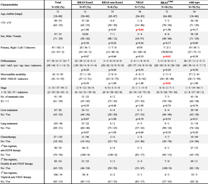
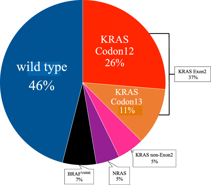
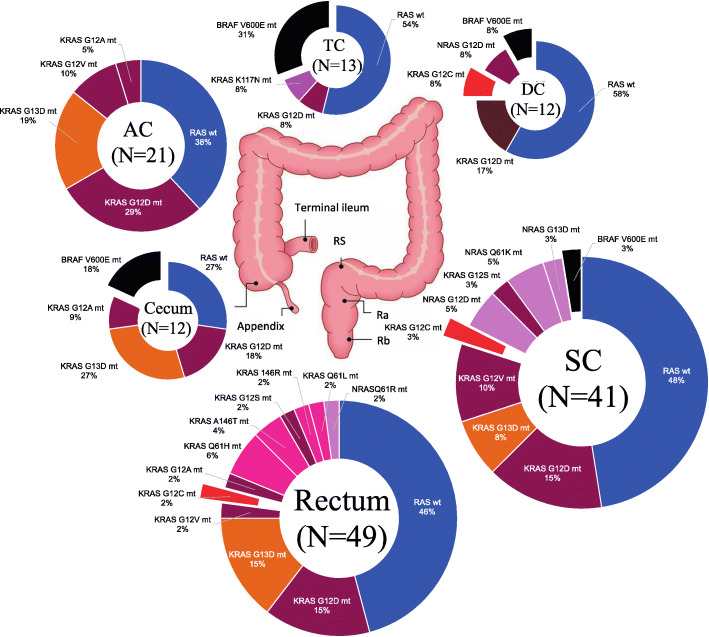
In total, 152 patients were investigated for RAS/BRAFV600E status from three institutions, and the median observation period was 378 days for censored cases (range, 46–2067 days). Table 1 shows the characteristics of the patients included in this study. Patients diagnosed with stage I to III disease were those who relapsed during the observation period and were enrolled in the study. The frequency of RAS mutations was 47% (n = 72), whereas that of the wild-type and BRAFV600E mutations was 46% (n = 70) and 7% (n = 10), respectively. KRAS mutations were found in codon 12 in 26% of cases and codon 13 in 11% of cases; therefore, we designated KRAS codons 12 and 13 as the KRAS Exon2 mutation groups. The other KRAS (non-KRAS codon 12 and non-KRAS codon 13) mutations were designated as the KRAS non-Exon2 mutation group, which included 5% of cases (N = 7; Fig. 1). The locations of the primary tumors in each RAS/BRAF mutation are shown in Fig. 2.
Table 1.
Clinical characteristics and concomitant mutations of patients with RAS/BRAFV600E mutant colorectal cancer
tub1/tub2 tubular adenocarcinoma, por poorly differentiated adenocarcinoma, sig signet-ring cell carcinoma, muc mucinous adenocarcinoma, MSS Microsatellite stable, MSI-H Microsatellite instability-high, EGFR Epidermal growth factor receptor, VEGF Vascular endothelial growth factor, HR Hazard ratio; P-value: Fisher’s exact test, and the value of the comparison between this group and other groups
Fig. 1.
Frequencies of RAS/BRAFV600E mutation subtypes. N = 152 (KRAS Exon2 group: KRAS codon 12 and codon 13; KRAS non-Exon2 group: other KRAS mutations; wild group: no mutations)
Fig. 2.
Frequency of RAS/BRAFV600E mutations by primary tumor site (AC, ascending colon; TC, transverse colon; DC, descending colon; SC, sigmoid colon)
Clinicopathological characteristics of each RAS/BRAF V600E group
We investigated the relationship between RAS/BRAFV600E mutation rate and age (< 75 and ≥ 75 years), sex, the location of the primary tumor, number of metastatic sites, liver metastasis, and lung metastasis (Table 1). NRAS mutations were more common in patients aged ≥75 years, whereas no correlation was observed between age and frequency in the other groups. KRAS non-Exon2 and NRAS mutations were predominantly present in the left colon, whereas BRAFV600E mutations were significantly more common in the right colon than in the left colon (p = 0.01). The MSI-H group was more common in BRAFV600E mutations (p = 0.01). We found no significant differences in other categories between the groups.
OS of patients with each RAS/BRAF V600E status
Among the 152 patients, 125 received systemic chemotherapy and were investigated for OS using the Kaplan-Meier method. The details of the patients are presented in Table 1. We analyzed the OS in each RAS/BRAFV600E mutation group (Fig. 3). The OS in the wild-type group was longer than that in the KRAS non-Exon2, NRAS, and BRAFV600E mutation groups; however, we did not observe significant differences between these groups (HR, 2.26; 95% CI, 0.64–8.03; p = 0.19, HR, 2.42; 95% CI, 0.68–8.61; p = 0.16; HR, 1.30; 95% CI, 0.29–5.83; p = 0.73, respectively). The OS was significantly longer in the KRAS Exon2 mutation group than in the KRAS non-Exon2 and NRAS mutation group (HR, 3.88; 95% CI, 0.92–16.3; p = 0.04; HR, 4.80; 95% CI, 1.14–20.2; p = 0.02). At the time of this analysis, the combination of encorafenib + binimetinib + cetuximab or encorafenib + cetuximab for the BRAFV600E mutant CRC had not been approved; therefore, no patients were treated with these combinations.
Fig. 3.
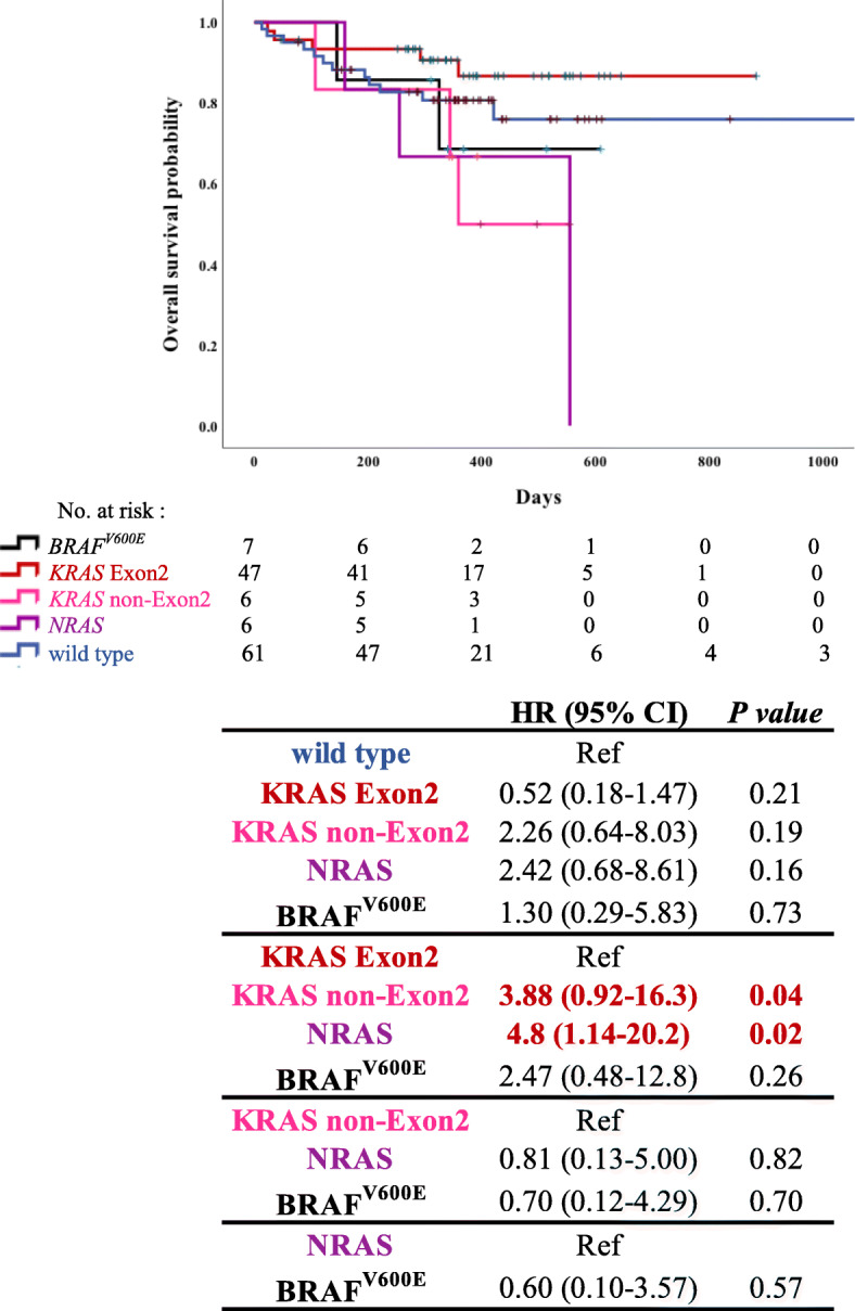
Overall survival (OS) of patients with different subtypes of RAS/BRAFV600E mutations. Analysis of hazard ratio of OS based on RAS/BRAFV600E mutation status in patients with colorectal cancer using Cox regression analysis (N = 152). P-value: Log-rank analysis
We conducted the analysis with sex, age (< 75 and ≥ 75 years), location of the primary tumor, number of metastatic sites, liver metastasis, and lung metastasis (Table 2). As shown, none of the categories showed any apparent significant differences.
Table 2.
Evaluation of clinicopathological characteristics in the subgroup analysis of OS
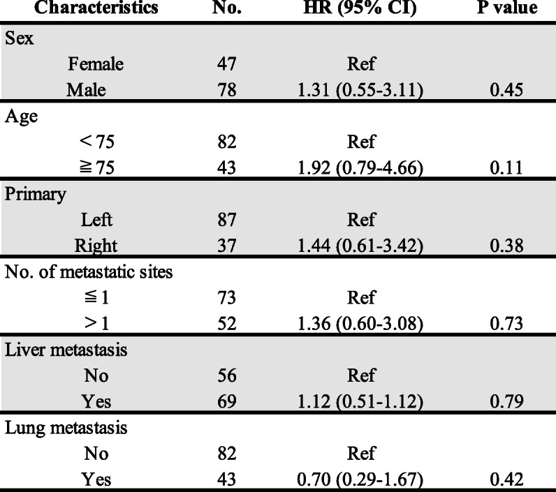
Ref Reference; P-value: Fisher’s exact test, and the value of the comparison between this group and other groups
OS and PFS in the patients treated with doublet therapy with anti-VEGF agents
Among the 125 patients who received systemic chemotherapy, 43 were treated with doublet therapy with anti-vascular endothelial growth factor (anti-VEGF) agents as primary treatment. To adjust for the treatment background, OS and PFS were evaluated only in this subgroup, excluding those who were treated with anti-EGFR antibodies against RAS wild-type. We investigated OS and PFS using the Kaplan-Meier method (Figs. 4 and 5). In the wild-type group, OS was longer than in the KRAS Exon2 and NRAS groups, and PFS was longer than that in the other groups. The median PFS was shorter in the NRAS group than in the other groups (187 days; 95% CI, 181–193 days).
Fig. 4.
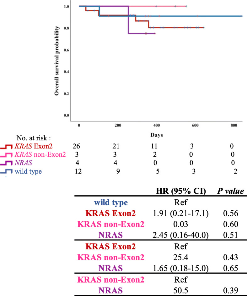
Overall survival (OS) of patients treated with doublet therapy with anti-vascular endothelial growth factor (anti-VEGF) agents. Analysis of hazard ratio of OS based on RAS mutation status in patients with colorectal cancer using Cox regression analysis (N = 43). P-value: Log-rank analysis
Fig. 5.
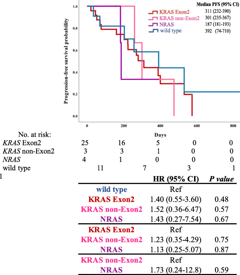
Progression-free survival (PFS) of patients treated with doublet therapy with anti-VEGF agents. Analysis of PFS of hazard ratio based on RAS mutation status in patients with colorectal cancer treated with doublet therapy with anti-VEGF agents using Cox regression analysis (N = 43). P-value: Log-rank analysis
Discussion
In the present study, we investigated the frequency of RAS/BRAFV600E mutations in 152 patients with mCRC in Japan and evaluated the association between each mutation and its clinical and pathological characteristics. We divided RAS mutations into the following three groups: KRAS codon 12 and KRAS codon 13 mutations as the KRAS Exon2 mutation group, KRAS mutations as the KRAS non-Exon2 mutation group, and NRAS mutation group. KRAS codon 12 and KRAS codon 13 mutations were observed in 26 and 11% of cases, respectively. In Japan, KRAS codon 12 and KRAS codon 13 mutations account for 29.9–34.1% and 3.8–7.7% of cases, respectively, which is consistent with the results of the present study [24, 26]. NRAS mutations have been reported in 2.5 –7.2% of cases, and in our study, NRAS mutations were detected in 5% of cases; thus, the frequency was generally consistent with previous reports [27]. BRAFV600E accounted for approximately 7% of cases in the present study, whereas previous studies have reported a range of 5–21% for the BRAFV600E mutation [18, 20]. The frequency of KRAS non-Exon2-mutant mCRC was the same as that in a previous study [25]; however, to date, no data have been published on the frequency of KRAS non-Exon2-mutant CRC in Asian populations, including Japanese patients.
We observed a relationship between the location of the primary lesion and distribution of RAS/BRAFV600E status. Some studies have reported that BRAFV600E mutations are more common in women over 60 years of age and in the right colon [9, 18, 19, 22]. In the present study, BRAFV600E mutations were also more common in the right colon, with statistical significance, and MSI-H patients more commonly contained the mutations. With regard to age and sex, we did not obtain the same results as those previously reported, although it tended to be higher in females than in males. KRAS non-Exon2 mutations were more common in the primary tumor of the left colon. Moreover, the frequency of KRAS non-Exon2 mutation gradually increased and the frequency of KRAS Exon2 mutation decreased, as the primary location was moved from the cecum to the rectum. However, due to the small number of cases, the statistical significance could not be demonstrated as a continuum model as reported [27–29]. There are many studies on the clinical findings of KRAS Exon2 mutations and RAS/BRAFV600E-wild types. However, literature focusing on KRAS non-Exon2 mutation is rare, and the present study may be representative of KRAS non-Exon2 mutation. When focusing on the NRAS mutation, it was more commonly detected in the elderly, with a significant difference; this was also consistent with a previous report [30].
We also investigated the relationship between prognosis and RAS/BRAFV600E status. In general, the prognosis of wild-type RAS was better than that of the RAS/BRAFV600E mutant; however, there were no significant differences in our study. Among the RAS mutations, KRAS Exon2 mutation was associated with the best prognosis. KRAS non-Exon2 and NRAS mutation was associated with a shorter OS than the wild type (HR, 2.26; 95% CI, 0.64–8.03; p = 0.19; HR, 2.42; 95% CI, 0.68–8.61; p = 0.16). Furthermore, patients with KRAS non-Exon2 and NRAS mutation had a significantly shorter OS than those with KRAS Exon2 mutation (HR, 3.88; 95% CI, 0.92–16.3; p = 0.04; HR, 4.80; 95% CI, 1.14–20.2; p = 0.02). It has been reported that NRAS mutation may be associated with better prognosis compared to KRAS mutation [30, 31]. We considered that the results were not consistent with those previously reported because of the small number of cases in this study. The classification of KRAS non-Exon2 mutation has not been widely reported; however, we hypothesized that this result could be supported by the differences in OS between patients with different KRAS mutations. In addition, multivariate analysis of OS in all patients was performed, but no significant differences were found.
Similarly, we examined PFS for each RAS mutation type. PFS was limited to primary treatment, and the treatment regimen was limited to doublet therapy with anti-VEGF agents. Doublet therapy was defined as oxaliplatin-based and irinotecan-based regimens, and the anti-VEGF agents included bevacizumab, ramucirumab, and aflibercept. Under these conditions, NRAS mutation tended to be associated with shorter PFS than KRAS mutations; however, there were no statistically significant differences between these groups. In the RAS mutation group, there were no signs of clear superiority or inferiority, although the regimen was limited in this study. Considering the present study, KRAS non-Exon2 mutation had a poor prognosis. Therefore, it is recommended that KRAS non-Exon2 mutation should be introduced from the first chemotherapy in patients who can be treated with potent therapy such as triplet therapy with anti-VEGF agents. It is hoped that this study on RAS mutation drugs other than KRAS G12C will contribute to future studies in the field.
Several limitations of this research warrant mention. First, this was a retrospective study with a relatively small sample size. Second, we did not follow up with most patients until death; therefore, follow-up data were insufficient.
Conclusion
This multicenter study revealed the detailed clinical and prognostic features of patients with RAS/BRAFV600E-mutant mCRC in Japan; each mutation had a different character. In the present study, the KRAS non-Exon2 and NRAS mutants were primarily in the left colon; to the best of our knowledge, this is the first study to reveal such findings in a Japanese population. The prognosis of patients in the KRAS non-Exon2 and NRAS mutation groups was worse than that of patients in the KRAS Exon2 group. Although the present study involved a relatively small number of patients, the results provide a basis for the development of specific drugs for RAS mutants, which have a poor prognosis.
Acknowledgments
Not applicable.
Abbreviations
- CRC
Colorectal cancer
- EGFR
Epidermal growth factor receptor
- mCRC
Metastatic colorectal cancer
- CI
Confidence interval
- CT
Computed tomography
- RAF
Rapidly accelerated fibrosarcoma
- RAS
Rat sarcoma
- MSS
Microsatellite stable
- MSI-H
Microsatellite instability-high
- VEGF
Vascular endothelial growth factor
- HR
Hazard ratio
- dMMR
Mismatch repair deficiency
- PFS
Progression-free survival
- OS
Overall survival
Authors’ contributions
Study concepts: SH, IT. Study design: SH, IT. Data acquisition: SH, KM, IT, MT, NH, BS, SN, YH. Data analysis and interpretation: SH, IT, SM. Statistical analysis: SM. Manuscript preparation: SH, IT. Manuscript editing: SH, IT. Manuscript review: All authors. The author(s) read and approved the final manuscript.
Author’s information
None.
Funding
None.
Availability of data and materials
The datasets generated and/or analyzed during the current study are not publicly available, but are available from the corresponding author on reasonable request.
Declarations
Ethics approval and consent to participate
This study was carried out in accordance with the Helsinki Declaration and Ethical Guidelines for Clinical Studies and was approved by the institutional review boards of all three participating hospitals, Kobe City Medical Center General Hospital, Kansai Medical University Hospital, and Sano Hospital. Written informed consent was obtained from all the participating patients before entering the study.
Consent for publication
Not applicable.
Competing interests
HS has received research funding from Ono Pharmaceutical Co., Ltd., Taiho Pharmaceutical Co., Ltd., Takeda Pharmaceutical Co., Ltd., and honoraria from Bayer, Bristol-Myers Squibb, Chugai Pharmaceutical Co., Ltd., Daiichi Sankyo, Eli Lilly Japan, Merck Bio Pharma, MSD, Ono Pharmaceutical, Sanofi, Taiho Pharmaceutical Co., Ltd., Takeda, and Yakult Honsha.
MK has received honoraria from Chugai Pharmaceutical Co., Ltd., Takeda Pharmaceutical Co., Ltd., Yakult, Taiho Pharma, and Merck Biopharma Co., Ltd.
The other physicians have no COI.
Footnotes
Publisher’s Note
Springer Nature remains neutral with regard to jurisdictional claims in published maps and institutional affiliations.
Contributor Information
Tatsuki Ikoma, Email: quouqbnonb@gmail.com.
Mototsugu Shimokawa, Email: mototsugu.shimokawa@gmail.com.
Masahito Kotaka, Email: tomomakotaka6410@yahoo.co.jp.
Toshihiko Matsumoto, Email: toshihiko_matsumoto@kcho.jp.
Hiroki Nagai, Email: hiroki_nagai@kcho.jp.
Shogen Boku, Email: shogen0820@gmail.com.
Nobuhiro Shibata, Email: shibanob.kmu@gmail.com.
Hisateru Yasui, Email: hyasui@kcho.jp.
Hironaga Satake, Email: takeh1977@gmail.com.
References
- 1.Lièvre A, Bachet JB, Le Corre D, Boige V, Landi B, Emile J-F, et al. KRAS mutation status is predictive of response to cetuximab therapy in colorectal cancer. Cancer Res. 2006;66(8):3992–3995. doi: 10.1158/0008-5472.CAN-06-0191. [DOI] [PubMed] [Google Scholar]
- 2.Douillard JY, Oliner KS, Siena S, Tabernero J, Burkes R, Barugel M, Humblet Y, Bodoky G, Cunningham D, Jassem J, Rivera F, Kocákova I, Ruff P, Błasińska-Morawiec M, Šmakal M, Canon JL, Rother M, Williams R, Rong A, Wiezorek J, Sidhu R, Patterson SD. Panitumumab-FOLFOX4 treatment and RAS mutations in colorectal cancer. New Engl J Med. 2013;369(11):1023–1034. doi: 10.1056/NEJMoa1305275. [DOI] [PubMed] [Google Scholar]
- 3.Schirripa M, Cohen SA, Battaglin F, Lenz H-J. Biomarker-driven and molecular targeted therapies for colorectal cancers. Semin Oncol. 2018;45(3):124–132. doi: 10.1053/j.seminoncol.2017.06.003. [DOI] [PMC free article] [PubMed] [Google Scholar]
- 4.Jonker DJ, O’Callaghan CJ, Karapetis CS, Zalcberg JR, Tu D, Au H-J, et al. Cetuximab for the treatment of colorectal cancer. New Engl J Med. 2007;357(20):2040–2048. doi: 10.1056/NEJMoa071834. [DOI] [PubMed] [Google Scholar]
- 5.Karapetis CS, Khambata-Ford S, Jonker DJ, O’Callaghan CJ, Tu D, Tebbutt NC, et al. K-ras mutations and benefit from cetuximab in advanced colorectal cancer. New Engl J Med. 2008;359(17):1757–1765. doi: 10.1056/NEJMoa0804385. [DOI] [PubMed] [Google Scholar]
- 6.Amado RG, Wolf M, Peeters M, Cutsem EV, Siena S, Freeman DJ, et al. Wild-type KRAS is required for panitumumab efficacy in patients with metastatic colorectal cancer. J Clin Oncol. 2008;26(10):1626–1634. doi: 10.1200/JCO.2007.14.7116. [DOI] [PubMed] [Google Scholar]
- 7.Van Cutsem E, Lenz HJ, Köhne CH, Heinemann V, Tejpar S, Melezínek I, et al. Fluorouracil, leucovorin, and irinotecan plus cetuximab treatment and RAS mutations in colorectal cancer. J Clin Oncol. 2015;33(7):692–700. doi: 10.1200/JCO.2014.59.4812. [DOI] [PubMed] [Google Scholar]
- 8.Van Cutsem E, Köhne CH, Hitre E, Zaluski J, Chien CC, Makhson A, et al. Cetuximab and chemotherapy as initial treatment for metastatic colorectal cancer. New Engl J Med. 2009;360(14):1408–1417. doi: 10.1056/NEJMoa0805019. [DOI] [PubMed] [Google Scholar]
- 9.Jones JC, Renfro LA, Al-Shamsi HO, Schrock AB, Rankin A, Zhang BY, et al. (non-V600) BRAF mutations define a clinically distinct molecular subtype of metastatic colorectal cancer. J Clin Oncol. 2017;35(23):2624–2630. doi: 10.1200/JCO.2016.71.4394. [DOI] [PMC free article] [PubMed] [Google Scholar]
- 10.Peeters M, Oliner KS, Price TJ, Cervantes A, Sobrero AF, Ducreux M, Hotko Y, André T, Chan E, Lordick F, Punt CJA, Strickland AH, Wilson G, Ciuleanu TE, Roman L, van Cutsem E, He P, Yu H, Koukakis R, Terwey JH, Jung AS, Sidhu R, Patterson SD. Analysis of KRAS/NRAS mutations in a phase III study of panitumumab with FOLFIRI compared with FOLFIRI alone as second-line treatment for metastatic colorectal cancer. Clin Cancer Res. 2015;21(24):5469–5479. doi: 10.1158/1078-0432.CCR-15-0526. [DOI] [PubMed] [Google Scholar]
- 11.Venook AP, Niedzwiecki D, Lenz HJ, Innocenti F, Fruth B, Meyerhardt JA, Schrag D, Greene C, O'Neil BH, Atkins JN, Berry S, Polite BN, O'Reilly EM, Goldberg RM, Hochster HS, Schilsky RL, Bertagnolli MM, el-Khoueiry AB, Watson P, Benson AB, III, Mulkerin DL, Mayer RJ, Blanke C. Effect of first-line chemotherapy combined with cetuximab or bevacizumab on overall survival in patients with KRAS wild-type advanced or metastatic colorectal cancer: a randomized clinical trial. J Am Med Assoc. 2017;317(23):2392–2401. doi: 10.1001/jama.2017.7105. [DOI] [PMC free article] [PubMed] [Google Scholar]
- 12.Yoshino T, Muro K, Yamaguchi K, Nishina T, Denda T, Kudo T, Okamoto W, Taniguchi H, Akagi K, Kajiwara T, Hironaka S, Satoh T. Clinical validation of a multiplex kit for RAS mutations in colorectal cancer: results of the RASKET (RAS KEy testing) prospective, multicenter study. EBioMedicine. 2015;2(4):317–323. doi: 10.1016/j.ebiom.2015.02.007. [DOI] [PMC free article] [PubMed] [Google Scholar]
- 13.Kim TW, Elme A, Kusic Z, Park JO, Udrea AA, Kim SY, Ahn JB, Valencia RV, Krishnan S, Bilic A, Manojlovic N, Dong J, Guan X, Lofton-Day C, Jung AS, Vrdoljak E. A phase 3 trial evaluating panitumumab plus best supportive care vs best supportive care in chemorefractory wild-type KRAS or RAS metastatic colorectal cancer. Br J Cancer. 2016;115(10):1206–1214. doi: 10.1038/bjc.2016.309. [DOI] [PMC free article] [PubMed] [Google Scholar]
- 14.Bokemeyer C, Köhne CH, Ciardiello F, Lenz H-J, Heinemann V, Klinkhardt U, Beier F, Duecker K, van Krieken JH, Tejpar S. FOLFOX4 plus cetuximab treatment and RAS mutations in colorectal cancer. Eur J Cancer. 2015;51(10):1243–1252. doi: 10.1016/j.ejca.2015.04.007. [DOI] [PMC free article] [PubMed] [Google Scholar]
- 15.Jones RP, Sutton PA, Evans JP, Clifford R, McAvoy A, Lewis J, Rousseau A, Mountford R, McWhirter D, Malik HZ. Specific mutations in KRAS codon 12 are associated with worse overall survival in patients with advanced and recurrent colorectal cancer. Br J Cancer. 2017;116(7):923–929. doi: 10.1038/bjc.2017.37. [DOI] [PMC free article] [PubMed] [Google Scholar]
- 16.Hong DS, Fakih MG, Strickler JH, Desai J, Durm GA, Shapiro GI, Falchook GS, Price TJ, Sacher A, Denlinger CS, Bang YJ, Dy GK, Krauss JC, Kuboki Y, Kuo JC, Coveler AL, Park K, Kim TW, Barlesi F, Munster PN, Ramalingam SS, Burns TF, Meric-Bernstam F, Henary H, Ngang J, Ngarmchamnanrith G, Kim J, Houk BE, Canon J, Lipford JR, Friberg G, Lito P, Govindan R, Li BT. KRAS(G12C) inhibition with sotorasib in advanced solid tumors. New Engl J Med. 2020;383(13):1207–1217. doi: 10.1056/NEJMoa1917239. [DOI] [PMC free article] [PubMed] [Google Scholar]
- 17.Van Cutsem E, Kohne CH, Lang I, Folprecht G, Nowacki MP, Cascinu S, et al. Cetuximab plus irinotecan, fluorouracil, and leucovorin as first-line treatment for metastatic colorectal cancer: updated analysis of overall survival according to tumor KRAS and BRAF mutation status. J Clin Oncol. 2011;29(15):2011–2019. doi: 10.1200/JCO.2010.33.5091. [DOI] [PubMed] [Google Scholar]
- 18.Sanz-Garcia E, Argiles G, Elez E, Tabernero J. BRAF mutant colorectal cancer: prognosis, treatment, and new perspectives. Ann Oncol. 2017;28(11):2648–2657. doi: 10.1093/annonc/mdx401. [DOI] [PubMed] [Google Scholar]
- 19.Yokota T, Ura T, Shibata N, Takahari D, Shitara K, Nomura M, Kondo C, Mizota A, Utsunomiya S, Muro K, Yatabe Y. BRAF mutation is a powerful prognostic factor in advanced and recurrent colorectal cancer. Br J Cancer. 2011;104(5):856–862. doi: 10.1038/bjc.2011.19. [DOI] [PMC free article] [PubMed] [Google Scholar]
- 20.De Roock W, Claes B, Bernasconi D, Schutter JD, Biesmans B, Fountzilas G, et al. Effects of KRAS, BRAF, NRAS, and PIK3CA mutations on the efficacy of cetuximab plus chemotherapy in chemotherapy-refractory metastatic colorectal cancer: a retrospective consortium analysis. Lancet Oncol. 2010;11(8):753–762. doi: 10.1016/S1470-2045(10)70130-3. [DOI] [PubMed] [Google Scholar]
- 21.Rowland A, Dias MM, Wiese MD, Kichenadasse G, McKinnon RA, Karapetis CS, et al. Meta-analysis of BRAF mutation as a predictive biomarker of benefit from anti-EGFR monoclonal antibody therapy for RAS wild-type metastatic colorectal cancer. Br J Cancer. 2015;112(12):1888–1894. doi: 10.1038/bjc.2015.173. [DOI] [PMC free article] [PubMed] [Google Scholar]
- 22.Kopetz S, Grothey A, Yaeger R, Cutsem EV, Desai J, Yoshino T, et al. Encorafenib, binimetinib, and cetuximab in BRAF V600E-mutated colorectal cancer. New Engl J Med. 2019;381(17):1632–1643. doi: 10.1056/NEJMoa1908075. [DOI] [PubMed] [Google Scholar]
- 23.André T, Shiu KK, Kim TW, Jensen BV, Jensen LH, Punt C, Smith D, Garcia-Carbonero R, Benavides M, Gibbs P, de la Fouchardiere C, Rivera F, Elez E, Bendell J, le DT, Yoshino T, van Cutsem E, Yang P, Farooqui MZH, Marinello P, Diaz LA., Jr Pembrolizumab in microsatellite-instability-high advanced colorectal cancer. N Engl J Med. 2020;383(23):2207–2218. doi: 10.1056/NEJMoa2017699. [DOI] [PubMed] [Google Scholar]
- 24.Watanabe T, Yoshino T, Uetake H, Yamazaki K, Ishiguro M, Kurokawa T, Saijo N, Ohashi Y, Sugihara K. KRAS mutational status in Japanese patients with colorectal cancer: results from a nationwide, multicenter, cross-sectional study. Jpn J Clin Oncol. 2013;43(7):706–712. doi: 10.1093/jjco/hyt062. [DOI] [PubMed] [Google Scholar]
- 25.Taniguchi H, Okamoto W, Muro K, Akagi K, Hara H, Nishina T, Kajiwara T, Denda T, Hironaka S, Kudo T, Satoh T, Yamanaka T, Abe Y, Fukushima Y, Yoshino T. Clinical validation of newly developed multiplex kit using luminex xMAP technology for detecting simultaneous RAS and BRAF mutations in colorectal cancer: results of the RASKET-B study. Neoplasia. 2018;20(12):1219–1226. doi: 10.1016/j.neo.2018.10.004. [DOI] [PMC free article] [PubMed] [Google Scholar]
- 26.Kawazoe A, Shitara K, Fukuoka S, Kuboki Y, Bando H, Okamoto W, Kojima T, Fuse N, Yamanaka T, Doi T, Ohtsu A, Yoshino T. A retrospective observational study of clinicopathological features of KRAS, NRAS, BRAF and PIK3CA mutations in Japanese patients with metastatic colorectal cancer. BMC Cancer. 2015;15(1):258. doi: 10.1186/s12885-015-1276-z. [DOI] [PMC free article] [PubMed] [Google Scholar]
- 27.Yamauchi M, Lochhead P, Morikawa T, Huttenhower C, Chan AT, Giovannucci E, Fuchs C, Ogino S. Colorectal cancer: a tale of two sides or a continuum? Gut. 2012;61(6):794–797. doi: 10.1136/gutjnl-2012-302014. [DOI] [PMC free article] [PubMed] [Google Scholar]
- 28.Yamauchi M, Morikawa T, Kuchiba A, Imamura Y, Qian ZR, Nishihara R, Liao X, Waldron L, Hoshida Y, Huttenhower C, Chan AT, Giovannucci E, Fuchs C, Ogino S. Assessment of colorectal cancer molecular features along bowel subsites challenges the conception of distinct dichotomy of proximal versus distal colorectum. Gut. 2012;61(6):847–854. doi: 10.1136/gutjnl-2011-300865. [DOI] [PMC free article] [PubMed] [Google Scholar]
- 29.Mima K, Cao Y, Chan AT, Qian ZR, Nowak JA, Masugi Y, Shi Y, Song M, da Silva A, Gu M, Li W, Hamada T, Kosumi K, Hanyuda A, Liu L, Kostic AD, Giannakis M, Bullman S, Brennan CA, Milner DA, Baba H, Garraway LA, Meyerhardt JA, Garrett WS, Huttenhower C, Meyerson M, Giovannucci EL, Fuchs CS, Nishihara R, Ogino S. Fusobacterium nucleatum in colorectal carcinoma tissue according to tumor location. Clin Transl Gastroenterol. 2016;7(11):e200. doi: 10.1038/ctg.2016.53. [DOI] [PMC free article] [PubMed] [Google Scholar]
- 30.Takane K, Akagi K, Fukuyo M, Yagi K, Takayama T, Kaneda A. DNA methylation epigenotype and clinical features of NRAS-mutation(+) colorectal cancer. Cancer Med. 2017;6(5):1023–1035. doi: 10.1002/cam4.1061. [DOI] [PMC free article] [PubMed] [Google Scholar]
- 31.Ogura T, Kakuta M, Yatsuoka T, Nishimura Y, Sakamoto H, Yamaguchi K, et al. Clinicopathological characteristics and prognostic impact of colorectal cancers with NRAS mutations. Oncol Rep. 2014;32(1):50–56. doi: 10.3892/or.2014.3165. [DOI] [PubMed] [Google Scholar]
Associated Data
This section collects any data citations, data availability statements, or supplementary materials included in this article.
Data Availability Statement
The datasets generated and/or analyzed during the current study are not publicly available, but are available from the corresponding author on reasonable request.


