

Abstract
Background
Most patients with oesophageal and gastro‐oesophageal carcinoma are diagnosed at an advanced stage and require palliative intervention. Although there are many kinds of interventions, the optimal one for the palliation of dysphagia remains unclear. This review updates the previous version published in 2009.
Objectives
The aim of this review was to systematically analyse and summarise the efficacy of different interventions used in the palliation of dysphagia in primary oesophageal and gastro‐oesophageal carcinoma.
Search methods
To find new studies for this updated review, in January 2014 we searched, according to the Cochrane Upper Gastrointestinal and Pancreatic Diseases model, the Cochrane Central Register of Controlled Trials (CENTRAL) (The Cochrane Library), MEDLINE, EMBASE and CINAHL; and major conference proceedings (up to January 2014).
Selection criteria
Only randomised controlled trials (RCTs) were included in which patients with inoperable or unresectable primary oesophageal cancer underwent palliative treatment. Different interventions like rigid plastic intubation, self‐expanding metallic stent (SEMS) insertion, brachytherapy, external beam radiotherapy, chemotherapy, oesophageal bypass surgery, chemical and thermal ablation therapy, either head‐to‐head or in combination, were included. The primary outcome was dysphagia improvement. Secondary outcomes included recurrent dysphagia, technical success, procedure related mortality, 30‐day mortality, adverse effects and quality of life.
Data collection and analysis
Data collection and analysis were performed in accordance with the methods of the Cochrane Upper Gastrointestinal and Pancreatic Diseases Review Group.
Main results
We included 3684 patients from 53 studies. SEMS insertion was safer and more effective than plastic tube insertion. Thermal and chemical ablative therapy provided comparable dysphagia palliation but had an increased requirement for re‐interventions and for adverse effects. Anti‐reflux stents provided comparable dysphagia palliation to conventional metal stents. Some anti‐reflux stents might have reduced gastro‐oesophageal reflux and complications. Newly‐designed double‐layered nitinol (Niti‐S) stents were preferable due to longer survival time and fewer complications compared to simple Niti‐S stents. Brachytherapy might be a suitable alternative to SEMS in providing a survival advantage and possibly a better quality of life, and might provide better results when combined with argon plasma coagulation or external beam radiation therapy.
Authors' conclusions
Self‐expanding metal stent insertion is safe, effective and quicker in palliating dysphagia compared to other modalities. However, high‐dose intraluminal brachytherapy is a suitable alternative and might provide additional survival benefit with a better quality of life. Some anti‐reflux stents and newly‐designed stents lead to longer survival and fewer complications compared to conventional stents. Combinations of brachytherapy with self‐expanding metal stent insertion or radiotherapy are preferable due to the reduced requirement for re‐interventions. Rigid plastic tube insertion, dilatation alone or in combination with other modalities, and chemotherapy alone are not recommended for palliation of dysphagia due to a high incidence of delayed complications and recurrent dysphagia.
Plain language summary
Interventions for reducing difficulty in swallowing in people with oesophageal cancer
Review question
For most patients with unresectable or inoperable oesophageal cancer, providing clinical benefit with palliative treatment is highly desirable. However, the optimal palliative technique for dysphagia improvement and better quality of life is not established.
Background
Dysphagia (difficulty or discomfort in swallowing) is common among patients with unresectable or inoperable oesophageal cancer. There are five levels of dysphagia, ranging from the ability to eat a normal diet to some solids, semisolids, liquids and to complete dysphagia.
Study characteristics
The review included randomised controlled studies comparing the use of different interventions to improve dysphagia among patients with inoperable or unresectable primary oesophageal cancer. To find new studies for this updated review, in January 2014 we searched, according to the Cochrane Upper Gastrointestinal and Pancreatic Diseases model, the Cochrane Central Register of Controlled Trials (CENTRAL) (The Cochrane Library), MEDLINE, EMBASE and CINAHL; and major conference proceedings (up to January 2014).
Key results
The review updates the previous version but still fails to present any obvious superiority of one technique over another among the different kinds of interventions. Self‐expanding metal stents provided safer and more effective relief of dysphagia compared to rigid plastic stents. Other techniques like radiotherapy or brachytherapy were also suitable alternatives and might be favourable in improving quality of life and prolonging survival. Individual differences should be emphasised when the intervention type was determined.
Quality of the evidence
Half of the studies included in this review were of high quality. Most studies did not state the methods used to seek and report quality of life outcomes and adverse effects.
Summary of findings
Background
Description of the condition
Oesophageal cancer was the sixth most common cause of cancer death in 2008, which led to 406,000 deaths worldwide (IARC 2008). More than 80% of the oesophageal cancer cases occur in developing countries (IARC 2008). The prognosis of oesophageal cancers is poor. Patients with such a tumour have a five‐year survival rate less than 20%, with the presence of locally advanced and undetected metastatic disease at the time of diagnosis (Shibata 2011). Basically, treatment with curative intention was excluded and palliation became the most suitable option (Weigel 2002; Yang 2012). Dysphagia is the predominant symptom in more than 70% of patients with oesophageal cancer (Brierley 1998). Many types of intervention have emerged in recent years such as newly‐designed stent insertion, external beam radiation, brachytherapy, chemotherapy, chemoradiotherapy, laser treatment and photodynamic therapy. Desipte recent progress in therapeutic methods, the optimal intervention has not been established (Allum 2002; Weigel 2002; Yang 2012).
Description of the intervention
Technique developments have led to a number of interventions including self‐expanding metal stent (SEMS) insertion, thermal laser therapy, photodynamic therapy (PDT) and argon plasma coagulation (APC), while conventional oesophageal bypass surgery, dilatation and chemoradiotherapy have been phased out (Acunaş 2002; Sur 2002b; Yang 2012).
How the intervention might work
Randomised controlled trials (RCTs) have presented evidence that SEMSs are effective, and safer compared to plastic tubes (Roseveare 1998; Shenfine 2009; Siersema 1998; Verschuur 2008). However, at the same time there were complications. Stent migration, tumour ingrowth and overgrowth may require re‐intervention for recurrent dysphagia. The use of the conventional SEMS stimulated the development of the anti‐reflux SEMS (Dua 2001; Laasch 2002) and retrievable SEMS (Song 2002). High‐dose intraluminal brachytherapy is considered as a suitable alternative to SEMS insertion (Sur 2002), while laser treatment, despite effective dysphagia improvement, has introduced adverse effects like perforation (Maciel 1996). Combinations of laser and other treatments have provided better quality of life compared to laser alone (Rupinski 2011).
Why it is important to do this review
With progress in the development of techniques, many new interventions have been used for palliative treatment, and more RCTs has been conducted to identify the optimal intervention for dysphagia in oesophageal cancer. This Cochrane Review aims to update the previous version published in 2009.
Objectives
To systematically analyse and summarise the efficacy of the different interventions used in the palliation of dysphagia in patients with primary oesophageal cancer and gastro‐oesophageal carcinoma.
Methods
Criteria for considering studies for this review
Types of studies
Randomised controlled trials (RCTs) comparing the different interventions mentioned below were included in this review. Both blinded and unblinded trials were included. Both published and unpublished studies, full articles and abstracts were included.
Types of participants
Patients with inoperable or unresectable primary oesophageal cancer who were to undergo palliative treatment. We included patients with primary squamous or adenocarcinoma of the oesophagus or the gastro‐oesophageal junction. Where available, we initially planned to consider these patient subgroups for separate subgroup analysis and comparison. We did not consider patients with extrinsic compression of the oesophagus from other tumours, or patients with recurrence of dysphagia or recurrence of tumour after previous surgery in this review.
Types of interventions
We included the following interventions:
self‐expanding metal stent insertion;
thermal ablative therapy, laser therapy, argon plasma coagulation, bipolar probe electrocoagulation (BICAP);
plastic stent insertion;
intraluminal brachytherapy;
photodynamic therapy;
external beam radiotherapy;
chemoradiotherapy;
chemotherapy;
chemical ablative therapy, alcohol injection, chemotherapeutic agent injection; and
oesophageal bypass surgery.
Comparisons included one or more of the interventions mentioned above or oesophageal dilatation alone. A combination of interventions was acceptable if one of the interventions was included in both arms of the study. Different types of conventional, anti‐reflux SEMS and new‐designed stents have been used in various studies (Table 10; Table 11; Table 12). We also considered studies comparing different types of SEMSs for inclusion in the review to evaluate and compare the outcomes among the different brands or types of stents. These included comparisons between:
1. Characteristics of conventional SEMS.
| SEMS characteristics | Ultraflex | Z stent | Wallstent |
| Material | Nitinol with polyurethane sheath | Stainless steel with polyurethane covering | Elgiloy with polyurethane sheath |
| Length | 10, 12 and 15 cm with 7, 9 and 12cm covered segments respectively | Available in 8, 10, 12, 14 cm lengths | Available as 10 cm with 8 cm covered segment and 15 cm with 13 cm covered segment |
| Inner diameter | 18 mm with 23 mm proximal flare or 23 mm larger stent with 28 mm proximal flare | 18 mm centre with 25 mm bidirectional flare | 20 mm centre with 23 mm bidirectional flare |
| Reconstrainability | Constrained by braided nylon wire. Not reconstrainable when partially deployed | Constrained with polyethylene sheath. Reconstrainable when partially deployed | Constrained in a polyethylene sheath and is reconstrainable when partially deployed |
| Foreshortening | 20% to 40% | None | Up to 28% foreshortening |
| Special characteristics | Available as covered and uncovered types. Distal and proximal release types available | — | — |
2. Characteristics of SEMS with an anti‐reflux mechanism.
| Type of SEMS | Anti‐reflux mechanism |
| Dua Z‐stent | Z‐stent with a 7.5 cm polyurethane sleeve that collapses with pressure in the stomach |
| DO stent | Tricuspid valve, 1 cm long with 30 mm diameter |
| Fer‐X‐Ella stent | Stainless steel with polyethylene covering and windsock type valve. The reflux valve is 4.5 cm long and has an inner diameter of 20 mm |
| Modified S‐type anti‐reflux stent | Polyurethane S‐type valve, 7 cm long and inner diameter of 17.4 mm |
| Hanaro stent | a 70 mm long valve with a malleable silicone‐based membrane |
3. Characteristics of new‐designed stents.
| Type of Stents | Characteristics |
| 125I radioactive stent | sheaths (4.8 mm long 0.8 mm wide) that contained 125I radioactive seeds attached to the outer surface of uncovered stent |
| Niti‐S stent | with braided nickel titanium alloy (nitinol) wire covered with a polyurethane membrane layer over the entire length. available in three lengths: 9, 12, and 15 cm |
| double‐layered Niti‐S stent | uncovered nitinol wire meshes on the outer layer of Niti‐S stent |
| Polyflex stent | silicone device with an encapsulated monofilament braid made of polyester. available in three lengths: 9, 12, and 15 cm |
| Iodine‐eluting stent | nitinol stent and a polyurethane membrane that was uniformly covered with 125l, 5‐13.5 mCi |
covered and uncovered stents;
cuffed and uncuffed stents, to prevent gastro‐oesophageal reflux; and
different commercially available brands of stents.
Types of outcome measures
Primary outcomes
The primary outcome of interest was improvement in dysphagia. Several dysphagia scales have been used to assess improvement in dysphagia grade across the studies (Bown 1987; Mellow 1985; O'Rourke 1988) (Table 13). Recent studies have used these scales with modifications. We compared dichotomous outcomes extracted from the studies using 1‐point and 2‐point improvement in dysphagia grade for each intervention and between the studies. We expressed and compared continuous data using the mean and standard deviation. We used standardised mean difference for studies using different scales.
4. Dysphagia grading systems.
| Dysphagia grade | Bown | Mellow and Pinkas | O'Rourke |
| Grade 0 | Normal swallow | No dysphagia | Normal swallow |
| Grade 1 | Occasional sticking of solids | Dysphagia to normal solids | Able to swallow some solids |
| Grade 2 | Able to swallow semi‐solid or pureed diet | Dysphagia to soft solids | Able to swallow semi‐solids |
| Grade 3 | Able to swallow liquids only | Dysphagia to solids and liquids | Able to swallow liquids only |
| Grade 4 | Unable to swallow liquids | Inability to swallow saliva | Unable to swallow liquids |
Secondary outcomes
Overall survival
Time period from intervention to improvement or relief of dysphagia
Recurrence of dysphagia
Time period from intervention to recurrence of dysphagia
Requirement for further interventions for recurrence of dysphagia
Procedure related mortality
Major and minor adverse effects of each intervention
Quality of life
We excluded trials including interventions with a curative intent and trials that looked at dysphagia improvement as a secondary outcome. This was to avoid the potential underestimation of improvement in dysphagia when this is not the primary outcome. We did not address cost‐effectiveness in this review.
Search methods for identification of studies
Electronic searches
We undertook the principal electronic search according to the guidance in the Cochrane Upper Gastrointestinal and Pancreatic Diseases Review Group module (published in The Cochrane Library).
We retrieved relevant studies from CENTRAL using the broad search terms used in the title of the review and the interventions mentioned above (Appendix 1).
We also searched MEDLINE (1966 to January 2014) (Appendix 2), EMBASE (1988 to January 2014) and CancerLIT (1985 to January 2014) (Appendix 3) using a combination of subject headings and text words related to the title of the review and the interventions mentioned above. We applied standard methodological filters to identify RCTs.
The search strategy was re‐run in August 2006, November 2012 and January 2014.
The Cochrane Upper Gastrointestinal and Pancreatic Diseases Review Group search strategy can be found in Appendix 4. And the study research flow diagram can be found in Figure 1.
1.

Study flow diagram.
Searching other resources
We contacted experts in the field registered with the Cochrane Upper Gastrointestinal and Pancreatic Diseases (CC UGPD) Group for leads on unpublished studies. We searched the UGPD Trials Register for the relevant completed, registered and ongoing trials.
We handsearched Digestive Disease Week (DDW) and United European Gastroenterology Week (UEGW) abstract books between 1994 and 2005. We contacted authors of trial reports published only as abstracts and asked them to contribute full data sets or completed papers. We also handsearched the reference lists of identified articles for further relevant trials.
Data collection and analysis
Selection of studies
One author (AS) assessed the articles identified by the search for eligibility. A second author adjudicated in the event of uncertainty and a consensus view was taken. The reasons for exclusion were documented. Trials satisfying the inclusion criteria were included in the review.
Data extraction and management
Two authors extracted the data using data extraction sheets designed a priori. The following features were recorded when available:
setting, single centre versus multicentre;
method of randomisation, true versus pseudo, stated versus not stated;
adequacy of allocation concealment, stated versus not stated;
inclusion and exclusion criteria used;
baseline comparability between treatment groups;
dysphagia grade used, 4‐point grade versus 5‐point grade;
location of cancer, upper, mid, lower oesophagus or gastro‐oesophageal junction;
type of cancer, squamous carcinoma versus adenocarcinoma;
length of cancer, stated versus not stated;
proportion of inoperable patients versus unresectable cancer, locally advanced versus metastatic cancer;
proportion of patients that had received previous chemoradiotherapy with curative intent;
adverse effects of the intervention, individual adverse effects versus subclassification as major and minor effects.
Assessment of risk of bias in included studies
We followed the instructions given in the Cochrane Handbook for Systematic Reviews of Interventions (Higgins 2011).
Sequence generation
Low risk of bias: the method used was either adequate (e.g., computer‐generated random numbers, table of random numbers) or unlikely to introduce confounding.
Uncertain risk of bias: there was insufficient information to assess whether the method used was likely to introduce confounding.
High risk of bias: the method used (e.g., quasi‐randomised studies) was improper and likely to introduce confounding. Such studies were excluded.
Allocation concealment
Low risk of bias: the method used (e.g., central allocation) was unlikely to induce bias in the final observed effect.
Uncertain risk of bias: there was insufficient information to assess whether the method used was likely to induce bias in the estimate of effect.
High risk of bias: the method used (e.g., open random allocation schedule) was likely to induce bias in the final observed effect.
Blinding of participants, personnel, and outcome assessors
Low risk of bias: blinding was performed adequately, or the outcome measurement was not likely to be influenced by lack of blinding.
Uncertain risk of bias: there was insufficient information to assess whether the type of blinding used was likely to induce bias in the estimate of effect.
High risk of bias: no blinding or incomplete blinding, and the outcome or the outcome measurement was likely to be influenced by lack of blinding.
Incomplete outcome data
Low risk of bias: the underlying reasons for missing data were unlikely to make treatment effects depart from plausible values, or proper methods had been employed to handle missing data.
Uncertain risk of bias: there was insufficient information to assess whether the missing data mechanism in combination with the method used to handle missing data was likely to induce bias in the estimate of effect.
High risk of bias: the crude estimate of effects (e.g., complete case estimate) will clearly be biased due to the underlying reasons for missing data, and the methods used to handle missing data were unsatisfactory.
Selective outcome reporting
Low risk of bias: the trial protocol was available and all of the trial's pre‐specified outcomes that were of interest in the review have been reported, or similar; if the trial protocol was not available, all the primary outcomes in this review that were likely to be measured in such a trial were reported.
Uncertain risk of bias: there was insufficient information to assess whether the magnitude and direction of the observed effect was related to selective outcome reporting.
High risk of bias: not all of the trial's pre‐specified primary outcomes had been reported, or similar.
We considered trials which were classified as low risk of bias in all the above domains as low bias‐risk trials.
Dealing with missing data
We discussed the strategy to deal with missing data in the studies a priori and we explored the probable reasons for missing data. We envisaged that the most common reason for missing data would be attrition contributed to by the withdrawal of patients due to the progression of their disease or general condition. These were actively looked for during data collection and compared between the intervention groups. Where appropriate, we used the 'last observation carried forward' procedure. Efforts were made to explore whether this would introduce bias.
Assessment of heterogeneity
We assessed statistical heterogeneity using the Chi2 test along with visual inspection of the graph. We interpreted a significance level less than 0.10 as evidence of heterogeneity. We looked for an explanation for statistical heterogeneity, discussed clinical heterogeneity, and reported this appropriately. We performed sensitivity analysis using the potential sources of heterogeneity to test the robustness of the overall results. We used the fixed‐effect model when no significant heterogeneity was observed between study results. We used the random‐effects model when variation between studies was observed. The potential reasons for heterogeneity, hypothesised a priori, include:
study quality;
study setting (multicentre versus single centre);
dysphagia grade used (4‐point versus 5‐point grades);
location of cancer, upper, mid, lower oesophagus or gastro‐oesophageal junction;
type of cancer, squamous carcinoma versus adenocarcinoma;
length of cancer;
radiotherapy dose fractionation;
different types of chemotherapy;
different types of stents used;
duration of laser treatment and photodynamic therapy (PDT);
proportion of inoperable patients versus unresectable cancer, locally advanced versus metastatic cancer;
unplanned additional treatment modalities occurring in the intervention groups.
Assessment of reporting biases
The review was designed to include published and unpublished studies, and studies published in all languages. Specialist translation help was sought to obtain data during data collection. We anticipated selective reporting bias at the protocol stage due to the presence of a variety of interventions and patient populations to be covered in this review. We decided to include only studies with dysphagia related outcomes as their primary outcomes. We actively looked for selective reporting bias during the data collection and described this in the description of studies section. We contacted authors, with individualised request forms using non‐leading questions, to provide further information where appropriate.
Data synthesis
We entered all trials included in the systematic review into Review Manager 5.0 (RevMan 2008). We used an intention‐to‐treat approach in all analyses. We performed meta‐analysis only if sufficient trials with similar comparisons and outcome measures were found.
Primary outcome
The primary outcome was the improvement in dysphagia grades. We encountered both dichotomous and continuous data in the trials assessing dysphagia improvement. We expressed dichotomous data (1‐point and 2‐point or more improvement in dysphagia grade) as odds ratios (OR) with 95% confidence intervals (CI). If only continuous data were reported, these were expressed as mean improvement in dysphagia grade with standard deviation following each intervention and were compared between the groups at specific follow‐up periods. We used the standardised mean difference (SMD) to compare dysphagia improvement between studies using different grades of measurement of dysphagia.
Secondary outcomes
We expressed dichotomous data for secondary outcome measures as ORs with 95% CIs. We expressed continuous data for each outcome as means and compared the means between the intervention groups. Where quality of life indices were available, we intended to document the method used, difference observed between the compared interventions and, if appropriate, compare these using group means. However, there was a paucity of data for this outcome.
Subgroup analysis and investigation of heterogeneity
We performed subgroup analysis only for the outcomes that were envisaged a priori. We performed subgroup analysis for all outcomes based on the level of allocation of concealment. We planned at the protocol stage to perform subgroup analysis between the different commercial brands of SEMSs used in the individual studies but there was a paucity of data. Where subgroup analyses were performed at the time of analysis these were described as post hoc analyses. We explored reasons for clinical heterogeneity for all the outcomes regardless of the presence of statistical heterogeneity and these were described in the Results section. We explored statistical heterogeneity using the Chi2 test for heterogeneity, with P < 0.1 being considered significant, and used a random‐effects model in the presence of statistical heterogeneity.
Sensitivity analysis
At the protocol stage we made a decision to conduct sensitivity analyses for all outcomes by excluding each study from the analysis one at a time to confirm the robustness of the results of the main analysis.
Results
Description of studies
Results of the search
The search of the CENTRAL, MEDLINE, EMBASE and CINAHL databases identified 639 articles, and searching major conference proceedings revealed eight further studies. Handsearching reference lists from these articles and repeated searching identified 18 further trials.
After reviewing the abstracts, 120 studies were obtained in full text and 664 studies were excluded because they were clearly not relevant. Four studies could not be obtained. Seven studies were only presented as abstracts and further data were requested but not received.
After going through the above studies, 64 studies were excluded and are described in the 'Characteristics of excluded studies' table. We included 51 full studies and two abstracts in this review. The 53 studies included 3684 patients in total.
Included studies
1. Self‐expanding metal stents (SEMS) versus plastic tube
Types of studies
Seven RCTs were included (De Palma 1996; Knyrim 1993; O'Donnell 2002; Roseveare 1998; Sanyika 1999; Shenfine 2009; Siersema 1998). Three were multicentre trials (Knyrim 1993; Roseveare 1998; Shenfine 2009), one trial (O'Donnell 2002) was performed in two centres and three other trials were from a single centre (De Palma 1996; Sanyika 1999; Siersema 1998). All but one (Sanyika 1999) of the studies were performed in Western Europe.
Types of participants
The seven included studies comprised 433 patients, ranging between 31 (Roseveare 1998) and 217 (Shenfine 2009) in each study. All the studies included patients with inoperable or unresectable oesophageal cancer. Three studies (Knyrim 1993; Roseveare 1998; Sanyika 1999) also included patients with secondary malignant involvement of the oesophagus. In the Roseveare study, four patients with bronchogenic carcinoma were included. The Knyrim study included two patients with bronchogenic carcinoma and one patient with pancreatic cancer. The Sanyika study included five patients with other cancers involving the oesophagus. All studies except one (De Palma 1996) reported the proportion of patients with adenocarcinoma and squamous carcinoma. The South African study (Sanyika 1999) included only patients with squamous carcinoma. In the remaining four studies (Knyrim 1993; O'Donnell 2002; Roseveare 1998; Siersema 1998) the proportion of participants with adenocarcinoma ranged from 40% (Knyrim 1993) to 68% (Roseveare 1998). There was a wide variation in the description of the age, location and length of cancer among the included studies. Three studies did not report the gender distribution (O'Donnell 2002; Roseveare 1998; Sanyika 1999). All the other studies showed a male preponderance, which ranged from 70% (Shenfine 2009) to 83% (Knyrim 1993). One study (Siersema 1998) reported the results with special reference to prior radiation and chemotherapy. Fifteen (39%) patients in the latex prosthesis group and 13 (35%) patients in the SEMS group had undergone prior chemotherapy or radiation, or both, in this study.
Types of interventions
Four studies used Wilson Cook plastic prostheses (De Palma 1996; Knyrim 1993; O'Donnell 2002; Shenfine 2009). One study used the Atkinson tube (Roseveare 1998), one study used the Celestin Pulsion tube (Siersema 1998) and one study used the Proctor Livingstone tube (Sanyika 1999). In three studies, (Knyrim 1993; Sanyika 1999; Siersema 1998) general anaesthesia (GA) was required to insert plastic tubes in all patients. In one study (De Palma 1996), GA was required in 60% of patients and in another (O'Donnell 2002) GA was required in 16% of patients. In the other two studies (Roseveare 1998; Shenfine 2009) only conscious sedation was used. Pre‐dilatations were required in all the studies.
Three studies (Roseveare 1998; Shenfine 2009; Siersema 1998) used the Gianturco Z SEMS, one study (De Palma 1996) used the uncovered Ultraflex SEMS, one study (Knyrim 1993) used the uncovered Wall SEMS and another (Sanyika 1999) used the covered Wall SEMS. O' Donnell et al (O'Donnell 2002) used the covered Wall SEMS and covered Ultraflex SEMS. All the studies performed SEMS insertion under conscious sedation. Three studies used only fluoroscopy to insert the SEMS (Knyrim 1993; O'Donnell 2002; Sanyika 1999). The maximal internal diameter of the SEMS ranged from 16 mm (Knyrim 1993) to 24 mm (Shenfine 2009). Scheduled concomitant co‐intervention was described and reported in only one trial (O'Donnell 2002). Five patients underwent chemotherapy in this study (O'Donnell 2002).
Types of outcomes
Primary outcome
There was a wide variation in reporting on the dysphagia improvement amongst the included studies.
De Palma 1996 described dysphagia with a modified Mellow and Pinkas (Mellow 1985) system of grading and reported median dysphagia score immediately before and after the interventions. No longer‐term follow up of the primary outcome was described.
Knyrim 1993 described dysphagia with a modified Mellow and Pinkas (Mellow 1985) system of grading and reported median dysphagia scores before the interventions and six weeks later. This study also reported the proportion of functional success at six weeks for oesophageal and gastro‐oesophageal junction cancers separately. The follow up was six‐weekly until death through the outpatients department alternating with telephone contact.
O'Donnell 2002 described dysphagia with a modified Mellow and Pinkas (Mellow 1985) system of grading and reported the proportion of patients with improvement in dysphagia without details of the grade of improvement at one and two months. The follow up was monthly until death.
Roseveare 1998 described dysphagia with a modified Mellow and Pinkas (Mellow 1985) system of grading and reported median dysphagia scores before and one week after the interventions. The long‐term results for the primary outcome were described as the proportion of patients with at worst grade 1 dysphagia at six weeks for the two groups.
Sanyika 1999 described dysphagia with a modified Mellow and Pinkas (Mellow 1985) system of grading and reported mean (range) dysphagia scores before and at 24 hours after the interventions. Long‐term improvement was described as the proportion of patients with patency at one month and at three months.
Shenfine 2009 described dysphagia with a modified Mellow and Pinkas (Mellow 1985) system of grading and reported mean (standard deviation) and median scores of dysphagia before and at six weeks after intervention.
Siersema 1998 described dysphagia with a modified Mellow and Pinkas (Mellow 1985) system of grading and reported mean (standard deviation) dysphagia scores before and at four weeks after the interventions.
Secondary outcomes
There was a wide variation amongst the studies in the evaluation and reporting of the different secondary outcomes including recurrence in dysphagia, interventions for recurrent dysphagia, adverse events, overall survival, procedure related mortality and quality of life.
2. SEMS versus laser
Types of studies
Two RCTs (Adam 1997; Dallal 2001) were included. Both studies were performed in a single centre in western Europe.
Types of participants
The studies included a total of 125 patients. Adam 1997 randomised 60 patients with inoperable primary oesophageal malignancy and Dallal 2001 randomised 65 patients with inoperable primary oesophageal cancer. Both studies excluded patients with cancer of the upper oesophagus when the cancer was less than 2 cm from the upper oesophageal sphincter. Adam 1997 also excluded patients who had received any form of previous treatment. Both studies included patients with adenocarcinoma and squamous carcinoma of the oesophagus and adenocarcinoma of the gastro‐oesophageal junction. Both studies reported the baseline comparability between the study groups.
Types of intervention
Both studies (Adam 1997; Dallal 2001) used Strecker or Ultraflex uncovered SEMSs and covered Wallstents of similar diameter. The stent insertions were carried out under sedation with fluoroscopy guidance in both trials.
Dallal 2001 used Nd YAG laser in the majority of patients and ERBE argon plasma coagulation in nine patients. Adam 1997 used Nd YAG laser in all 18 patients randomised to the laser arm. The procedures in both the studies were performed under conscious sedation and dilatation was used appropriately to pass the scope through the stricture. Adam 1997 repeated laser therapy in all patients at four to six‐week intervals whereas in Dallal 2001 ablation was performed at four to six‐week intervals depending upon the degree of dysphagia.
Types of outcomes
Primary outcome
Both studies (Adam 1997; Dallal 2001) assessed dysphagia with a modified Mellow and Pinkas grading system (Mellow 1985). The dysphagia grade before and after the interventions was expressed as the median dysphagia grade at monthly intervals until death in both the studies.
Secondary outcomes
Both studies (Adam 1997; Dallal 2001) reported dichotomous outcomes for recurrent dysphagia, interventions for recurrent dysphagia, re‐admission for recurrent dysphagia, and adverse events for both interventions. They also reported median hospital stay and median overall survival. Dallal 2001 also reported detailed quality of life assessment using the European Organization for Research and Treatment of Cancer (EORTC) core quality of life questionnaire (QLQ‐30) and with oesophageal cancer (EORTC QLQ‐OES 24) and Hospital Anxiety and Depression scale (HAD).
3. SEMS versus brachytherapy
Types of studies
Two RCTs (Bergquist 2005; Homs 2004a; Homs 2004b) were included. Both the studies were performed in Western Europe. One study (Homs 2004a; Homs 2004b) was a multicentre trial and the other (Bergquist 2005) was performed in two centres.
Types of participants
The two studies (Bergquist 2005; Homs 2004a) randomised 274 patients to either SEMS insertion or intraluminal brachytherapy. Homs 2004a randomised 209 patients in a multicentre trial and Bergquist 2005 randomised 65 patients in a trial performed in two centres. Both studies included squamous and adenocarcinoma of the oesophagus. Homs 2004a stratified their patients for tumour location and previous chemotherapy. Both the studies reported the location, type of carcinoma, mean age, gender proportion and proportion of unresectable tumours or inoperable patients.
Types of intervention
Both studies (Bergquist 2005; Homs 2004a) performed SEMS insertion endoscopically under conscious sedation with fluoroscopic guidance. Partially covered Ultraflex stents of similar diameter were used in both the trials.
Brachytherapy was performed with a similar technique in the both the trials. However, Homs 2004a used a Nucleotron applicator and iridium192 source to deliver a single dose of 12 Gy and Bergquist 2005 used an iridium192 source to deliver three fractions of 7 Gy at one to two‐week intervals.
Types of outcomes
Primary outcome
The primary outcome in Homs 2004a was dysphagia improvement. They used the O' Rourke grading system (O'Rourke 1988) before and monthly after the interventions. Bergquist 2005 assessed health related quality of life (HRQOL) as the primary outcome measure using the EORTC QLQ‐30 and EORTC QLQ‐OES 18 questionnaires.
Secondary outcomes
The Homs trial (Homs 2004a; Homs 2004b) reported dichotomous outcomes for recurrent dysphagia, interventions for recurrent dysphagia 30‐day mortality, six‐month mortality, early (< seven days) and late (> seven days) complications along with median survival rates and mean hospital stay. Quality of life was also studied in detail (Homs 2004b) using the EORTC QLQ‐30, EORTC OES‐23 and EQUAS questionnaires. Bergquist 2005 assessed dysphagia improvement using the Ogilvie (Ogilvie 1982) grading system before and at one, three, six, nine and 12 months after the interventions. The trial also assessed mean survival time, time to start and completion of treatment since inclusion, and Karnofsky performance status. This trial (Bergquist 2005) did not specifically address or report adverse effects of the interventions.
Uncovered versus covered SEMS
Types of studies
One RCT (Vakil 2001) was included. This study was a multicentre trial performed in western Europe and the United States.
Types of participants
The study (Vakil 2001) randomised 62 patients to covered and uncovered stents of identical design. Patients with gastro‐oesophageal junction or distal oesophageal adenocarcinoma with < 12 cm stricture were included. The authors reported mean age; length of tumour; proportion of patients with different location of tumour; Tumour, Node, Metastases (TNM) staging and comparability of baseline characteristics.
Types of intervention
Covered and uncovered stents were inserted endoscopically with or without fluoroscopic guidance. Co‐interventions, such as chemotherapy or chemoradiation, were reported.
Types of outcomes
Primary outcome
The primary outcome was reduction in the need for re‐intervention for recurrent dysphagia at follow‐up visits, at one week, one, two, three, four, five and six‐month follow up.
Secondary outcomes
These included mean dysphagia improvement using a modified Mellow and Pinkas (Mellow 1985) grading system, Karnofsky performance status, early and late complications and overall survival.
Different products of SEMS
Types of studies
Two RCTs (Sabharwal 2003; Siersema 2001) were included. Both were single centre studies performed in western Europe.
Types of participants
The studies randomised 153 patients to different commercial brands of SEMS. The Sabharwal study (Sabharwal 2003) included inoperable lower oesophageal carcinoma. Siersema 2001 randomised 100 consecutive patients with inoperable oesophageal or gastro‐oesophageal junctional cancer and patients with recurrent dysphagia following previous chemotherapy or radiotherapy. Both studies reported mean age, length of tumour, proportions of patients with different locations of tumour, and reasons for inoperability or unresectability.
Types of intervention
Sabharwal 2003 compared covered Flamingo Wallstents and covered Ultraflex stents. The Siersema study (Siersema 2001) used either covered Gianturco Z stents, partially covered Flamingo Wallstent or partially covered Ultraflex stents. Stents were inserted under conscious sedation in both the studies. Stents were inserted endoscopically under fluoroscopic guidance in the Siersema study (Siersema 2001) and under fluoroscopic guidance only in Sabharwal 2003. Both studies used small and large diameter stents and in the Siersema study all patients with gastro‐oesophageal junction tumours had large diameter stents (n = 29).
Types of outcomes
Primary outcome
Both studies (Sabharwal 2003; Siersema 2001) assessed dysphagia using the modified Mellow and Pinkas grading system (Mellow 1985). Siersema 2001 reported results at four weeks and Sabharwal 2003 reported on day one post‐insertion and at late follow up. However, the authors did not define the time scales for late follow up.
Secondary outcomes
Both studies (Sabharwal 2003; Siersema 2001) reported dichotomous data for recurrent dysphagia, interventions for recurrent dysphagia, overall survival, 30‐day mortality and complications. Sabharwal 2003 reported complications as early (< 30 days) and late (> 30 days). Detailed quality of life data were not assessed.
Anti‐reflux versus standard open stent
Types of studies
Six RCTs (Homs 2004c; Power 2007; Sabharwal 2008; Shim 2005; Wenger 2006; Wenger 2010) were included. Five studies were performed in Europe and one (Shim 2005) was from Asia. Homs 2004c was conducted in two centres and the Shim 2005 study was a single centre trial. Wenger 2006 conducted a multicentre trial involving nine centres in Sweden, and in Wenger 2010 there were 11 centres. Four studies (Homs 2004c; Power 2007; Wenger 2006; Wenger 2010) blinded the patients to the type of stent received.
Types of participant
Two hundred and seventy‐six patients were randomised in these five trials. The number of patients randomised in the individual trials ranged from 30 (Homs 2004c) to 72 (Wenger 2010 ). All trials included patients with inoperable distal oesophageal and gastro‐oesophageal junction tumours. All the studies collected baseline demographic data and reported baseline comparability.
Types of intervention
All studies (Homs 2004c; Power 2007; Sabharwal 2008; Shim 2005; Wenger 2006) placed the stents endoscopically under conscious sedation with fluoroscopic control. Homs 2004c used the FER X‐Ella stents with a windsock type valve foil and without anti‐reflux valves. Shim 2005 compared three different covered stents, that is the Open MI Tech Pyongtack stent, an early model anti‐reflux stent with a tricuspid valve (DO stent, MI Tech Pyongtack, Korea) and a modified anti‐reflux stent (MI Tech, Pyongtack, Korea) with an S‐type anti‐reflux valve with a 17 mm inner diameter, which is slightly less than the other two stents. Wenger 2006 used covered Z stents with a Dua anti‐reflux valve (Wilson Cook Medical, USA) and either an uncovered Ultraflex, Flamingo Wallstent or Standard open Z stent as controls. Power 2007 used a new anti‐reflux stent (Hanarostent, MI Tech, Seoul, Korea) and a standard covered Ultraflex stent. Sabharwal 2008 used an anti‐reflux stent (FerX‐Ella; Dr Karel Volenec, Ella CS, Hradec Králové, Czech Republic) and a combination of a standard open stent (Ultraflex covered stent; Boston Scientific, Natick, MA, USA) with an anti‐reflux medication (omerprazole). Wenger 2010 used a covered Esophageal Z‐Stent with a Dua Anti‐Reflux Valve (Wilson‐Cook Medical, Winston Salem, NC) and an Ultraflex single‐strand nitinol wire stent (Boston Scientific, Natick, MA), or a Wallstent (Boston Scientific).
Types of outcomes
Primary outcome
The primary outcome in Homs 2004c was gastro‐oesophageal reflux. This was assessed by interviews and 24‐hour pH monitoring. Shim et al (Shim 2005) measured dysphagia using the modified Mellow and Pinkas grading system (Mellow 1985) and reported mean dysphagia scores pre and post‐stent insertion. The main outcome of the Wenger study (Wenger 2006) was assessment of quality of life at baseline, one, three and six months after placement of the stents. This was evaluated using validated EORTC questionnaires, EORTC QLQ‐30 and EORTC QLQ‐OES 18. Power 2007 compared the relief of dysphagia before and after stent placement. The QLQ‐C30 and QLQ‐OES 24 were used to evaluate HRQOL in the study. Sabharwal 2008 took the occurrence of post‐procedure reflux as the primary outcome. Wenger 2010 reported the EORTC questionnaire results as the primary outcome.
Secondary outcomes
Homs et al assessed dysphagia using the O'Rourke (O'Rourke 1988) grading system and reported the median (range) scores at two weeks and then at two‐month intervals. Four studies (Homs 2004c; Sabharwal 2008; Wenger 2006; Wenger 2010) reported median survival and proportion of complications. Shim et al (Shim 2005) did not report the proportion of complications but collected the 30‐day mortality rate and median overall survival data. Twenty‐four‐hour pH studies were performed in all patients on day seven in this study and the total number of reflux episodes, mean longest duration of reflux, mean DeMeester scores and per cent total time with pH < 4 were compared amongst the five stent groups. Power 2007 reported the control of symptomatic gastro‐esophogeal reflux (GER) and impact on the PH profile of the oesophagus post‐intervention.
Irradiation stent versus covered stent
Types of studies
One RCT (Guo 2008) was included. This study was a single centre trial performed in China.
Types of participants
The study (Guo 2008) randomised 53 patients to either the irradiation stent group (stent loaded with 125I) or control group. Patients with unresectable tumours because of extensive lesions, metastatic disease, or poor medical condition were included. The author reported the mean age, gender, dysphagia grade, histologic type of cancer, location of strictures and metastatic disease condition in the baseline characteristics.
Types of intervention
Stent insertions were performed under fluoroscopic guidance; 125I was shielded into the sheaths on the irradiation stent by using a see‐loading gun before stent placement.
Types of outcomes
Primary outcome
The primary outcome was the relief of dysphagia. They used the O' Rourke grading system (O'Rourke 1988) before and monthly after the interventions.
Secondary outcomes
These included procedure related complications and median survival time.
Ultraflex stent versus Polyflex stent versus Niti‐S stent
Types of studies
One RCT (Verschuur 2008) was included. This study was a multicentre trial performed in the Netherlands and Italy.
Types of participants
The study (Verschuur 2008) randomised 125 patients into the Ultraflex stent group, Polyflex stent group and Niti‐S stent group. Patients with an inoperable malignant obstruction of the oesophagus or gastric cardia, or recurrent dysphagia after prior radiation with curative or palliative intent for oesophageal cancer were included. The authors reported the mean age, gender, median dysphagia grade before treatment, mean tumour length, tumour histology, location of tumour, prior radiation and chemotherapy in the baseline characteristics.
Types of intervention
During stent placement, deployment of the stent was performed endoscopically and radiographically assessed. When the tumour obstruction did not allow passage of a standard endoscope, the oesophagus was dilated to a maximum of 12 mm by a Savary‐Miller Esophageal Dilator, or the standard diameter endoscope was changed for a smaller one.
Types of outcomes
Primary outcome
The primary outcome was occurrence of recurrent dysphagia. The authors reported the number and per cent of patients with recurrent dysphagia.
Secondary outcomes
These included technical and functional outcomes, complications, and survival.
Different types of Niti‐S stents
Types of studies
One RCT (Kim 2009) was included. This study was a prospective,single centre study performed in Korea.
Types of participants
The study (Kim 2009) randomised 37 patients to either a double‐layered or covered Niti‐S stent. Patients with unresectable oesophageal or gastric cardia cancer, or oesophageal invasion of other malignancy were included. Patients who had received oesophageal surgery were excluded. The authors reported the mean age, gender, dysphagia score before treatment, tumour length, tumour location, histology, and radiotherapy or chemotherapy before treatment in the baseline characteristics.
Types of intervention
Both stents were placed under fluoroscopic visualisation. After stent deployment, the correct positioning of the stents was assessed endoscopically and radiographically.
Types of outcomes
Primary outcome
The primary outcome was dysphagia grade one week and one month after treatment.
Secondary outcomes
The secondary outcomes were technical success, complications and survival.
SEMS versus iodine‑eluting oesophageal stent
Types of studies
One RCT (Dai 2013) was included. This study was a prospective, single centre study performed in Korea.
Types of participants
The study (Dai 2013) randomised 36 patients to the conventional stent group and 31 to the iodine‑eluting oesophageal stent group. Patients with oesophageal cancer and an expected survival time of more than one month were included. The exclusion criteria were acute infection, severe cardiovascular or mental illness, and evidence of multiple small bowel obstructions. The authors reported age, gender, heart rate, respiratory rate, dysphagia grade, pain grade in the baseline characteristics.
Types of intervention
Both the stents were placed under fluoroscopic guidance.
Types of outcomes
Primary outcome
The primary outcome was dysphagia grade during the follow up.
Secondary outcomes
The secondary outcomes were survival time and side effects.
5. Other studies comparing SEMS with other modalities
Seven other RCTs (Canto 2002; Fu 2004; Horneaux 2001; Javed 2012; Konigsrainer 2000; Shenfine 2009; Turrisi 2002) compared SEMS insertion to various other modalities, head‐to‐head or in combination. Canto 2002 randomised 56 patients with inoperable, persistent or metastatic oesophageal and gastro‐oesophageal junction cancer to SEMS or photodynamic therapy (PDT). Fu 2004 randomised 53 patients to either covered SEMS or SEMS followed by chemotherapy or chemoradiotherapy. Horneaux 2001 randomised 40 patients to either SEMS insertion or oesophageal bypass surgery. Konigsrainer 2000 randomised 39 patients to either SEMS insertion alone, SEMS plus laser treatment or laser and radiotherapy. Shenfine 2009, in a multicentre study, randomised 209 patients to SEMS insertion, rigid plastic tube insertion or non‐stent therapy in a pragmatic RCT. The non‐stent therapy arm included external beam radiotherapy, brachytherapy, thermal ablation therapy and ethanol tumour necrosis, left at the discretion of the treating physicians as appropriate for the tumour characteristics and expertise available at each of the centres. This study also included detailed quality of life and cost‐effectiveness assessment. Turrisi 2002 randomised 32 patients to SEMS insertion or external beam radiotherapy in a multicentre trial. Javed 2012 randomised 79 patients either to SEMS alone or a combination of SEMS followed by external beam radiotherapy (EBRT). The details of the above studies are provided in the 'Characteristics of included studies' table.
6. Laser versus brachytherapy, laser versus laser augmented by external beam radiotherapy, laser versus laser augmented by brachytherapy
Two RCTs (Low 1992; Sargeant 1997) were included. All the studies were single centre trials performed in western Europe.
Low 1992 randomised 23 patients either to brachytherapy or laser. Fourteen patients had either squamous or adenocarcinoma of the oesophagus and five patients had small cell carcinoma. They reported dichotomous outcomes for dysphagia improvement and secondary outcomes including overall survival, complication rates and recurrent dysphagia at follow up at two‐monthly intervals.
Sargeant 1997 randomised 67 patients to either laser therapy or laser augmented by external beam radiotherapy. The primary outcome in this study was dysphagia improvement and this was reported as a dichotomous outcome. The authors also reported separate data for squamous and adenocarcinoma as dysphagia controlled. This was defined as the mean interval between the end of treatment to repeat intervention.
Four RCTs (Ries 1989; Sander 1991; Spencer 2002; Tan 1998) were included. All studies were conducted in western Europe. One study (Sander 1991) was a two centre study and the others were performed from a single centre.
Four trials randomised 128 patients with inoperable oesophageal cancer. One study (Spencer 2002) included only adenocarcinoma and the other three studies (Ries 1989; Sander 1991; Tan 1998) included both adenocarcinoma and squamous carcinoma. The intervention details of these studies are described in the table 'Characteristics of included studies'. All studies studied the dysphagia‐free interval as the primary outcome and also included the secondary outcomes considered for this review. One study (Spencer 2002) assessed quality of life indices using the Longitutinal Aging Study Amsterdam (LASA) questionnaire.
7. Laser versus photodynamic therapy (PDT)
Two RCTs (Heier 1995; Lightdale 1995) were included. Both studies were performed in North America. One study (Lightdale 1995) was a multicentre study. The studies randomised 278 patients with inoperable squamous and adenocarcinoma of the oesophagus to either laser therapy or PDT. The primary outcome in both studies was dysphagia improvement and was reported as dichotomous outcomes for the grades of dysphagia. All secondary outcomes were reported apart from quality of life data. The details of the studies are described in the table 'Characteristics of included studies'.
8. Laser versus plastic stent
Three RCTs were included (Alderson 1990; Carter 1992; Fuchs 1991). All these trials were single centre studies from western Europe. The trials randomised 80 patients with inoperable squamous and adenocarcinoma and compared Atkinson or Celestin tubes to laser therapy. The primary outcome of dysphagia improvement was presented as a dichotomous outcome at monthly follow up. The details of the studies are described in the table 'Characteristics of included studies'.
9. Laser versus chemical ablation
Two RCTs (Angelini 1991; Carrazone 1999) were included. Both studies were single centre trials from Western Europe. Patients with adenocarcinoma and squamous carcinoma were included. Angelini 1991 compared Nd YAG laser and 3% polidocanol. Carrazone 1999 compared 98% absolute alcohol to laser therapy. The details of these studies are described in the table 'Characteristics of included studies'.
10. Other studies including multiple comparisons or plastic tubes
Nine RCTs (Amdal 2013; Anghorn 1983; Barr 1990; Mannell 1986; Mehta 2008; Reed 1991; Rosenblatt 2010; Rupinski 2011; Sur 2004) included comparisons of a plastic tube, external beam radiotherapy, brachytherapy, laser and chemotherapy, head‐to‐head or in various combinations.
Barr 1990, in a single centre study from western Europe, randomised 40 patients with inoperable squamous or adenocarcinoma to either laser therapy alone or laser therapy followed by intubation in 10 to 14 days. In the laser therapy only group initial therapy was directed to increase the lumen size to that of a normal oesophagus and then performed monthly until death. In the combination group initial laser therapy was given to ensure placement of a guidewire and prosthetic tube. The primary outcome of this study was mean dysphagia score throughout the follow‐up period until death, overall survival, recurrent dysphagia and adverse effects. Quality of life was studied using the QL index and LASA questionnaire including a Visual Analogue Scale (VAS) for dysphagia.
Mannell 1986, in a single centre study from South Africa, randomised 170 patients with inoperable squamous cell carcinoma to plastic tube insertion or dilatation followed by bleomycin 30 mg intramuscularly for five days. Dysphagia was the primary outcome and mortality, adverse effects and recurrent dysphagia were the secondary outcomes.
Reed 1991 randomised 27 patients to plastic tube insertion only, plastic tube insertion followed by external beam radiotherapy, or laser therapy plus external beam radiotherapy in a single centre study from North America. The study included inoperable patients with squamous carcinoma.
Anghorn 1983 included 106 patients randomised to either oesophageal bypass surgery or plastic tube insertion in a single centre study from South Africa. The primary outcome was dysphagia improvement defined as successful swallow.
Rupinski 2011 randomised 93 patients to argon plasma coagulation (APC) combined with high dose rate (HDR) brachytherapy, APC combined with photodynamic therapy (PDT), and APC alone in a single centre study from Poland. The primary outcome was the dysphagia‐free period, meaning the time from randomisation to the recurrence of dysphagia requiring therapy. Secondary outcomes were overall survival, quality of life scores and complications.
Three RCTs (Mehta 2008; Rosenblatt 2010; Sur 2004) were included. Sur 2004, in a single centred study from South Africa, randomised 60 patients with squamous carcinoma to either brachytherapy alone or brachytherapy followed by external beam radiotherapy. The primary outcome of this study was dysphagia‐free survival (DFS). Secondary outcomes included overall survival and adverse effects. Rosenblatt 2010 was a multicentre clinical trial conducted in six countries (Brazil, China, Croatia, India, South Africa and Sudan); 219 patients were included. Dysphagia relief experience (DRE) was the primary outcome; additional outcomes were various scores, performance status, weight and adverse events. Mehta 2008 randomised 62 patients to either an external radiotherapy plus HDR brachytherapy group or different doses of radiotherapy; quality of life and dysphagia relief were the primary outcomes.
Excluded studies
Sixty‐four studies were excluded. Some of the studies did not randomly assign patients. In other studies, dysphagia improvement was not the primary outcome, but survival, complications or other symptoms were.
Risk of bias in included studies
Only RCTs were included in this review. Twenty‐six studies reported the method of randomisation (Adam 1997; Amdal 2013; Angelini 1991; Barr 1990; Bergquist 2005; Carter 1992; Dallal 2001; De Palma 1996; Guo 2008; Homs 2004a; Homs 2004c; Javed 2012; Knyrim 1993; Lightdale 1995; Mehta 2008; O'Donnell 2002; Power 2007; Rupinski 2011; Sabharwal 2003; Sabharwal 2008; Sargeant 1997; Shenfine 2009; Siersema 1998; Vakil 2001; Verschuur 2008; Wenger 2010).
Thirty‐four studies described and compared the baseline characteristics and important prognostic features in the study and control groups (Alderson 1990; Amdal 2013; Barr 1990; Bergquist 2005; Carrazone 1999; Dai 2013; Dallal 2001; De Palma 1996; Fu 2004; Guo 2008; Heier 1995; Homs 2004a; Homs 2004c; Javed 2012; Kim 2009; Lightdale 1995; Mehta 2008; Power 2007; Reed 1991; Rosenblatt 2010; Rupinski 2011; Sabharwal 2003; Sabharwal 2008; Sargeant 1997; Shenfine 2009; Siersema 1998; Siersema 2001; Spencer 2002; Sur 2004; Tan 1998; Vakil 2001; Verschuur 2008; Wenger 2006; Wenger 2010). Fifteen studies stated the baseline characteristics in the study and control groups but did not present the comparison data (Adam 1997; Angelini 1991; Anghorn 1983; Carter 1992; Fuchs 1991; Horneaux 2001; Knyrim 1993; Konigsrainer 2000; Low 1992; O'Donnell 2002; Power 2007; Roseveare 1998; Sabharwal 2008; Sander 1991; Shim 2005). In the two included studies published only in the abstract form (Canto 2002; Turrisi 2002) and in three other studies published in full (Mannell 1986; Ries 1989; Sanyika 1999) the baseline characteristics for the important prognostic features were not stated in detail or compared between the study and control groups.
Four studies had not clearly stated the inclusion and exclusion criteria (Roseveare 1998; Sabharwal 2003; Sabharwal 2008; Sanyika 1999). Twenty‐nine studies (Adam 1997; Amdal 2013; Barr 1990; Bergquist 2005; Dallal 2001; De Palma 1996; Guo 2008; Homs 2004a; Homs 2004c; Javed 2012; Kim 2009; Knyrim 1993; Lightdale 1995; O'Donnell 2002; Power 2007; Roseveare 1998; Rupinski 2011; Sabharwal 2003; Sabharwal 2008; Sanyika 1999; Shenfine 2009; Siersema 1998; Siersema 2001; Sur 2004; Turrisi 2002; Vakil 2001; Verschuur 2008; Wenger 2006; Wenger 2010) had reported on the completion, withdrawal and drop‐out rates. Twenty one studies (Adam 1997; Amdal 2013; Barr 1990; Bergquist 2005; Carter 1992; Dallal 2001; Fu 2004; Homs 2004a; Knyrim 1993; Lightdale 1995; O'Donnell 2002; Rosenblatt 2010; Sabharwal 2003; Sanyika 1999; Shenfine 2009; Siersema 1998; Siersema 2001; Sur 2004; Vakil 2001; Verschuur 2008) analysed the results on an intention‐to‐treat basis. Only nine studies reported the estimation of the sample size for the study (Dallal 2001; Homs 2004a; Homs 2004c; Rosenblatt 2010; Shenfine 2009; Siersema 1998; Siersema 2001; Vakil 2001; Verschuur 2008). One study (Bergquist 2005), however, presented the per protocol analysis only and reported that the findings were no different to the intention‐to‐treat analysis.
Allocation
Twenty‐six studies had reported adequate concealment of allocation (Figure 2, Figure 3) (Adam 1997; Amdal 2013; Angelini 1991; Barr 1990; Bergquist 2005; Dallal 2001; De Palma 1996; Guo 2008; Homs 2004c; Javed 2012; Knyrim 1993; Lightdale 1995; Mehta 2008; O'Donnell 2002; Power 2007; Rupinski 2011; Sabharwal 2003; Sabharwal 2008; Sargeant 1997; Shenfine 2009; Vakil 2001; Verschuur 2008; Wenger 2010).
2.

Risk of bias graph: review authors' judgements about each risk of bias item presented as percentages across all included studies.
3.

Risk of bias summary: review authors' judgements about each risk of bias item for each included study.
Blinding
Due to the nature of the interventions included in this review, blinding was not possible and all the studies were unblinded.
Incomplete outcome data
During the protocol stage of the review, it was envisaged that there would be incomplete data in the reporting of outcomes separately in groups such as for adenocarcinoma and squamous carcinoma. Incomplete outcome data were actively sought and were described, if present, in the 'Results' section.
Selective reporting
Selective reporting of outcome data was actively looked for and further information was requested from the authors. The potential for selective reporting to influence the results was explored and described, if present, in the 'Results' section.
Other potential sources of bias
Most studies had stratified their patients for tumour location and histological type during randomisation. However, there was a paucity of data reporting the outcomes separately for oesophageal versus gastro‐oesophageal junction tumours or squamous versus adenocarcinoma. Hence the outcomes could not be analysed in these subgroups. Most studies included in this review did not report the method used to collect the incidence of adverse effects with the intervention used, increasing the potential for reporting bias for the outcomes related to adverse effects.
Effects of interventions
See: Table 1; Table 2; Table 3; Table 4; Table 5; Table 6; Table 7; Table 8; Table 9
Summary of findings 1. SEMS compared to plastic tube (main analysis) for dysphagia in oesophageal cancer.
| SEMS compared to plastic tube (main analysis) for dysphagia in oesophageal cancer | ||||||
| Patient or population: patients with dysphagia in oesophageal cancer Settings: Intervention: SEMS Comparison: plastic tube (main analysis) | ||||||
| Outcomes | Illustrative comparative risks* (95% CI) | Relative effect (95% CI) | No of participants (studies) | Quality of the evidence (GRADE) | Comments | |
| Assumed risk | Corresponding risk | |||||
| Plastic tube (main analysis) | SEMS | |||||
| Dysphagia improvement | The mean dysphagia improvement in the intervention groups was 0.36 standard deviations lower (0.63 to 0.09 lower) | 231 (2 studies) | ⊕⊕⊕⊝ moderate | SMD ‐0.36 (‐0.63 to ‐0.09) | ||
| Subgroup analysis dysphagia improvement | The mean subgroup analysis dysphagia improvement in the intervention groups was 0.25 lower (0.5 lower to 0 higher) | 178 (2 studies) | ⊕⊕⊕⊝ moderate1 | |||
| Persistent or recurrent dysphagia | Study population | OR 0.41 (0.2 to 0.85) | 433 (7 studies) | ⊕⊕⊕⊕ high | ||
| 49 per 100 | 29 per 100 (16 to 45) | |||||
| Moderate | ||||||
| 55 per 100 | 33 per 100 (20 to 51) | |||||
| All major adverse effects | Study population | OR 0.25 (0.16 to 0.39) | 433 (7 studies) | ⊕⊕⊕⊝ moderate2 | ||
| 54 per 100 | 23 per 100 (16 to 32) | |||||
| Moderate | ||||||
| 48 per 100 | 19 per 100 (13 to 26) | |||||
| *The basis for the assumed risk (e.g. the median control group risk across studies) is provided in footnotes. The corresponding risk (and its 95% confidence interval) is based on the assumed risk in the comparison group and the relative effect of the intervention (and its 95% CI). CI: Confidence interval; OR: Odds ratio | ||||||
| GRADE Working Group grades of evidence High quality: Further research is very unlikely to change our confidence in the estimate of effect. Moderate quality: Further research is likely to have an important impact on our confidence in the estimate of effect and may change the estimate. Low quality: Further research is very likely to have an important impact on our confidence in the estimate of effect and is likely to change the estimate. Very low quality: We are very uncertain about the estimate. | ||||||
1 No explanation was provided 2 The heterogeneity was relatively high. OR the heterogeneity test was of no statistical significance. OR 95% CI covered 0.
Summary of findings 2. SEMS compared to laser for dysphagia in oesophageal cancer.
| SEMS compared to laser for dysphagia in oesophageal cancer | ||||||
| Patient or population: patients with dysphagia in oesophageal cancer Settings: Intervention: SEMS Comparison: laser | ||||||
| Outcomes | Illustrative comparative risks* (95% CI) | Relative effect (95% CI) | No of participants (studies) | Quality of the evidence (GRADE) | Comments | |
| Assumed risk | Corresponding risk | |||||
| Laser | SEMS | |||||
| Persistent or recurrent dysphagia | Study population | OR 0.67 (0.3 to 1.54) | 125 (2 studies) | ⊕⊕⊕⊕ high | ||
| 31 per 100 | 23 per 100 (12 to 41) | |||||
| Moderate | ||||||
| 29 per 100 | 21 per 100 (11 to 38) | |||||
| Interventions for recurrent dysphagia | Study population | OR 0.27 (0.12 to 0.6) | 125 (2 studies) | ⊕⊕⊕⊕ high | ||
| 60 per 100 | 28 per 100 (15 to 47) | |||||
| Moderate | ||||||
| 69 per 100 | 38 per 100 (21 to 57) | |||||
| Adverse effects ‐ All adverse effects | 19 per 100 | 35 per 100 (19 to 56) | OR 2.26 (0.96 to 5.33) | 125 (2 studies) | ⊕⊕⊕⊝ moderate1 | |
| *The basis for the assumed risk (e.g. the median control group risk across studies) is provided in footnotes. The corresponding risk (and its 95% confidence interval) is based on the assumed risk in the comparison group and the relative effect of the intervention (and its 95% CI). CI: Confidence interval; OR: Odds ratio | ||||||
| GRADE Working Group grades of evidence High quality: Further research is very unlikely to change our confidence in the estimate of effect. Moderate quality: Further research is likely to have an important impact on our confidence in the estimate of effect and may change the estimate. Low quality: Further research is very likely to have an important impact on our confidence in the estimate of effect and is likely to change the estimate. Very low quality: We are very uncertain about the estimate. | ||||||
1 The heterogeneity was relatively high. OR the heterogeneity test was of no statistical significance. OR 95% CI covered 0.
Summary of findings 3. Laser compared to plastic tube for dysphagia in oesophageal cancer.
| Laser compared to plastic tube for dysphagia in oesophageal cancer | ||||||
| Patient or population: patients with dysphagia in oesophageal cancer Settings: Intervention: laser Comparison: plastic tube | ||||||
| Outcomes | Illustrative comparative risks* (95% CI) | Relative effect (95% CI) | No of participants (studies) | Quality of the evidence (GRADE) | Comments | |
| Assumed risk | Corresponding risk | |||||
| Plastic tube | Laser | |||||
| Dysphagia improvement | 52 per 100 | 78 per 100 (46 to 94) | OR 3.22 (0.78 to 13.37) | 80 (2 studies) | ⊕⊕⊕⊝ moderate1 | |
| Recurrent dysphagia | 15 per 100 | 34 per 100 (0 to 99) | OR 2.89 (0.02 to 461.22) | 80 (2 studies) | ⊕⊕⊕⊕ high | |
| Technical success of procedure | 92 per 100 | 92 per 100 (72 to 98) | OR 1 (0.21 to 4.75) | 80 (2 studies) | ⊕⊕⊕⊝ moderate1 | |
| All adverse effects | 22 per 100 | 40 per 100 (20 to 64) | OR 2.33 (0.87 to 6.24) | 80 (2 studies) | ⊕⊕⊕⊝ moderate1 | |
| *The basis for the assumed risk (e.g. the median control group risk across studies) is provided in footnotes. The corresponding risk (and its 95% confidence interval) is based on the assumed risk in the comparison group and the relative effect of the intervention (and its 95% CI). CI: Confidence interval; OR: Odds ratio | ||||||
| GRADE Working Group grades of evidence High quality: Further research is very unlikely to change our confidence in the estimate of effect. Moderate quality: Further research is likely to have an important impact on our confidence in the estimate of effect and may change the estimate. Low quality: Further research is very likely to have an important impact on our confidence in the estimate of effect and is likely to change the estimate. Very low quality: We are very uncertain about the estimate. | ||||||
1 The heterogeneity was relatively high. OR the heterogeneity test was of no statistical significance. OR 95% CI covered 0.
Summary of findings 4. Laser compared to laser plus brachytherapy for dysphagia in oesophageal cancer.
| Laser compared to laser plus brachytherapy for dysphagia in oesophageal cancer | ||||||
| Patient or population: patients with dysphagia in oesophageal cancer Settings: Intervention: laser Comparison: laser plus brachytherapy | ||||||
| Outcomes | Illustrative comparative risks* (95% CI) | Relative effect (95% CI) | No of participants (studies) | Quality of the evidence (GRADE) | Comments | |
| Assumed risk | Corresponding risk | |||||
| Laser plus brachytherapy | Laser | |||||
| Recurrent dysphagia | Study population | OR 0.22 (0.06 to 0.87) | 87 (3 studies) | ⊕⊕⊕⊝ moderate1 | ||
| 84 per 100 | 54 per 100 (25 to 83) | |||||
| Moderate | ||||||
| 91 per 100 | 69 per 100 (37 to 90) | |||||
| Adverse effects ‐ All adverse effects | Study population | OR 0.74 (0.31 to 1.77) | 124 (4 studies) | ⊕⊕⊕⊝ moderate2 | ||
| 22 per 100 | 17 per 100 (8 to 33) | |||||
| Moderate | ||||||
| 20 per 100 | 15 per 100 (7 to 30) | |||||
| *The basis for the assumed risk (e.g. the median control group risk across studies) is provided in footnotes. The corresponding risk (and its 95% confidence interval) is based on the assumed risk in the comparison group and the relative effect of the intervention (and its 95% CI). CI: Confidence interval; OR: Odds ratio | ||||||
| GRADE Working Group grades of evidence High quality: Further research is very unlikely to change our confidence in the estimate of effect. Moderate quality: Further research is likely to have an important impact on our confidence in the estimate of effect and may change the estimate. Low quality: Further research is very likely to have an important impact on our confidence in the estimate of effect and is likely to change the estimate. Very low quality: We are very uncertain about the estimate. | ||||||
1 No explanation was provided 2 The heterogeneity was relatively high. OR the heterogeneity test was of no statistical significance. OR 95% CI covered 0.
Summary of findings 5. Laser compared to photodynamic therapy (PDT) for dysphagia in oesophageal cancer.
| Laser compared to photodynamic therapy (PDT) for dysphagia in oesophageal cancer | ||||||
| Patient or population: patients with dysphagia in oesophageal cancer Settings: Intervention: laser Comparison: photodynamic therapy (PDT) | ||||||
| Outcomes | Illustrative comparative risks* (95% CI) | Relative effect (95% CI) | No of participants (studies) | Quality of the evidence (GRADE) | Comments | |
| Assumed risk | Corresponding risk | |||||
| Photodynamic therapy (PDT) | Laser | |||||
| Dysphagia improvement (2‐point grade or more) | Study population | OR 0.92 (0.57 to 1.5) | 278 (2 studies) | ⊕⊕⊕⊝ moderate | ||
| 52 per 100 | 50 per 100 (38 to 62) | |||||
| Moderate | ||||||
| 62 per 100 | 60 per 100 (48 to 71) | |||||
| Adverse effects ‐ All adverse effects | Study population | OR 0.6 (0.33 to 1.07) | 278 (2 studies) | ⊕⊕⊕⊝ moderate1 | ||
| 82 per 100 | 73 per 100 (60 to 83) | |||||
| Moderate | ||||||
| 76 per 100 | 66 per 100 (52 to 78) | |||||
| *The basis for the assumed risk (e.g. the median control group risk across studies) is provided in footnotes. The corresponding risk (and its 95% confidence interval) is based on the assumed risk in the comparison group and the relative effect of the intervention (and its 95% CI). CI: Confidence interval; OR: Odds ratio | ||||||
| GRADE Working Group grades of evidence High quality: Further research is very unlikely to change our confidence in the estimate of effect. Moderate quality: Further research is likely to have an important impact on our confidence in the estimate of effect and may change the estimate. Low quality: Further research is very likely to have an important impact on our confidence in the estimate of effect and is likely to change the estimate. Very low quality: We are very uncertain about the estimate. | ||||||
1 The heterogeneity was relatively high. OR the heterogeneity test was of no statistical significance. OR 95% CI covered 0.
Summary of findings 6. Covered Ultraflex SEMS compared to covered Wallstent for dysphagia in oesophageal cancer.
| Covered Ultraflex SEMS compared to covered Wallstent for dysphagia in oesophageal cancer | ||||||
| Patient or population: patients with dysphagia in oesophageal cancer Settings: Intervention: covered Ultraflex SEMS Comparison: covered Wallstent | ||||||
| Outcomes | Illustrative comparative risks* (95% CI) | Relative effect (95% CI) | No of participants (studies) | Quality of the evidence (GRADE) | Comments | |
| Assumed risk | Corresponding risk | |||||
| Covered Wallstent | Covered Ultraflex SEMS | |||||
| Dysphagia improvement | The mean dysphagia improvement in the intervention groups was 0.15 higher (0.04 lower to 0.33 higher) | 120 (2 studies) | ⊕⊕⊕⊝ moderate | |||
| Persistent or recurrent dysphagia | Study population | OR 1.27 (0.49 to 3.31) | 120 (2 studies) | ⊕⊕⊕⊝ moderate1 | ||
| 18 per 100 | 22 per 100 (10 to 42) | |||||
| Moderate | ||||||
| 16 per 100 | 19 per 100 (8 to 38) | |||||
| All adverse effects | Study population | OR 0.61 (0.27 to 1.38) | 120 (2 studies) | ⊕⊕⊕⊝ moderate1 | ||
| 56 per 100 | 44 per 100 (26 to 64) | |||||
| Moderate | ||||||
| 51 per 100 | 39 per 100 (22 to 59) | |||||
| *The basis for the assumed risk (e.g. the median control group risk across studies) is provided in footnotes. The corresponding risk (and its 95% confidence interval) is based on the assumed risk in the comparison group and the relative effect of the intervention (and its 95% CI). CI: Confidence interval; OR: Odds ratio | ||||||
| GRADE Working Group grades of evidence High quality: Further research is very unlikely to change our confidence in the estimate of effect. Moderate quality: Further research is likely to have an important impact on our confidence in the estimate of effect and may change the estimate. Low quality: Further research is very likely to have an important impact on our confidence in the estimate of effect and is likely to change the estimate. Very low quality: We are very uncertain about the estimate. | ||||||
1 The heterogeneity was relatively high. OR the heterogeneity test was of no statistical significance. OR 95% CI covered 0.
Summary of findings 7. SEMS compared to plastic tube (degree of concealment) for dysphagia in oesophageal cancer.
| SEMS compared to plastic tube (degree of concealment) for dysphagia in oesophageal cancer | ||||||
| Patient or population: patients with dysphagia in oesophageal cancer Settings: Intervention: SEMS Comparison: plastic tube (degree of concealment) | ||||||
| Outcomes | Illustrative comparative risks* (95% CI) | Relative effect (95% CI) | No of participants (studies) | Quality of the evidence (GRADE) | Comments | |
| Assumed risk | Corresponding risk | |||||
| Plastic tube (degree of concealment) | SEMS | |||||
| Persistent or recurrent dysphagia (analysis by concealment of allocation) ‐ Concealment of allocation A | 49 per 100 | 32 per 100 (16 to 55) | OR 0.49 (0.19 to 1.28) | 323 (4 studies) | ⊕⊕⊕⊕ high | |
| Persistent or recurrent dysphagia (analysis by concealment of allocation) ‐ Concealment of allocation non‐A | 50 per 100 | 22 per 100 (7 to 55) | OR 0.29 (0.07 to 1.21) | 110 (3 studies) | ⊕⊕⊕⊕ high | |
| All major side effects (analysis by concealment of allocation) ‐ Concealment of allocation A | 63 per 100 | 29 per 100 (18 to 40) | OR 0.23 (0.13 to 0.39) | 303 (4 studies) | ⊕⊕⊕⊝ moderate | |
| All major side effects (analysis by concealment of allocation) ‐ Concealment of allocation non‐A | Study population | OR 0.25 (0.1 to 0.65) | 110 (3 studies) | ⊕⊕⊕⊝ moderate1 | ||
| 38 per 100 | 13 per 100 (6 to 28) | |||||
| Moderate | ||||||
| 45 per 100 | 17 per 100 (8 to 35) | |||||
| *The basis for the assumed risk (e.g. the median control group risk across studies) is provided in footnotes. The corresponding risk (and its 95% confidence interval) is based on the assumed risk in the comparison group and the relative effect of the intervention (and its 95% CI). CI: Confidence interval; OR: Odds ratio | ||||||
| GRADE Working Group grades of evidence High quality: Further research is very unlikely to change our confidence in the estimate of effect. Moderate quality: Further research is likely to have an important impact on our confidence in the estimate of effect and may change the estimate. Low quality: Further research is very likely to have an important impact on our confidence in the estimate of effect and is likely to change the estimate. Very low quality: We are very uncertain about the estimate. | ||||||
1 The heterogeneity was relatively high. OR the heterogeneity test was of no statistical significance. OR 95% CI covered 0.
Summary of findings 8. Anti‐reflux compared to standard open stent for dysphagia in oesophageal cancer.
| Anti‐reflux compared to standard open stent for dysphagia in oesophageal cancer | ||||||
| Patient or population: patients with dysphagia in oesophageal cancer Settings: Intervention: anti‐reflux Comparison: standard open | ||||||
| Outcomes | Illustrative comparative risks* (95% CI) | Relative effect (95% CI) | No of participants (studies) | Quality of the evidence (GRADE) | Comments | |
| Assumed risk | Corresponding risk | |||||
| Standard open | Antireflux | |||||
| Dysphagia improvement | The mean dysphagia improvement in the intervention groups was 0.47 standard deviations higher (0.08 to 0.86 higher) | 106 (2 studies) | ⊕⊕⊕⊝ moderate | SMD 0.47 (0.08 to 0.86) | ||
| All adverse effects | 36 per 100 | 32 per 100 (17 to 52) | OR 0.86 (0.38 to 1.94) | 106 (2 studies) | ⊕⊕⊕⊝ moderate1 | |
| *The basis for the assumed risk (e.g. the median control group risk across studies) is provided in footnotes. The corresponding risk (and its 95% confidence interval) is based on the assumed risk in the comparison group and the relative effect of the intervention (and its 95% CI). CI: Confidence interval; OR: Odds ratio | ||||||
| GRADE Working Group grades of evidence High quality: Further research is very unlikely to change our confidence in the estimate of effect. Moderate quality: Further research is likely to have an important impact on our confidence in the estimate of effect and may change the estimate. Low quality: Further research is very likely to have an important impact on our confidence in the estimate of effect and is likely to change the estimate. Very low quality: We are very uncertain about the estimate. | ||||||
1 The heterogeneity was relatively high. OR the heterogeneity test was of no statistical significance. OR 95% CI covered 0.
Summary of findings 9. Brachytherapy compared to brachytherapy plus radiotherapy for dysphagia in oesophageal cancer.
| Brachytherapy compared to brachytherapy plus radiotherapy for dysphagia in oesophageal cancer | ||||||
| Patient or population: patients with dysphagia in oesophageal cancer Settings: Intervention: brachytherapy Comparison: brachytherapy plus radiotherapy | ||||||
| Outcomes | Illustrative comparative risks* (95% CI) | Relative effect (95% CI) | No of participants (studies) | Quality of the evidence (GRADE) | Comments | |
| Assumed risk | Corresponding risk | |||||
| Brachytherapy plus radiotherapy | Brachytherapy | |||||
| Adverse effects | Study population | OR 1.17 (0.44 to 3.15) | 554 (2 studies) | ⊕⊕⊕⊝ moderate | ||
| 6 per 100 | 7 per 100 (3 to 18) | |||||
| Moderate | ||||||
| 8 per 100 | 9 per 100 (4 to 22) | |||||
| Adverse effects ‐ stricture | Study population | OR 1.43 (0.16 to 12.85) | 277 (2 studies) | ⊕⊕⊕⊕ high | ||
| 6 per 100 | 8 per 100 (1 to 44) | |||||
| Moderate | ||||||
| 12 per 100 | 16 per 100 (2 to 64) | |||||
| *The basis for the assumed risk (e.g. the median control group risk across studies) is provided in footnotes. The corresponding risk (and its 95% confidence interval) is based on the assumed risk in the comparison group and the relative effect of the intervention (and its 95% CI). CI: Confidence interval; OR: Odds ratio | ||||||
| GRADE Working Group grades of evidence High quality: Further research is very unlikely to change our confidence in the estimate of effect. Moderate quality: Further research is likely to have an important impact on our confidence in the estimate of effect and may change the estimate. Low quality: Further research is very likely to have an important impact on our confidence in the estimate of effect and is likely to change the estimate. Very low quality: We are very uncertain about the estimate. | ||||||
1 The heterogeneity was relatively high. OR the heterogeneity test was of no statistical significance. OR 95% CI covered 0.
Forty RCTs were included in this review. Twenty‐two compared two of the available modalities head‐to‐head and 12 compared different treatment combinations or had more than two groups for comparison. Six studies compared one or more SEMS groups including different commercially available brands of SEMS, covered and uncovered SEMSs, or open and anti‐reflux SEMSs.
1. SEMS versus plastic tube
Seven RCTs comparing the use of plastic tubes and SEMS were included (De Palma 1996; Knyrim 1993; O'Donnell 2002; Roseveare 1998; Sanyika 1999; Shenfine 2009; Siersema 1998).
One study (Shenfine 2009) had four treatment arms comprising 18 mm SEMS (51 patients), 24 mm SEMS (53 patients), plastic tube (52 patients) and a non‐stent group (47 patients). The two SEMS groups in this study (Shenfine 2009) were combined and compared with the plastic tube group. The non‐stent group was excluded for this comparison and hence 433 patients from the above seven trials were included. Only one study (Knyrim 1993) reported outcomes separately for gastro‐oesophageal junction cancer. None of the studies reported outcomes separately for the different histologies.
Primary outcome
All trials reported dysphagia improvement as the primary outcome but this was evaluated and reported in different ways precluding quantitative assessment of the data to obtain a pooled effect. Only two studies (Shenfine 2009; Siersema 1998) could be included in the quantitative analysis for the primary outcome. In a total of 231 patients, the standardised mean difference (SMD) of the dysphagia grade at four or more weeks post‐intervention was ‐0.36 (95% CI ‐0.63 to ‐0.09, P = 0.009, Analysis 1.1), favouring SEMS. There was no statistical heterogeneity between the two trials. However, the Shenfine study had included two separate types of stents (18 mm and 24 mm stents). In a post hoc sensitivity analysis performed by excluding either group the result remained robust.
1.1. Analysis.

Comparison 1: SEMS versus plastic tube (main analysis), Outcome 1: Dysphagia improvement
Of the other studies, two (Roseveare 1998; Sanyika 1999) reported significantly greater improvement of dysphagia with SEMS insertion. The Roseveare study observed a median dysphagia grade of 1 (1 to 4) at one week in the SEMS group compared to 2 (1 to 3) in the plastic tube group (P = 0.03). They also reported that 89% of patients treated with SEMS had at worst grade 1 dysphagia compared to 50% in the plastic tube group. In Sanyika 1999 the mean dysphagia score was 0 at 24 hours after SEMS insertion compared to 2 after plastic tube insertion. Also the patency (dysphagia grade 0 to 2) was 90% compared to 66% in the plastic tube group. The three remaining studies (De Palma 1996; Knyrim 1993; O'Donnell 2002) did not find a significant difference in dysphagia improvement between the two groups. The evidence from the above analyses indicates that SEMS insertion is superior to plastic tube insertion in improving dysphagia in these patients.
Secondary outcomes
Overall survival
There was considerable variation in the reporting of overall survival among the seven studies. De Palma 1996 found no difference in the median overall survival between patients treated with SEMS and a plastic tube (6.6 months and 6.2 months, respectively). Knyrim 1993 found no difference in the mean overall survival between patients treated with SEMS and plastic tubes (167 (standard deviation (SD) 28) days and 146 (SD 29) days respectively). O'Donnell 2002 reported a trend towards an increased overall survival in the SEMS group (median 107 days) compared to the plastic tube group (median 62 days). Roseveare 1998 observed a significant difference in the overall survival favouring the SEMS insertion (median 96 days) compared to plastic tube insertion (41 days) (P = 0.003). Sanyika 1999 did not report the survival figures in their study but found 10% in each group had died at three‐months follow up with a further 10% of patients lost to follow up in the plastic tube group. Shenfine 2009 reported a median survival of 13.29 weeks in the SEMS group, which was significantly less than in the non‐SEMS group (median 18.86 weeks). This non‐SEMS group comprised 57 patients with plastic tube insertion and 52 patients undergoing a variety of other palliative modalities. No separate survival data were presented in figures for the plastic tube group. Siersema 1998 reported no difference in the survival figures between patients treated with SEMS or plastic tubes (median 81 days and 69 days respectively).
Persistent or recurrent dysphagia
The data for persistent or recurrent dysphagia could be extracted from all trials (De Palma 1996; Knyrim 1993; O'Donnell 2002; Roseveare 1998; Sanyika 1999; Shenfine 2009; Siersema 1998). Recurrent dysphagia was reported in 64 patients out of 241 in the SEMS group and 95 of 192 patients in the plastic tube group. There was significant statistical heterogeneity among the studies (Chi2 = 15.97, df = 6, I2 = 62.4%). Hence, we used a random‐effects model to obtain the pooled summary statistic. The pooled OR by the random‐effects model was 0.41 (95% CI 0.20 to 0.85, Analysis 1.3), which was statistically significant (P = 0.02). When the individual studies were excluded in sensitivity analysis the results remained robust but the statistical heterogeneity remained.
1.3. Analysis.

Comparison 1: SEMS versus plastic tube (main analysis), Outcome 3: Persistent or recurrent dysphagia
We performed a secondary analysis using a random‐effects model for studies with concealment of allocation grade A (Knyrim 1993; O'Donnell 2002; Shenfine 2009; Siersema 1998). This showed no significant difference between the groups for this outcome and persistence of statistical heterogeneity (P = 0.01, I2 = 72.3%). Secondary analysis of studies with concealment of allocation not grade A (De Palma 1996; Roseveare 1998; Sanyika 1999) showed no significant difference between the two groups for this outcome and persistence of statistical heterogeneity (P = 0.08, I2 = 60%). This was probably due to the clinical heterogeneity amongst the studies due to the extremely low rate of recurrent dysphagia in the SEMS group (10%) as reported in the Sanyika study (Sanyika 1999) and the high rate of recurrent dysphagia in the plastic tube group (66%) in the Shenfine study (Shenfine 2009).
Technical success of intervention
This outcome was reported by all studies. Technical success was observed in 237 out of 241 patients in the SEMS group and 181 out of 192 in the plastic tube group. This resulted in a pooled OR of 2.42 (95% CI 0.92 to 6.38). The result was not statistically significant and there was no statistical heterogeneity among the studies, (P = 0.74, I2 = 0%). The result was robust during the sensitivity analysis performed by deselecting each of the studies. Secondary analysis of studies with concealment of allocation grade A (Knyrim 1993; O'Donnell 2002; Shenfine 2009; Siersema 1998) showed no statistically significant difference between the groups but analysis of studies with concealment of allocation not grade A (De Palma 1996; Roseveare 1998; Sanyika 1999) showed a pooled OR of 4.9 (95% CI 1.03 to 23.24). This was statistically significant (P = 0.05). This was primarily due to the extremely low success rate with plastic tube insertion (75%) in the Sanyika study (Sanyika 1999).
Initial hospital stay
Three studies (Knyrim 1993; Roseveare 1998; Siersema 1998) reported the mean initial hospital stay in days. However, the Roseveare study did not present the SD for the mean and hence the effect could not be estimated. From the other two studies, the MD for initial length of stay was ‐3.05 days (95% CI ‐5.86 to ‐0.25, Analysis 1.7) in favour of SEMS and the result was statistically significant (P = 0.03). No statistical heterogeneity was demonstrated between the studies. Shenfine et al (Shenfine 2009) also reported median initial length of stay, but only in graphical format. This was not statistically different between the SEMS group and the plastic tube group.
1.7. Analysis.

Comparison 1: SEMS versus plastic tube (main analysis), Outcome 7: Initial hospital stay in mean days
Procedure related mortality
This outcome was described by all studies. The procedure related mortality was 9 out of 241 patients in the SEMS group and 16 out of 192 patients in the plastic stent group. This resulted in a pooled OR of 0.36 (95% CI 0.15 to 0.84, Analysis 1.5), which was statistically significant in favour of SEMS insertion. Although there was no statistical heterogeneity among the studies (P = 0.56, I2 = 0%), the results probably reflect the variation among the older and the more recent studies in the methodology of the procedures. General anaesthesia and pre‐stent dilatation up to 20 mm were routinely used in the studies showing higher mortality with plastic stents (De Palma 1996; Knyrim 1993; Siersema 1998). The other recent studies (O'Donnell 2002; Roseveare 1998; Shenfine 2005) attempted to restore methodological comparability by standardising dilatation and the use of conscious sedation. No increased mortality was evident in the pooled OR from these studies. Concealment of allocation analysis for high quality studies (grade A) confirmed the overall result but this difference was not demonstrated in the analysis of low quality studies (not grade A).
1.5. Analysis.

Comparison 1: SEMS versus plastic tube (main analysis), Outcome 5: Procedure mortality
Thirty‐day mortality
Thirty‐day mortality was reported by four studies (Knyrim 1993; Roseveare 1998; Shenfine 2009; Siersema 1998). This was observed to be 33/177 in the SEMS group compared to 34/127 in the plastic tube group. The pooled OR was 0.67 (95% CI 0.38 to 1.18). The result was not statistically significant and there was no statistical heterogeneity among the studies (P = 0.63, I2 = 0%). This result remained robust for high and low quality studies.
Adverse events
All studies reported on adverse outcomes but only one study (De Palma 1996) reported this outcome in relation to a defined timescale. In the SEMS group 152 patients had adverse events out of 241 and in the plastic group 152 out of 192 patients, resulting in a pooled OR of 0.25 (95% CI 0.16 to 0.39, Analysis 1.8). The results were highly statistically significant (P < 0.0001). Although there was no significant statistical heterogeneity among the studies, we found clinical heterogeneity among the studies in terms of actively seeking for and reporting this outcome. In particular, in Shenfine 2009 there was a very high rate of adverse events in the plastic tube group but a review of the study indicated the meticulous reporting of all minor and major adverse events, and several patients might have had more than one adverse event in the early as well as the late period of reporting. However, on excluding this study in the sensitivity analyses the results remained robust. The other possible reason for the heterogeneity was the lack of uniformity in reporting minor adverse effects like chest pain and reflux, which were reported in only four studies (O'Donnell 2002; Sanyika 1999; Shenfine 2009; Siersema 1998). One study (Siersema 1998) included 37% of patients who had undergone previous chemoradiotherapy. It was possible that this might have skewed the adverse effects data. However, on excluding this study and performing a sensitivity analysis all results remained robust. This result also remained robust with sensitivity analysis performed by deselecting each of the studies and on secondary analysis for high and low quality studies.
1.8. Analysis.

Comparison 1: SEMS versus plastic tube (main analysis), Outcome 8: All major adverse effects
Quantitative analyses on individual side effects was undertaken in this review. All studies reported on perforation rates. Three perforations were reported in 241 patients in the SEMS group compared to 14 in 192 patients in the plastic tube group. The pooled estimate was OR 0.22 (95% CI 0.07 to 0.67, Analysis 1.9), which was statistically significant (P = 0.007) in favour of SEMS.
1.9. Analysis.

Comparison 1: SEMS versus plastic tube (main analysis), Outcome 9: Adverse effects
Migration of the prosthesis was reported in all studies. This occurred in 16 of 245 patients in the SEMS group compared to 37 of 195 in the plastic tube group. The pooled OR was 0.24 (95% CI 0.13 to 0.46, Analysis 1.9) favouring SEMS insertion (P < 0.0001).
Bolus obstruction was noted in 13 of 241 patients in the SEMS group compared to 25 of 192 in the plastic group, with a pooled OR of 0.41 (95% CI 0.21 to 0.80, Analysis 1.9), which was statistically significant in favour of SEMS (P = 0.01).
Tumour ingrowth was noted in 8 of 137 patients in the SEMS group and 2 of 139 in the plastic stent group. The pooled OR was 3.81 (95% CI 0.89 to 16.30), which was not statistically significant (P = 0.07). Although there was no statistical heterogeneity among the studies, the higher incidence of tumour ingrowth was a result of the use of uncovered SEMS in the older studies (De Palma 1996; Knyrim 1993). The more recent studies used a covered SEMS, which appeared to have offset this adverse event. The Shenfine study did not report any tumour ingrowth.
There was no statistical difference between the groups for other adverse events such as tumour overgrowth, chest pain, haemorrhage, fistula formation, aspiration pneumonia, sepsis, reflux or stent malfunction.
All these results remained robust on sensitivity analysis performed by deselecting each of the studies. Secondary analysis performed on high quality studies (grade A) demonstrated the same result as the overall analysis, however analysis of low quality (not grade A) studies demonstrated a statistically significant difference in favour of SEMS insertion only for the migration outcome and not for perforation or bolus obstruction.
Quality of life (QOL)
Only four studies reported performance scale or QOL data (Knyrim 1993; O'Donnell 2002; Roseveare 1998; Shenfine 2009).
Knyrim 1993 found no difference in the Karnofsky performance scales of patients before and after treatment between the SEMS and plastic tube treated patients. O'Donnell 2002 measured QOL using the EORTC QLQ‐30, a multidimensional cancer‐specific QOL questionnaire, and included a oesophageal cancer‐specific component. The authors found no statistical difference in any of the 26 components but reported that a trend was seen in favour of SEMSs in 21 of the 26 measured components. Roseveare 1998 studied the enjoyment of swallowing between the groups at six weeks and found a significant difference between the SEMS group and the plastic tube group (89% versus 33%). They also studied QOL with the Nottingham health profile and Spitzer QL questionnaire and found no difference between the two groups.
Shenfine et al (Shenfine 2009) studied QOL in detail using four different questionnaires including the Spitzer QL index, Karnowsky performance scale, EuroQol EQ‐5D and EORTC QLQ‐30. They also used proxy and self‐administered questionnaires. They reported differences in the baseline QOL index favouring the non‐SEMS group and reported one‐week and six‐week QOL data for the different treatment groups. The mean QL index for the SEMS group at six weeks was 6.27 (SD 2.25), which was significantly lower than the QL index at baseline for the same group. The QL index in the plastic tube group at baseline reduced to a lesser extent, from 7.23 (SD 1.65) to 7.08 (SD 2.12). The authors concluded that decreased QOL in the SEMS group at six weeks, although not statistically significant, reflected the presence of pain following the intervention and that the effect of pain on QOL had significant implications for treatment with SEMS.
In summary, this review showed evidence that SEMS provides greater dysphagia improvement and reduced recurrent dysphagia rates compared to plastic tube insertion. Although the analyses demonstrated less procedure related mortality, this had to be viewed in perspective considering the lack of evidence to support this in the recent trials which performed plastic tube insertion under conscious sedation rather than using GA. We could not find any evidence to suggest that SEMSs are better than plastic tubes in improving overall survival or QOL. However, there was evidence to show that SEMS insertion had a lower incidence of overall adverse events and major adverse events such as perforation, migration and bolus obstruction. From the above analyses there appeared to be evidence to show that the initial hospital stay for SEMS insertion was less than that for plastic tube insertion. Overall, we concluded that SEMS insertion was safer and more effective than plastic tube insertion.
2. SEMS versus laser
Two RCTs were included (Adam 1997; Dallal 2001). One hundred and twenty‐five patients in total were included in the studies comparing SEMS insertion with thermoablative therapy (predominantly laser). Neither study reported the outcomes separately for different location or histology.
Primary outcome
Both the studies reported dysphagia improvement as the primary outcome. Quantitative analysis to estimate the pooled effect was not possible due to the variation in the reporting of this outcome. Adam 1997 reported a median (range) improvement in dysphagia score of 2 (1 to 3) in the covered SEMS group and 2 (2 to 4) in the uncovered SEMS group, both of which were significantly better (P = 0.03) compared to laser treatment (1 (0 to 2)) at one month. They also observed that similar results were noted at one week, two months and three months. Dallal 2001 reported the median improvement of dysphagia grade in both groups, with a score of 0 in both, hence concluding that the procedures were similar in improving the dysphagia. They also reported that the results were similar at two and three months.
Secondary outcomes
Overall survival
Dallal 2001 reported a significant increase in median overall survival of 125 days (17 to 546) in the thermal ablation group compared to 68 (8 to 602) in the SEMS group. However, in Adam 1997 there was no significant difference in the SEMS groups compared to the laser group. The median (range) survival in days was 48 (7 to 200) in the uncovered SEMS group, 60 (1 to 300) in the covered SEMS group and 56 (1 to 200) in the laser group.
Persistent or recurrent dysphagia
Persistent or recurrent dysphagia occurred in 18 patients out of 73 in the SEMS group compared to 16 patients out of 52. The pooled OR was not statistically significant (OR 0.67, 95% CI 0.30 to 1.54).
Technical success of intervention
SEMS was successful in all 73 patients in the studies compared to 45 patients out of 52 in the laser group. The pooled OR was 12.17 (95% CI 1.40 to 106.18), which was statistically significant (P = 0.02). No statistical heterogeneity was noted between the studies.
Interventions for recurrent dysphagia
Twenty‐five of 73 patients required intervention in the SEMS group and 31/52 patients received repeated intervention in the laser group. The pooled OR was 0.27 (95% CI 0.12 to 0.60, Analysis 2.2), which was statistically significant in favour of SEMS insertion (P = 0.001). However, there was considerable heterogeneity in reporting between the trials (Chi2 P = 0.005). This was likely to be due to the design of the studies. In Adam 1997 all patients required more than one laser treatment and these were reported as re‐interventions resulting in a 100% re‐intervention rate. However, in Dallal 2001 thermal ablation was performed on a four to six‐weekly interval as required, but this was not reported as an additional unscheduled intervention. Overall, it was clear that repeated laser treatment was required to provide adequate palliation for patients treated by this method. Neither study measured or reported the duration of effective palliation before additional intervention was required.
2.2. Analysis.

Comparison 2: SEMS versus laser, Outcome 2: Interventions for recurrent dysphagia
Hospital stay
In Adam 1997 there was no difference in median hospital stay between the SEMS and the laser groups (two days), and in Dallal 2001 the median (range) hospital stay was considerably longer (23 (2 to 117)) compared to the SEMS group (12 (0 to 44)). It was not clear in the Dallal study if this was the total hospital stay rather than the initial hospital stay, but it would appear that the former was more likely.
Procedure related mortality
Procedure related mortality was reported for 6 out of 73 patients in the SEMS group compared to 2 out of 52 patients in the laser group. The pooled OR was 2.20 (95% CI 0.43 to 11.31) and was not statistically significant. No statistical heterogeneity was noted between the studies.
Thirty‐day mortality
Neither study reported 30‐day mortality data.
Adverse events
Twenty‐eight patients had adverse effects out of 73 in the SEMS group and 10/52 in the laser group. The pooled OR for all adverse effects was 2.26 (95% CI 0.96 to 5.33, Analysis 2.3). This was not statistically significant (P = 0.06). There was no statistical heterogeneity between the studies. Data could be extracted from both studies for all other important adverse effects including perforation, fistula, haemorrhage, bolus obstruction, tumour regrowth and overgrowth. The pooled effect was not significantly different for any of these adverse effects although tumour regrowth, perforation and fistula formation were only observed in the laser group and haemorrhage and migration were only noted in the SEMS group.
2.3. Analysis.

Comparison 2: SEMS versus laser, Outcome 3: Adverse effects
Quality of life (QOL)
Dallal 2001 evaluated and reported comprehensive data on QOL. The authors evaluated cancer‐specific and oesophageal cancer‐specific questionnaires (EORTC QLQ‐30 and EORTC QLQ‐OES 24) along with a generic questionnaire (SF‐36) and psychometric questionnaire (Hospital Anxiety and Depression (HAD) scale). The baseline QOL data were reported to be similar in the two groups. However, at one month the SEMS group was significantly worse in physical function, physical health, pain and emotional health. Results of the cancer‐specific questionnaires were reported to be significantly worse in the SEMS group for fatigue, emotional, cognitive and social function, and troublesome taste. No difference was noted in dysphagia, deglutition and eating scores.
In summary, from the above analyses no evidence was found to suggest that either of these modalities was different to the other in improving dysphagia, recurrent dysphagia and procedure related mortality. It was uncertain if patients treated with laser had a better overall survival and QOL. There was evidence that SEMS insertion has a better technical success rate and also reduced the number of repeat interventions. We did not find any evidence from the above analysis to suggest an increase in overall adverse effect in the SEMS group although it was evident that certain adverse effects were more common in each group.
3. SEMS versus brachytherapy
Two RCTs (Bergquist 2005; Homs 2004a; Homs 2004b) randomised 274 patients with inoperable oesophageal or gastro‐oesophageal junction tumours to either SEMS or brachytherapy. Neither study reported outcomes separately with respect to the location or histology of the tumour. Quantitative analysis was not undertaken due to the variation in outcomes and reporting between the two studies. The primary outcome in Bergquist 2005 was assessment of HRQOL. This assessment also incorporated patient assessed dysphagia grade. The study did not assess or report other secondary outcomes defined in this review. Homs 2004a, in their multicentre study, observed and reported dysphagia improvement and other secondary outcomes defined in this review and, in addition, also assessed HRQOL in detail using generic and disease‐specific validated questionnaires to address various aspects of health. This was reported in a separate publication (Homs 2004b).
Primary outcome
Homs 2004a assessed dysphagia scores at 14 days, one, three, six, nine and 12 months and reported at least 1‐point dysphagia grade at one month for the two groups. Sixty‐four (73%) patients in the brachytherapy group compared to 70 (76%) in the SEMS group achieved at least a 1‐point improvement in dysphagia grade. This result was not statistically significant. However, they also graphically reported the trend in dysphagia grades between the two groups. A statistically significant difference in improvement of dysphagia was noted at two weeks post‐intervention and better dysphagia scores were noted in the brachytherapy group from six to 12 months. The differences diminished gradually after 12 months. The median dysphagia‐free survival in the brachytherapy group was 115 days compared to 82 days in the SEMS group (difference 33, 95% CI 1 to 64, P < 0.05).
Secondary outcomes
Only the Homs (Homs 2004a; Homs 2004b) study reported persistent or recurrent dysphagia but did not report 30‐day mortality or technical success. Bergquist 2005 reported only QOL data.
Overall survival
The median (95% CI) overall survival in the brachytherapy group was 155 (127 to 183) days compared to 145 (103 to 187) days in the SEMS group. This was not statistically significant. In Bergquist 2005 the median overall survival in the brachytherapy group was 106 (17 to 538) days compared to 132 (5 to 668) days in the SEMS group. This result was not statistically significant.
Persistent or recurrent dysphagia
Homs et al (Homs 2004a) reported recurrent dysphagia in 43 (43%) patients in the brachytherapy group compared to 43 (40%) patients in the SEMS group. This result was not statistically significant.
Technical success
This outcome was not reported in either study.
Procedure related mortality
No procedure related mortality was noted in either study.
Thirty‐day mortality
This outcome was not reported in either study.
Adverse effects
Only the Homs study (Homs 2004a) reported this outcome in detail. Twenty‐one (21%) patients had complications in the brachytherapy group compared to 36 (33%) in the SEMS group. This was statistically significant (P = 0.02). Thirteen (13%) had major complications in the brachytherapy group compared to 27 (25%) in the SEMS group. This was statistically significant (P = 0.02). This was mainly due to the incidence of late haemorrhage (> seven days) in 14 patients with stents compared to five having brachytherapy.
Quality of life (QOL)
Both studies (Bergquist 2005; Homs 2004b) assessed and reported QOL in detail. In the Homs study (Homs 2004b) the disease‐specific EORTC OES‐23 scale scores showed overall significant differences in favour of brachytherapy on the dysphagia (P = 0.009) and eating scales (P = 0.003). Other scales including deglutition, indigestion, retrosternal pain, emotional scales and single symptom scales showed no statistically significant differences between the two groups. In all patients dysphagia scales improved in both the groups until one month and gradually deteriorated subsequently. The scores on deglutition, indigestion and pain scales remained stable during follow up. The emotional scale and single symptom scale deteriorated to a moderate degree during follow up. The VAS for pain increased slightly during follow up in both groups, with a trend favouring brachytherapy (P = 0.07).
General HRQOL was measured using the EORTC QLQ‐30 scale. This showed an overall significant difference favouring brachytherapy on four out of five scales including role functioning (P = 0.05), emotional functioning (P = 0.04), cognitive functioning (P = 0.006) and social functioning (P = 0.03). The self‐rated health questionnaire EQ‐5D and EQ‐VAS for general health status were not significantly different between either of the groups. Overall, the general health quality deteriorated in both groups for all functional and individual symptom scales, particularly on physical and role functioning scales. These were comparatively more pronounced at six months, ‐28 and ‐30 in the stent group compared to ‐18 and ‐19 in the brachytherapy group, respectively.
Bergquist 2005 assessed dysphagia improvement as part of the EORTC OES‐23 and found a statistically significant improvement (P < 0.05) in dysphagia grade, ability to swallow saliva, choking and coughing with SEMS compared to baseline scores. There was no improvement in these outcomes for patients treated with brachytherapy. In an interim inter‐group analysis at one month, improvement in dysphagia scale favoured the SEMS group. This result was statistically significant (P < 0.05). At three months some of the dysphagia related parameters continued to show clinical improvement in the SEMS group but these did not achieve statistical significance. In the brachytherapy group clinically significant improvement was noted in some of the parameters related to dysphagia at three months and were maintained at six months. However, these did not achieve statistical significance.
General health QOL was measured using the EORTC QLQ‐30 scale. In the stent group all functional scales and single symptom scales deteriorated compared to the mean scores at inclusion. The largest deterioration was found for social function, followed by pain, role function and insomnia. In the brachytherapy group, a clinically relevant deterioration was found for most variables on the function and single symptom scales, with physical function, global quality of life and pain scales reaching statistical significance (P < 0.05). Only six patients of the randomised 23 patients at inclusion provided these data so the result has to be viewed in perspective.
In summary, the above analysis of two well‐designed studies confirmed that SEMS insertion provided a swift palliation of dysphagia compared to brachytherapy. However, this difference gradually diminished over time and brachytherapy appeared to provide better dysphagia improvement and related QOL scores along with better general HRQOL scores in these gradually deteriorating patients. Also, the lower incidence of major complications substantiated the role of high‐dose rate brachytherapy as a suitable alternative to SEMS insertion in the palliation of patients with advanced oesophageal and gastro‐oesophageal junction cancers.
4. Laser versus plastic tube
Three RCTs (Alderson 1990; Carter 1992; Fuchs 1991) comprising 120 patients were included.
Primary outcome
All the studies reported the dysphagia grade of individual patients before and after intervention, although slightly differently. Data could be obtained from only two studies (Alderson 1990; Carter 1992) to perform a quantitative analysis (Analysis 3.1). After treatment 26/40 patients had a dysphagia grade of 0 or 1 in the laser group compared to 21/40 in the plastic tube group. The pooled OR was 3.22 (95% CI 0.78 to 13.37). This was not statistically significant. Although, there was no statistical heterogeneity, there was clinical heterogeneity in the reporting of this outcome. Carter 1992 reported that 19 patients out of 20 in the laser group had no dysphagia (grade 0) and one patient had dysphagia grade 1. In the plastic group they observed one patient with dysphagia grade 0 and 18 patients with grade 1. Hence they concluded significant dysphagia improvement with laser compared to a plastic tube. This was not reflected by the above analysis due to the pooling of grades 0 and 1 to reach a summary effect. Carter 1992 also reported that the best median dysphagia and the median dysphagia before death were significantly worse in the plastic tube group.
3.1. Analysis.

Comparison 3: Laser versus plastic tube, Outcome 1: Recurrent dysphagia
Secondary outcomes
Overall survival
In Carter 1992 the median overall survival was 45 (7 to 102) days in the plastic tube group compared to 45 (4 to 62) days in the laser group. In Alderson 1990 the median survivals were 16 days and 12 days in the respective groups. This was not statistically significant.
Persistent or recurrent dysphagia
Recurrent dysphagia occurred in 17 of the 40 patients treated with laser compared to 6 of 40 patients treated with a plastic tube. However, there was significant statistical heterogeneity between the studies (P = 0.005, I2 = 91.7%); looking through the studies, the patients requiring further laser treatment were reported to have recurrent dysphagia in Alderson 1990. The result was not statistically significant using a random‐effects model.
Technical success of intervention
Technical success was reported in 37 patients in both the laser group (n = 40) and plastic tube group (n = 40).
Procedure related mortality
Two instances of procedure related mortality occurred in 40 patients treated with laser compared to none in the 40 patients treated with a plastic tube. The pooled OR was 3.15 (95% CI 0.31 to 31.62, P = 0.33). There was no statistical heterogeneity between the studies.
Thirty‐day mortality
Neither study reported this outcome.
Adverse events
Sixteen adverse effects were seen in 40 patients in the laser group compared to 9/40 in the plastic tube group. The pooled OR was 2.33 (95% CI 0.87 to 6.24, P = 0.09). The difference was not statistically significant. None of the adverse effects, including perforation, haemorrhage, sepsis or bolus obstruction, were significantly different between the two groups.
Fuchs 1991 randomised 23 patients to laser therapy and 17 to plastic tube insertion. Nineteen (86%) in the laser group and 15 (89%) in the plastic tube group had at least one grade improvement in dysphagia after the intervention. The median overall survival was 12 weeks in both the groups. Five patients in the laser group and eight patients in the plastic tube group had complications. This difference was not statistically significant. This study could not be included in the meta‐analysis as the outcomes were reported as medians.
Quality of life (QOL)
Neither study evaluated or reported this outcome.
In summary, from the above analysis there was some evidence that showed laser treatment to be better than plastic tube insertion in relieving dysphagia. We could not find any evidence to suggest laser reduced procedure related mortality, 30‐day mortality or adverse effects, or improved overall survival, compared to plastic tube insertion. The increased incidence of recurrent dysphagia in the laser group was likely to be a reporting bias although it reflected on the requirement for repeat endoscopic interventions to achieve effective palliation.
5. Laser versus brachytherapy, laser versus laser augmented by external beam radiotherapy, laser versus laser augmented by brachytherapy
Laser versus brachytherapy
Low 1992 randomised 11 patients to laser therapy and 12 patients to brachytherapy. Nine (81%) patients in the laser group showed a 2‐grade improvement at two months compared to nine (75%) in the brachytherapy group. This difference was not statistically significant. Recurrent dysphagia requiring re‐treatment occurred in three patients in the laser group compared to one in the brachytherapy group. Complication rates were similar and no QOL data were assessed.
Laser versus laser augmented by external beam radiotherapy
Sargeant 1997 randomised 30 patients to laser therapy and 37 patients to laser augmented by external beam radiotherapy. The median dysphagia grade improved from 3 to 1 in both groups after therapy. In patients with squamous carcinoma, the median dysphagia controlled interval (range,) defined as the interval (weeks) between end of treatment to re‐treatment, was 5 (0 to 10) compared to 9 (0 to 24) respectively. This difference was statistically significant (P < 0.05). In patients with adenocarcinoma the median dysphagia controlled interval was five weeks (0 to 15) in the laser group compared to 9 (0 to 48) in the laser plus radiotherapy group. This difference was statistically significant (P < 0.01). Overall survival and complication rates were similar in both groups.
Laser versus laser augmented by brachytherapy
Four RCTs comparing laser with laser augmented by brachytherapy (Ries 1989; Sander 1991; Spencer 2002; Tan 1998) included 128 patients with inoperable oesophageal or gastro‐oesophageal junction cancer. One study (Spencer 2002) included only adenocarcinoma of the gastro‐oesophageal junction and another study (Tan 1998) excluded primary cancer of the cardia. One study (Sander 1991) reported the primary outcome separately for squamous carcinoma and adenocarcinoma. None of the other studies reported the results separately in relation to the location or histology of the carcinoma.
All four RCTs (Ries 1989; Sander 1991; Spencer 2002; Tan 1998) reported this outcome as dysphagia‐free survival, first interval. However, qualitative analysis could not be performed due to the variation in the statistical method of reporting. Ries et al found a significant mean dysphagia‐free survival of 67 days in the laser and brachytherapy group (N = 15) compared to 28 days in the laser only group (N = 15).
Sander 1991 reported this outcome separately for squamous carcinoma and adenocarcinoma. In 17 patients with squamous carcinoma, the mean dysphagia‐free survival was 65.2 days in the laser and brachytherapy group compared to 29.8 days in the laser only group (P = 0.001). However, in 22 patients with adenocarcinoma there was no statistical difference in dysphagia‐free first interval between the two groups (68 and 32 days respectively).
In Spencer 2002 the median dysphagia scores for all patients (N = 22) were 3 before index treatment and 1 at two weeks. This remained stable at four, six and 10 weeks. The follow up was reported to be complete in all but one patient. The authors also reported a median dysphagia‐free interval of 19 weeks (4 to 152) in the laser and brachytherapy group (n = 11) compared to 5 weeks (2 to 11) in the laser only group (N = 11) (P < 0.0001).
Tan 1998 reported a mean dysphagia‐free interval of 83 days (range 14 to 277) in the laser and brachytherapy group (N = 14) compared to 35.6 days (6 to 90) in the laser only group. The mean improvements in grade of dysphagia were 2.2 and 1.8 respectively.
The above data suggested that addition of brachytherapy to laser therapy certainly improved the dysphagia‐free interval after the first treatment.
Secondary outcomes
Overall survival
All studies reported this outcome. Ries 1989 reported a mean survival of 131 days in the laser group compared to 123 days in the laser and brachytherapy group. This was not statistically significant.
Sander 1991 reported an overall mean survival of 165 days (25 to 616) in the laser group compared to 126 (11 to 380) days in the laser and brachytherapy group. This was not statistically significant. In patients with squamous carcinoma the respective mean survivals were 110 (25 to 252) and 139 (25 to 269) days. Interestingly, in the adenocarcinoma group the mean survival in the laser alone group was 196 days (31 to 616) compared to 112 (11 to 380) days in the laser and brachytherapy group. This was not statistically significant.
Persistent or recurrent dysphagia
Data could only be extracted from three studies (Sander 1991; Spencer 2002; Tan 1998) for quantitative analysis (Analysis 4.1). Thirty‐eight of 45 patients had recurrent dysphagia in the laser group compared to 29/42 patients in the laser and brachytherapy group. The pooled OR was 0.22 (95% CI 0.06 to 0.87) favouring the combination treatment (P = 0.03). There was no statistical heterogeneity between the studies.
4.1. Analysis.

Comparison 4: Laser versus laser plus brachytherapy, Outcome 1: Recurrent dysphagia
Requirement for endoscopic intervention
Three studies (Sander 1991; Spencer 2002; Tan 1998) reported this outcome in different ways.
Tan 1998 reported intervention for recurrent dysphagia in 61.2% of patients in the laser group compared to 32.3% in the laser and brachytherapy group (P = 0.03). However, the mean number of endoscopies (range) was 3.2 (1 to 7) and 2.9 (2 to 4) in the respective groups. This difference was not significantly significant (P = 0.29).
In Spencer 2002 the median number of endoscopies during follow up per patient was 5 (1 to 11) in the laser group compared to 2 (1 to 23) in the laser and brachytherapy group.
In Sander 1991 the mean number of endoscopies was 1.8 (0.3 to 3.3) in the laser group compared to 3 (0.7 to 7.5) in the combination group. However, this was not statistically significant. Interestingly, the authors reported a significant increase in the requirement for endoscopy in the combination group in patients with adenocarcinoma.
Technical success of intervention
Three studies (Sander 1991; Spencer 2002; Tan 1998) reported this outcome (Analysis 4.4). All 45 patients in the laser group were treated successfully compared to 38 out of 42 patients undergoing laser and brachytherapy. The pooled OR was 15.35 (95% CI 0.73 to 321.58), which was not statistically significant (P = 0.08).
4.4. Analysis.

Comparison 4: Laser versus laser plus brachytherapy, Outcome 4: Technical success of procedure
Procedure related mortality
Three studies (Ries 1989; Sander 1991; Spencer 2002) had no procedure related mortality for either groups. In Tan 1998, there were two patients with laser treatment related mortality.
Thirty‐day mortality
None of the studies reported this outcome.
Initial hospital stay
Only one study (Ries 1989) reported this outcome. The mean stay in hospital was reported to be 42 days in the laser group compared to 25 days in the laser and brachytherapy group.
Adverse events
Eleven patients had adverse effects out of 65 patients in the laser group compared to 13 patients out of 59 in the laser and brachytherapy group. The pooled OR was 0.74 (95% CI 0.31 to 1.77). This was not statistically significant. No statistical heterogeneity was noted among the studies. No difference was noted in the occurrence of individual adverse effects between the two groups across the studies.
Quality of life (QOL)
One study (Spencer 2002) evaluated and reported the QOL in these patients. The authors used a linear analogue self‐assessment score (LASA) at zero, two, four, six and 10 weeks. The authors reported an improvement in QOL after the index laser treatment in both the groups, and no deterioration in either group until six weeks follow up. They reported a deterioration in both groups at 10 weeks follow up without any difference between the laser and combination groups.
In summary, from the above analyses, laser treatment and brachytherapy were comparable in palliating dysphagia in these patients. The above analyses provided evidence to support the augmentation of external beam radiotherapy and brachytherapy to laser treatment to improve the dysphagia‐free interval and decrease recurrence of dysphagia. There was equivocal evidence that addition of brachytherapy to laser treatment reduced the need for repeat intervention. We did not find any evidence to suggest that adding brachytherapy to laser improved overall survival and QOL or reduced the incidence of adverse effects.
6. Laser versus photodynamic therapy (PDT)
Two RCTs (Heier 1995) comprising 278 patients were included. The studies included inoperable or previously failed patients with oesophageal cancer and excluded patients with tracheal involvement, complete obstruction and extremely poor performance status. Neither study reported the details of outcomes separately in relation to the tumour location or histology but reported no difference in response between adenocarcinoma and squamous carcinoma.
Primary outcome
Both the studies (Heier 1995; Lightdale 1995) reported this outcome in a dichotomous way with minor differences. Quantitative analysis was performed for at least a 2‐point improvement in dysphagia grade at one week following the interventions. Seventy‐two patients out of 138 showed a 2‐point improvement in grade of dysphagia in the laser group compared to 71 of 140 in the PDT group. The pooled OR was 0.92 (95% CI 0.57 to 1.50), which was not statistically significant. There was no statistical heterogeneity between the two studies (Analysis 5.1).
5.1. Analysis.

Comparison 5: Laser versus photodynamic therapy (PDT), Outcome 1: Dysphagia improvement (2‐point grade or more)
Secondary outcomes
Overall survival
In Lightdale 1995, the median overall survival was 123 days compared to 140 days in the laser group. This difference was not statistically significant. In Heier 1995, mean survival was 145 days in the PDT group compared to 128 days in the laser group. This difference was not statistically significant.
Persistent or recurrent dysphagia
Both studies (Heier 1995; Lightdale 1995) reported this outcome as mean time to recurrence in dysphagia. In the Heier study, the mean time for recurrence was 84 days in the PDT group compared to 52.5 days in the laser group. This was statistically significant (P = 0.008). In Lightdale 1995 no significant difference was found and the mean time to recurrence was 34 days in the PDT group compared to 42 days in the laser group. Quantitative analysis could not be performed due to the paucity of statistical data.
Technical success of intervention
One study (Heier 1995) reported 100% technical success for both procedures.
Requirement for additional endoscopic intervention
One study (Lightdale 1995) reported this outcome. The mean number of endoscopic treatments to achieve adequate palliation was 1.5 in the PDT group compared to 2.4 in the laser group. This was statistically significant (P < 0.05).
Procedure related mortality
One study (Heier 1995) reported no procedure related mortality with either procedures.
Thirty‐day mortality
One study (Lightdale 1995) reported 20% 30‐day mortality in the PDT group (n = 118) compared to 18% in the laser group (n = 118). This difference was not statistically significant.
Initial hospital stay
Neither study reported this outcome.
Adverse events
One hundred and fifteen patients had adverse effects out of 140 treated with PDT compared to 102 out of 138 treated with laser. The pooled OR was 0.60 (95% CI 0.33 to 1.07, P = 0.08). This was not statistically significant. The above result was mainly due to the incidence of significant sunburn in the PDT group, which was noted in 25 of 140 patients treated with PDT. The pooled OR was 0.03 (95% CI 0.00 to 0.24, P = 0.00008, Analysis 5.2). The authors also reported that most patients became photosensitive for one to two months. This was also reflected in the occurrence of fever in 22 of 140 patients treated with PDT compared to 7 of 137 treated with laser. The pooled OR for this outcome was 0.29 (95% CI 0.12 to 0.70, P = 0.006, Analysis 5.2). However, there was an increased incidence of perforation in the laser group, occurring in 10/137 patients compared to 2/140 patients. The pooled OR was 5.55 (95% CI 1.18 to 26.20, P = 0.03, Analysis 5.2). Although there was no statistical heterogeneity between the studies, reviewing the study revealed meticulous seeking and reporting of all minor and major adverse effects in Lightdale 1995, which might have resulted in the increased incidence of all adverse effects. However, in the Lightdale study termination of treatment due to an adverse event was significantly greater in the laser arm (19/108 compared to 3/110).
5.2. Analysis.
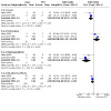
Comparison 5: Laser versus photodynamic therapy (PDT), Outcome 2: Adverse effects
Quality of life (QOL)
Neither study reported this outcome, but one study (Heier 1995) evaluated and reported the Karnofsky performance status (KPS) and dietary performance. The mean change (SD) in KPS at one month was 7.2 (14.5) in the PDT group compared to ‐7.2 (14.3) in the laser group. This was statistically significant (P = 0.001). The mean change in dietary performance at one month was reported to be 1.8 (1.2) compared to 1 (1.5) in the laser group. This difference was statistically significant (P = 0.006).
In summary, from the above analysis we found no evidence to suggest any difference between PDT and laser treatment in improving dysphagia and procedure related or 30‐day mortality. There was equivocal evidence to suggest that PDT decreased the need for repeated endoscopic interventions or improved QOL and dietary performance compared to laser treatment. Although the overall adverse effects were no different, it was apparent that certain adverse effects were more common in patients treated with either procedure.
7. Laser versus chemical ablation
Two RCTs included 84 patients and compared laser treatment with chemical ablation treatment. Angelini 1991 performed chemical ablation with 3% polidocanol and Carrazone 1999 performed chemical ablation with 98% ethanol. One study (Angelini 1991) described the primary outcome for adenocarcinoma and squamous cell carcinoma separately and also concluded that the length and location of the tumour did not influence the results.
Primary outcome
Angelini 1991 reported that at least grade 1 dysphagia was achieved in 16 /18 patients in the laser group compared to 13/16 patients in the polidocanol group. The difference was not statistically significant.
Carrazone 1999 reported a 2‐point improvement in the grade of dysphagia in 21/24 patients in the laser group compared to 18/21 in the ethanol group. The difference was not statistically significant.
Secondary outcomes
Overall survival
In Angelini 1991 4 out of 18 patients were alive at 6 months in the laser group compared to 5/16 in the polidocanol group. Carrazone 1999 reported death in 15/24 patients over a mean interval of 6 months (2 to 15) in the laser group compared to 18/23 in the ethanol group (mean interval 6 (2 to 30)).
Persistent or recurrent dysphagia
In Angelini 1991 12/18 patients had persistent or recurrent dysphagia. Ten patients required 47 sessions of further laser therapy compared to 10 sessions in 2/16 patients receiving polidocanol injections. Three patients were lost to follow up and no data were presented for these patients.
In Carrazone 1999 patients treated with laser had a mean dysphagia‐free survival of 30 days. Three out of 24 patients needed stent insertion for persistent dysphagia. In the ethanol group, the mean dysphagia‐free interval was 37 days and 5 out of 23 patients needed a stent insertion.
Technical success
All procedures in the 34 patients were successful in the Angelini study. Technical success was not reported in Carrazone 1999.
Procedure related mortality
No procedure related mortality was observed in the 84 patients treated in both studies.
Thirty‐day mortality
Neither study reported this outcome.
Adverse effects
In Angelini 1991 7/18 patients had mild pain compared to 1/16 in the polidocanol group. One patient had a fistula in the polidocanol group.
In Carrazone 1999 1 perforation was noted and 18/23 (78%) patients had mild pain in the ethanol group. No adverse effect was noted in the laser group and the authors reported a significantly improved compliance in this group.
Quality of life (QOL)
Neither study reported this outcome.
In summary, from the above analyses we have reported evidence that chemical ablation was as effective as laser treatment in improving dysphagia. However, the occurrence of pain in the vast majority of patients receiving ethanol injection might preclude its widespread use.
8. Covered Ultrafelx SEMS versus covered Wallstent
Two studies (Sabharwal 2003; Siersema 2001) included 153 patients comparing different commercially available covered stents. Sabharwal 2003 compared Ultraflex and Flamingo Wallstent. Siersema 2001 compared Ultraflex, Flamingo and Gianturco Z‐stents. The Siersema study reported the primary outcome for oesophageal and cardia cancers separately. Quantitative analysis could be undertaken only for the comparison of the former two stent, with 120 patients in the comparison of covered Ultraflex and covered Flamingo Wallstents.
Primary outcome
Both studies (Sabharwal 2003; Siersema 2001) reported a significant improvement in the dysphagia grade with the Ultraflex and Flamingo Wallstent arms. However, the pooled weighted mean difference (WMD) was not significant for the two stent groups (n = 120) (WMD 0.15, 95 CI ‐0.04 to 0.33, Analysis 6.1).
6.1. Analysis.

Comparison 6: Covered Ultraflex SEMS versus covered Wallstent, Outcome 1: Dysphagia improvement
Secondary outcomes
Overall survival
In Siersema 2001, median survival in the Ultraflex group was 104 days compared to 113 days in the Wallstent group. Sabharwal 2003 did not report on overall survival data.
Persistent or recurrent dysphagia
Recurrent dysphagia was reported in 13 out of 65 patients in the Ultraflex group compared to 10 out of 55 patients in the Wallstent group. The pooled OR was 1.27 (95% CI 0.49 to 3.31) and was not statistically significant (P = 0.62). No statistical heterogeneity was noted between the studies (Analysis 6.2).
6.2. Analysis.

Comparison 6: Covered Ultraflex SEMS versus covered Wallstent, Outcome 2: Persistent or recurrent dysphagia
Technical success
All but one Ultraflex stent were successfully deployed in Siersema 2001 (n = 67). All stents were deployed successfully in Sabharwal 2003 (n = 53) (Analysis 6.3).
6.3. Analysis.

Comparison 6: Covered Ultraflex SEMS versus covered Wallstent, Outcome 3: Technical success
Procedure related mortality
One death was related to Ultraflex stent insertion in the Siersema study. No procedure related mortality was noted in the Sabharwal study.
Thirty‐day mortality
Eleven patients of 65 died within 30 days in the Ultraflex group compared to 8 out of 55 in the Wallstent group. The pooled OR was not significant for this outcome (OR 1.18, 95% CI 0.44 to 3.18, P = 0.74). No statistical heterogeneity was noted between the studies.
Adverse effects
Twenty‐eight patients had adverse events out of 65 in the Ultraflex group compared to 31 out of 55 in the Wallstent group. The pooled OR was 0.61 (95% CI 0.27 to 1.38, P = 0.23) and was not statistically significant. No statistical heterogeneity was noted between the studies. However, the proportion of adverse effects in Siersema 2001 was mainly because of the meticulous presentation of minor side effects, such as chest pain and fever.
There was no significant difference in the occurrence of major side effects including perforation, haemorrhage, migration and food bolus obstruction between the stent groups. In Siersema 2001, migration was more common mainly in the Ultraflex group although this did not reach statistical significance and the authors noted that eight of the nine migrations involved smaller diameter stents.
Siersema 2001 also included a group of 33 patients with Gianturco Z‐stents in their study, which was not used for the qualitative analysis. The dysphagia palliation and overall survival were similar to the Ultraflex and Wallstents. However, major and minor complications were more frequent in the Z‐stent group although this did not reach statistical significance. The authors also reported that recurrent dysphagia was not influenced by age, gender, tumour length or, more importantly, prior radiation or chemotherapy.
Quality of life (QOL)
Siersema 2001 used World Health Organization (WHO) performance grading to evaluate QOL and found this to be similar in both stent groups at zero and four weeks. Sabharwal 2003 did not report this outcome.
In summary, from the above analyses, there was no evidence to support any difference in dysphagia palliation or incidence of adverse effects between the covered Ultraflex or covered Flamingo Wallstent and it appeared that the Gianturco Z‐stent was comparable to the other two.
9. Comparisons of different types of SEMS
Seven randomised studies (Homs 2004c; Sabharwal 2003; Shim 2005; Siersema 2001; Vakil 2001; Wenger 2006; Wenger 2010) compared different types of SEMS. Two studies (Sabharwal 2003; Siersema 2001) included 153 patients comparing different commercially available covered stents. Sabharwal et al compared the Ultraflex and Flamingo Wallstent. Siersema et al (Siersema 2001) compared Ultraflex, Flamingo and Gianturco Z stents. Quantitative analysis could be undertaken only for the comparison of the former two stents from these two studies. Four studies (Homs 2004c; Shim 2005; Wenger 2006; Wenger 2010) compared open stents and anti‐reflux stents. One study (Vakil 2001) compared uncovered and covered stents.
Covered Ultraflex versus covered Wallstent SEMS
One RCT (Vakil 2001) included 62 patients, 32 of them randomised to covered stents and 30 patients to uncovered stents.
Primary outcome
Interventions for recurrent dysphagia and migration of stents were the primary outcomes in this trial. Nine (27%) re‐interventions were performed in patients with covered stents compared to 23 (77%) in patients with an uncovered stent. This was statistically significant (P < 0.001).
Secondary outcomes
Overall survival
A survival plot was graphically reported for this outcome. The authors reported no significant difference in survival between the two stent groups (P = 0.378, log rank test).
Dysphagia improvement
At one week after stent insertion, the mean (SD) dysphagia score in the covered stent group improved from 3 (0.1) to 1 (0.2) compared to 3 (0.1) to 1 (0.1) in the uncovered stent group. This result was not statistically significant between the two groups. Three months after initial stenting, dysphagia scores were higher in the uncovered stent group but this did not achieve statistical significance.
Technical success
Twenty‐nine (91%) patients in the covered stent group and 30 (100%) in the uncovered stent group had successful stent insertion. The difference was not statistically significant.
Procedure related mortality
There was no procedure related mortality in this study.
Thirty‐day mortality
This outcome was not reported in this study.
Adverse effects
The authors (Vakil 2001) reported early and late complications but did not define the time scales for these outcomes. Overall, 24 early complications and 22 late complications were noted in the covered stent group and 18 early complications and 25 late complications in the uncovered group. The difference was not statistically significant. Tumour ingrowth occurred in one patient in the covered stent group compared to nine in the uncovered stent group. This difference was statistically significant (P = 0.005). Four of the covered stents migrated compared to two of the uncovered stents but this difference was not statistically significant.
Quality of life (QOL)
This outcome was not assessed in this study.
In summary, covered metallic stents improved dysphagia effectively and rapidly with reduced requirement for repeat intervention for recurrent dysphagia compared to uncovered metallic stents.
Irradiation stent versus covered stent
One RCT (Guo 2008) included 53 patients, 27 of them randomised to irradiation stents and 26 patients to covered stents.
Primary outcome
At one month after stent insertion, the mean (SD) dysphagia score in the covered stent group improved from 3.12 ± 0.326 to 1.17 ± 0.38 compared to 3.22 ± 0.424 to 1.22 ± 0.42 in the irradiation stent group. This result was not statistically significant. One month after initial stenting, dysphagia scores increased in both groups but more substantially in the control group than in the irradiation stent group. After two months, there was a significant difference (P = 0.05).
Secondary outcomes
Overall survival
The median survival in the irradiation stent group was 7 months (95% CI 5.0 to 10.0), with a mean of 8.3 months (95% CI 6.36 to 10.21), versus a median survival in the control group of 4 months (95% CI 2.0 to 4.0), with a mean of 3.5 months (95% CI 2.72 to 4.16). The differences between both measures of survival in the two groups were significant (P < 0.001, log‐rank test).
Persistent or recurrent dysphagia
No recurrent dysphagia was noted in the study.
Technical success
Stent insertion was performed successfully in all patients.
Procedure related mortality
This outcome was not reported in the study.
Thirty‐day mortality
This outcome was not reported in the study.
Adverse effects
Authors (Guo 2008) reported side effects and complications including dull chest, haemorrhage, tracheoesophageal fistula and partial stent migration. There were 15 patients (8 in the irradiation stent group and 7 in the control group) complaining of severe chest pain, but the degree of chest pain in the two groups was not significantly different.
Haemorrhage occurred in 16 patients (9 in the irradiation group and 7 in the control group) during follow up. No significant difference was found in the incidence of haemorrhage between the two groups.
A tracheoesophageal fistula occurred in one patient in the irradiation group. There was no complete stent migration but partial stent migration was found in five patients (two in the irradiation group and three in the control group) at one month after stent insertion.
Quality of life (QOL)
No details about QOL were reported in the study.
In summary, compared to conventional covered stents, stents loaded with 125I had potential benefit in that they provided slightly longer relief of dysphagia and extended survival.
Ultraflex stent versus Polyflex stent versus Niti‐S stent
One RCT (Verschuur 2008) included 125 patients, 42 of them were randomised to the Ultraflex stent group, 41 to the Polyflex stent group, and 42 to the Niti‐S stent group. As no further detail could be obtained the study was placed under 'Comparisons of different types of SEMS'.
Primary outcome
At four weeks after stent placement the dysphagia score had improved from a median of 3 to 0 in the Ultraflex stent group, 3 to 1 in the Polyflex stent group, and 3 to 0 in the Niti‐S stent group; and no significant difference was found in the degree of improvement among the three groups over four weeks (P = 0.22).
Secondary outcomes
Overall survival
Median survival was 132 days in patients with Ultraflex stents, 102 days in those with Polyflex stents, and 159 days in those with Niti‐S stents (P = 0.13). Most patients died from tumour progression, while three patients, two with a Polyflex stent and one with an Ultraflex stent, died from stent related complications.
Persistent or recurrent dysphagia
At a median of 79 days after stent placement, recurrent dysphagia occurred in 22/42 (52%) patients with an Ultraflex stent, 15/41 (37%) with a Polyflex stent, and 17/42 (31%) with a Niti‐S stent. The difference was of statistical significance (P = 0.03). Recurrent dysphagia was caused by tissue ingrowth or overgrowth, stent migration, or food obstruction.
Technical success
Stent placement was technically successful in 42/42 (100%) patients with an Ultraflex stent, in 34/41 (83%) patients with a Polyflex stent, and in 40/42 (95%) patients with a Niti‐S stent (P = 0.008). The reasons for technical failures were that they were too proximal (Polyflex stent N = 4) or too distal (Polyflex stent N = 3, Niti‐S stent N = 2). In six patients the stent was successfully repositioned with grasping forceps. In two patients the Polyflex stent was again loaded in the introducer sheath and placed successfully, while another patient was randomised to another type of Polyflex stent.
Procedure related mortality
This outcome was not reported in the study.
Thirty‐day mortality
A total of 2/42 (5%) patients with an Ultraflex stent died in 30 days, and the 30‐day mortality for the Polyflex stent group and Niti‐S stent group was 17% and 5% respectively (P = 0.07).
Adverse effects
Complications occurred in 9 (21%) patients with Ultraflex stents, in 10 (24%) with Polyflex stents, and in 9 (21%) with Niti‐S stents (P = 0.89). Perforations were observed in two patients with Polyflex stents, and one of them died from septic complications. Haemorrhage occurred in five patients with Ultraflex stents and five with Polyflex stents; nobody died as a consequence of haemorrhage.
Stent migration occurred more frequently in patients with Polyflex stents (12/41, 29%) compared to Ultraflex stents (7/42,17%) and Niti‐S stents (5/42,12%), and was mainly treated with a second stent or repositioning of the stent.
Quality of life (QOL)
At four weeks after stent placement, the median WHO performance scores remained the same as those before treatment, and no differences in WHO performance score were observed (P = 0.31). Following stent placement, 24/125 (19%) patients, most of whom had Niti‐S stents (N = 15) or Ultraflex stents (N = 7), received six courses of additional palliative chemotherapy. After that the tumour was considered to be resectable in five patients. The surgery with curative intent was performed.
In summary, all three stents offered adequate palliation of dysphagia, but Polyflex stents seemed to be the least preferable as placement of the stent was technically demanding and associated with a high rate of stent migrations and haemorrhage.
Different types of Niti‐S stents
One RCT (Kim 2009) included 37 patients; 19 of them were assigned to the covered Niti‐S stent group, and 18 to the double‐layered Niti‐S stent group.
Primary outcome
The mean dysphagia score improved from 2.95 ± 0.52 to 1.00 ± 0.47 (P < 0.001) in the covered group, and from 2.88 ± 0.33 to 1.06 ± 0.24 (P < 0.001) in the double‐layered group at one week after stent insertion. At one month, the mean dysphasia score improved to 1.18 ± 0.64 and 1.08 ± 0.49 in the covered and double‐layered groups respectively (P < 0.001), compared to the baseline data. However, the degree of improvement was not different between the groups (P = 0.365).
Secondary outcomes
Overall survival
The median survival was 62 days in patients with covered stents and 74 days in those with double‐layered stents. There was no difference in overall survival between the groups.
Persistent or recurrent dysphagia
No recurrent dysphagia was noted in the study.
Technical success
Except in one patient with a double‐layered stent, all the stents were successfully placed.
Procedure related mortality
This outcome was not reported in the study.
Thirty‐day mortality
This outcome was not reported in the study.
Adverse effects
Complications occurred more frequently in the covered stent group (11/19, 58%) than in the double‐layered stent group (2/17, 12%) (P = 0.006), such as tumour overgrowth, stent migration, gastro‐oesophageal reflux and haemorrhage. Based on adjustment for age, location of tumour, tumour length, baseline dysphagia score, and previous radiotherapy or chemotherapy, the differences remained significant (adjusted OR 18.2, 95% CI 1.9 to 171.4, P = 0.01).
Quality of life
This outcome was not reported in the study.
In summary, Niti‐S stents were newly‐developed self‐expanding metal stents. The two types (covered and double‐layered stents) were both effective for malignant dysphagia. Nonetheless, double‐layered Niti‐S stents were preferable due to longer survival time and fewer complications.
10. Other studies comparing SEMS to various modalities
Seven RCTs (Canto 2002; Fu 2004; Horneaux 2001; Javed 2012; Konigsrainer 2000; Shenfine 2005; Turrisi 2002) compared SEMS to various modalities, head‐to‐head or in combination.
Canto et al (Canto 2002) randomised 56 patients to either SEMS insertion or PDT. Dysphagia improved significantly for both groups but more patients in the SEMS group had normal swallowing at three weeks (P = 0.03). However, at three months the dysphagia grade was similar in both groups. Patients treated with PDT underwent re‐intervention more frequently (P = 0.04). QOL decreased significantly in the stent group at three weeks (P = 0.01) but not in the PDT group.
Fu 2004 randomised 53 patients with inoperable oesophageal carcinoma to either SEMS insertion alone (n = 27) or to SEMS insertion followed by chemotherapy or chemoradiotherapy (n = 26). The median dysphagia score improved from 3 to 0 in both groups immediately after the intervention. The overall survival at six months was 52% in both groups and 19% at one year in the SEMS only group compared to 22% in the combination treatment group. This difference was not statistically significant. Sixteen patients in the SEMS only group had complications compared to 14 in the combination group. This difference was not statistically significant. However, more SEMS only patients encountered tumour regrowth: nine (30%) compared to one (4%) in those receiving chemotherapy or chemoradiotherapy after SEMS insertion (P = 0.007). Other secondary outcomes were not reported in this study.
Horneaux 2001 randomised 40 patients with advanced squamous carcinoma to either SEMS insertion (n = 20) or oesophageal bypass surgery using the Posthelwaite technique (n = 20). The mean dysphagia score (SD) decreased significantly in both groups following the intervention. In the SEMS group dysphagia improved from 3.3 (0.9) to 1.2 (0.8) at one month, and 1.7 (0.5) at four months (P < 0.001). In the surgical group, dysphagia improved from 2.5 (1.2) to 1.4 (1) at one month, and 1.1 (0.9) at four months (P = 0.002). The difference in improvement between the groups was not statistically significant. The median Karnofsky performance status improved from 50 to 75 in the SEMS group compared to the surgical group (68 to 70). No procedure related mortality was noted in either group. The complications rate, QOL and overall survival were similar between the groups. The median hospital stay in the surgical group was 15.5 days compared to three days in the SEMS group. This difference was statistically significant (P < 0.001).
Konigsrainer 2000 randomised 39 patients with inoperable advanced oesophageal carcinoma to either laser treatment and radiotherapy (n = 21), laser therapy followed by SEMS insertion (n = 8) or SEMS insertion alone (n = 10). The mean dysphagia scores (SD) at the end of treatment were 0.48 (0.12) in the laser and radiotherapy group compared to 0.40 (0.13) in the SEMS only group. This difference was not statistically significant. Recurrent dysphagia occurred in nine (43%) patients in the laser and radiotherapy group compared to 3 (16.6) in the SEMS plus laser group and one (5.5%) in the SEMS only group. This difference was statistically significant (P = 0.001). Mean survival was comparable between the two groups. However, the mean stay in hospital was 30 (5.4) days in the laser and radiotherapy group compared to 18.9 (4.2) in the laser plus SEMS group and 7.1 (3.1) in the SEMS only group. This difference was statistically significant (P = 0.001). No complications were seen in the SEMS groups compared to four (20%) in the laser plus radiotherapy group.
In a multicentre crossover RCT, Turrisi et al (Turrisi 2002) randomised 32 patients to either SEMS insertion or external beam radiotherapy. The median dysphagia‐free survival in the stent group was 32 days compared to four days in the radiotherapy group. This difference was not statistically significant. However, median overall survival in the radiotherapy group was longer (141 days, 95% CI 188 to 209) compared to the SEMS group (101 days, 95% CI 146 to 127). Further details of this study were not available at the time of this analysis. Communication with the authors is ongoing and further details will be added at the next update of this review.
Javed 2012 randomised 37 patients to the SEMS group and 42 to SEMS plus external beam radiation therapy (EBRT). The mean dysphagia scores at baseline were 3.22 ± 0.48 in SEMS group and 3.10 ± 0.3 in the SEMS plus EBRT group. The dysphagia improvement was significantly different after stent placement (P < 0.001). Until three months after treatment the dysphagia score was significantly lower in the SEMS plus EBRT group (P < 0.002), and then the dysphagia scores remained comparable between groups. The median survival was 120 days in the SEMS group and 180 days in the SEMS plus EBRT group (P = 0.009). The authors also reported a mean dysphagia‐free survival of 96.8 ± 43 days in the SEMS group, and 118.6 ± 55.8 days in the SEMS plus EBRT group (P = 0.054). Major complications occurred in 27 patients (35%). The authors did not report on how many minor complications there were, but no significant difference was found in the incidence of major and minor complications between the two groups (P = 0.26). Javed 2012 also compared the QOL in the two groups at baseline, one week after stenting, and one week after completion of EBRT. Significant improvements were found in all QOL indices in both groups at one week. However, patients in the SEMS plus EBRT group showed significant worsening in all QOL parameters except physical functioning at one week after EBRT.
In summary, SEMS insertion was effective, safe and swift for palliating dysphagia compared to other modalities and avoided delays in effectively treating these patients. However, from the above analysis there appeared to be some evidence that other modalities including brachytherapy and external beam radiotherapy might provide a survival advantage and possibly a better QOL compared to SEMS treatment.
11. Anti‐reflux versus standard open stent
Six studies (Homs 2004c; Power 2007; Sabharwal 2008; Shim 2005; Wenger 2006; Wenger 2010) compared open and anti‐reflux stents in 276 patients with inoperable lower oesophageal or gastro‐oesophageal junction tumours. Due to the difference in the reporting of the primary and secondary outcomes amongst the trials, quantitative analysis was not undertaken and the results of the studies were described separately.
Primary outcome
Homs et al (Homs 2004c) randomised 15 patients to open stents and 15 patients to anti‐reflux stents. Only 12 patients underwent 24‐hour pH monitoring, nine in the anti‐reflux stent group and three in the open stent group. The median (range) total reflux time was 23% (0% to 65%) in the anti‐reflux stent group compared to 10% (0.1% to 19%) in the open stent group, and this difference was not statistically significant. The median (interquartile range) number of reflux episodes longer than five minutes was 14 (2 to 19) in the anti‐reflux stent group compared to 5 (0 to 10) in the open stent group. This result was not statistically significant. The authors reported indigestion scores assessed using the EORTC OES‐23 questionnaire at 14 days. The median (interquartile range) indigestion score (0 (best) to 100 (worst)) in the anti‐reflux stent group was 22 (11 to 33) compared to 11 (11 to 28) in the open stent group. This result was not statistically significant.
Shim 2005 randomised 12 patients to an open stent, 12 patients to the DO anti‐reflux stent and 12 patients to the newly designed S‐type anti‐reflux stent. The mean (SD) score in the open stent group improved from 3.25 (0.45) at the baseline to 1 (0.6) post‐procedure (P = 0.002). In the DO stent group, dysphagia improved from 3.08 (0.69) to 1.08 (0.69) after the stent insertion (P = 0.001). In the newly designed S‐type anti‐reflux stent group dysphagia improved from 2.83 (0.58) to 0.91 (0.51) post‐procedure (P = 0.001). However, the differences in improvement amongst the three stent groups was not statistically significant.
Shim 2005 assessed the reflux symptom score in their patients using a simple Likert scale for heartburn, acid reflux, chest pain, foreign body sensation and hoarseness (scored 0 (best) to 4 (worst)). The mean (SD) reflux symptom score in the open stent group was 4.42 (3.4) at baseline and increased to 6.25 (2.7) after stent insertion. This was statistically significant (P = 0.049). In the DO stent group, the mean (SD) reflux symptom score at baseline was 5.58 (3.4) and increased to 5.75 (6.15). This result was not statistically significant (P = 0.798). In the newly designed S‐type stent group, the baseline mean (SD) reflux symptom score was 5.42 (3.4) and this decreased to 2.5 (1.78) after stent insertion, which was statistically significant (P = 0.005). The difference in dysphagia improvement was superior to both the open stent and the DO stent (P = 0.005). On 24‐hour pH monitoring, the DeMeester score in the open stent group was 60.44 (48.66), 105.29 (51.96) in the DO stent group, and 12 (21.51) in the newly‐designed S‐type anti‐reflux stent group. The difference between the‐newly designed S‐type stent group and the other two stent groups was statistically significant in favour of the newly‐designed S‐type stents (P < 0.001). Other outcomes during the 24‐hour pH monitoring, including total number of reflux episodes, longest duration of reflux and total time with pH < 4, were all less in the newly‐designed S‐type stent group compared to the open stent group and the DO stent group. There was no significant difference between the open stent group and the DO stent group.
Power 2007 randomised 24 patients to anti‐reflux stents and 25 to open stents. According to the EORTC QLQ‐C30, both stents brought significant improvement in patients’ HRQoL and their perception of their health status. Similar findings were presented with the QLQ‐OES 24 cumulative score and patients' dysphagia situation. Two months later the two stents were still associated with a statistically significant benefit. At two months the standard stent provided better passage for soft food; however, patients did not attain any benefit from either stent with regard to the passage of liquid or saliva.
Sabharwal 2008 randomised 22 patients to anti‐reflux stents and 26 to a combination of a standard open stent and omerprazole. Compared with the baseline score, both groups had a significant improvement in their dysphagia score on the day following the stent placement (P < 0.05) and at later follow up (P < 0.05). There was no difference in the degree of dysphagia improvement between the two groups (P = 0.62).
Wenger 2006 randomised 19 patients to anti‐reflux stents and 22 patients to open stent insertion; Wenger 2010 randomised 28 patients to anti‐reflux stents and 37 patients to open stent insertion. The primary outcome of the two studies was the assessment of global QOL and specific symptoms using the EORTC QLQ‐30 and EORTC QLQ‐OES 18 validated questionnaires. Wenger 2006 reported the mean dysphagia score as 51 ± 32 in the anti‐reflux stent group and 36 ± 21 in the standard open stent group at one month after stent insertion; Wenger 2010 reported 56 ± 30 and 45 ± 22 respectively. Higher scores in the symptom scales from the EORTC QLQ‐OES 18 questionnaire represented more severe symptoms, with the full score of 100. The two studies (Wenger 2006; Wenger 2010) could be included in a quantitative analysis for dysphagia improvement. In a total of 106 patients, the SMD in the dysphagia score at one month after stent insertion was 0.47 (95% CI 0.08 to 0.86, P = 0.02, Analysis 8.1) favouring the standard open stent. No statistical heterogeneity between the two trials was found.
8.1. Analysis.

Comparison 8: Anti‐reflux versus standard open stent, Outcome 1: Dysphagia improvement
Secondary outcomes
In one study (Homs 2004c), the median (range) dysphagia score improved from 3 (3 to 3) at baseline to 1 (0 to 2) at day 14 in the anti‐reflux stent group (P = 0.002). In the open stent group the median (range) dysphagia grade improved from 3 (3 to 4) to 0 (0 to 2.5) (P = 0.005). The difference in improvement between the two stent groups was not statistically significant.
Overall survival
In the Homs study (Homs 2004c) the median overall survival was 107 days (95% CI 11 to 203) in the anti‐reflux stent group compared to 87 days (95% CI 58 to 116) in the open stent group. This result was not statistically significant. Shim 2005 reported a median survival period of 114 days in the open stent group, 107 days in the DO stent group, and 109 days in the newly‐designed S‐type stent group. The difference between the stent groups was not statistically significant. Wenger 2006 reported a median (range) survival of 58 days (9 to 154) in the anti‐reflux stent group compared to 68 days (4 to 511) in the open stent group. This difference was not statistically significant. Sabharwal 2008 did not report the exact survival in days, but the log‐rank test showed no statistically significance between the two groups and the overall survival was quite similar. Wenger 2006 reported complications in eight patients in the anti‐reflux stent group compared to 12 in the open stent group. This result was not statistically significant. Wenger 2010 reported a median survival of 63 days in the anti‐reflux stent group and 70 days in the open stent group (P = 0.75).
Persistent or recurrent dysphagia
Only one study reported this outcome. In the Homs study (Homs 2004c), recurrent dysphagia occurred in six (40%) patients with anti‐reflux stents compared to two (13%) patients in the open stent group. This result was not statistically significant.
Technical success
In the Homs study (Homs 2004c) 13 (87%) of the anti‐reflux stent insertions were successful compared to 14 (93%) of the open stent insertions. This result was not statistically significant. Power 2007; Sabharwal 2008; Shim 2005; Wenger 2006; and Wenger 2010 reported technical success in all patients.
Procedure related mortality
No procedure related mortality was noted in any of the studies.
Thirty‐day mortality
Shim 2005 observed an 8% 30‐day mortality in the open stent group, 9% mortality in the DO stent group, and 12% in the newly‐designed S‐type anti‐reflux stent group. The differences between the three groups were not statistically significant. Sabharwal 2008 reported an 18% 30‐day mortality in the FerX Ella group and 12% in the Ultraflex‐omeprazole group. No significant difference was found (P > 0.1).
Adverse effects
Homs et al (Homs 2004c) reported early (< 7 days) and late (> 7 days) major complications and minor complications separately. Overall seven (47%) patients had complications in the anti‐reflux stent group and five (33%) had complications in the open stent group. In the anti‐reflux stent group, one (7%) patient had severe pain, two (13%) patients had haemorrhage, three (20%) patients had gastro‐oesophageal reflux and one (7%) had mild retrosternal pain. In the open stent group, one (7%) patient had severe pain, one (7%) patient had haemorrhage, two (13%) patients had gastro‐oesophageal reflux and one (7%) patient had aspiration pneumonia. Five (33%) anti‐reflux stents migrated compared to two (13%) open stents. These results were not statistically significant. Shim 2005 observed no detectable differences with regard to complications or need for repeat interventions, but did not report these outcomes in detail. Sabharwal 2008 reported different types of complications: 3/22 (13.6%) patients with FerX Ella stents complained of reflux compared to 2/26 (7.7%) in those with Ultraflex stents (P = 0.649). There were 2/22 (9.1%) with significant pains in the FerX Ella stent group and 9/26 (34.6%) in the Ultraflex group (P = 0.036), 2/22 (9.1%) obstructions in the FerX Ella group and 7/26 (26.9%) in the Ultraflex group (P = 0.151). Power 2007 reported, at a median follow up of 10 months, that there were 2/25 (8%) complications in the anti‐reflux stent group and 2/24 (8.3%) in the standard stent group, such as severe pain and food bolus obstruction. No difference was found to be significant.
Wenger 2006 and Wenger 2010 reported 45 complications in total. The OR for all adverse effects was 0.86 (95% CI 0.38 to 1.94, P = 0.72, Analysis 8.5). With regard to the types of complications, authors reported stent migration (OR 0.68, 95% CI 0.19 to 2.50, P = 0.56, Analysis 8.6), stent occlusion (OR 1.10, 95% CI 0.35 to 3.49, P = 0.87, Analysis 8.6), bleeding (OR 1.93, 95% CI 0.31 to 11.98, P = 0.48, Analysis 8.6), oesophageal perforation (OR 0.40, 95% CI 0.04 to 3.94, P = 0.43, Analysis 8.6) and gastric perforation (OR 0.40, 95% CI 0.04 to 3.94, P = 0.43, Analysis 8.6). No statistical heterogeneity between the two studies was found. Anti‐reflux stents had an advantage over standard open stents in causing stent migration and perforation.
8.5. Analysis.

Comparison 8: Anti‐reflux versus standard open stent, Outcome 5: All adverse effects
8.6. Analysis.

Comparison 8: Anti‐reflux versus standard open stent, Outcome 6: Adverse effects
Quality of life (QOL)
Wenger 2006 and Wenger 2010 assessed the QOL with EORTC QLQ‐30 and EORTC QLQ‐EOS 18 questionnaires. The authors reported comparable QOL scores in the anti‐reflux and open stent groups. The SMD was ‐0.04 (95% CI ‐0.42 to 0.35, P = 0.85, Analysis 8.2) and there was no statistical heterogeneity. The SMDs for the reflux score and dyspnoea score were 0.36 (95% CI ‐0.02 to 0.75, P = 0.07, Analysis 8.3) and ‐0.62 (95% CI ‐1.02 to ‐0.23, P = 0.002, Analysis 8.4) respectively. No statistical heterogeneity was found. Therefore, there was no difference between the two treatments in improving QOL and reflux, but the anti‐reflux stent had an advantage over the standard open stent in decreasing dyspnoea.
8.2. Analysis.

Comparison 8: Anti‐reflux versus standard open stent, Outcome 2: Quality of life
8.3. Analysis.

Comparison 8: Anti‐reflux versus standard open stent, Outcome 3: Reflux score
8.4. Analysis.

Comparison 8: Anti‐reflux versus standard open stent, Outcome 4: Dyspnea score
According to Power 2007, at one week patients with the anti‐reflux stent reported fewer supine gastro‐oesophageal reflux (GER) and heartburn symptoms. The difference was found to be significant. At two months the heartburn severity and modified DeMeester symptom score remained significantly less in the anti‐reflux stent group (P < 0.01). The authors also measured 24‐hour pH. The acid reflux score in the anti‐reflux stent group was significantly lower than that in the standard stent group. Moreover, the percentage of recording time, time in the upright position, and time in the supine position when the oesophageal pH was < 4 was significantly less in the anti‐reflux stent group.
In summary, a variety of anti‐reflux stents were available and were effective in rapidly palliating dysphagia with comparable complication rates and QOL to conventional SEMSs. Although some of the stents appeared effective in reducing GER, further research is required to confirm this favourable outcome.
12. Other studies including multiple comparisons or plastic stents
Nine RCTs (Amdal 2013; Anghorn 1983; Barr 1990; Mannell 1986; Mehta 2008; Reed 1991; Rosenblatt 2010; Rupinski 2011; Sur 2004) included comparisons of plastic tube insertion, external beam radiotherapy, brachytherapy, laser or chemotherapy and dilatation, head‐to‐head or in various combinations.
Barr 1990 randomised 20 patients to laser therapy only and 20 patients to laser therapy followed by plastic tube intubation. The mean (SD) dysphagia grade in the laser group during follow up was 1.6 (1) compared to 1.7 (1) in the laser plus intubation group. This difference was not statistically significant. Recurrent dysphagia occurred in five (25%) patients in the laser group compared to nine (45%) patients in the combination group. Mean overall survival was 18.3 weeks in the laser only group compared to 16.1 in the combination group. These differences were not statistically significant. Overall complications occurred in two (10%) of the laser only group compared to eight (40%) in the laser plus intubation group. This was statistically significant (P < 0.05). The QL index and LASA scores improved significantly in both groups but no significant difference was noted between the two groups.
Mannell 1986 compared dilatation plus bleomycin to plastic tube insertion. On discharge from hospital, 80% of patients in the dilatation plus bleomycin group had improved eating soft foods compared to 52% in the intubation group. Procedure mortality for intubation was 18% compared to 6% for the dilatation and bleomycin treatment. These differences were statistically significant. The overall complication rate was 28 (40%) in the intubation group compared to seven (9%) in the bleomycin and dilatation group. This difference was statistically significant (P < 0.001). The mean length (SD) of palliation was 61.59 days (46.19) in the intubation group compared to 83.35 days (76.37) in the bleomycin and dilatation group.
Reed 1991 randomised 10 patients to plastic tube insertion, eight patients to plastic tube insertion plus radiotherapy, and nine patients to laser therapy plus radiotherapy. The mean improvement in dysphagia post‐treatment was 2.3 (1.1) in the plastic tube only group, 1.8 (1) in the plastic tube and radiotherapy group, and 1.4 (0.5) in the laser and radiotherapy group. These differences were not statistically significant. Complications occurred in eight (100%) of the plastic tube and radiotherapy group compared to five (50%) of the plastic tube only group and none in the laser and radiotherapy group. This difference between the laser group and plastic tube only group was statistically significant (P < 0.02).
The overall survival was similar in the three groups.
Anghorn 1983 allocated 106 patients to either oesophageal bypass surgery or plastic tube insertion in a single centre study from South Africa. Fifty‐seven (93%) showed dysphagia improvement in the plastic tube group compared to 47 (92%) in the surgical group. This difference was not statistically significant. Procedure mortality was three (5.5%) in the plastic tube group compared to four (7.8%) in the surgical group. Complications occurred in 18 (34%) patients in the plastic tube group compared to 31 (60%) patients in the surgical group.
Rupinski 2011 randomised 27 patients to high‐dose rate brachytherapy (HDR) plus argon plasma coagulation (APC), 26 to photodynamic therapy (PDT) plus APC, and 27 to APC alone. The dysphagia grades changed from 2.81 ± 0.56 before APC treatment to 0.41 ± 0.50 in the HDR plus APC group, from 2.69 ± 0.55 to 0.38 ± 0.50 in the PDT plus APC group, and from 2.67 ± 0.62 to 0.44 ± 0.51 in the APC group; no significant differences were found between the groups. The median dysphagia‐free period was 88 days in the HDR plus APC group, 59 days in the PDT plus APC group, and 35 days in the APC group (P = 0.006).
In one month there were four deaths, one from the HDR plus APC group, one from the PDT plus APC group, and two from the APC group. No difference was found to be significant. The median survival was 6.2 months (4.4 to 9.9) in the HDR plus APC group, 5.2 months (4.4 to 9.9) in the PDT plus APC group, and 6.0 months (2.0 to 9.2) in the APC group (P = 0.27).
At the 30‐day follow up after completing treatment the QOL (Spitzer Quality of Life Index (SQLI)) had declined in all three groups compared to immediately after recanalisation. Nonetheless, the SQLI in the HDR plus APC group was significantly higher than that in the APC group (P = 0.0067) or PDT plus APC group (P = 0.022). The only major complication was fever, which occurred in three patients in the HDR plus APC group. Minor complications were observed significantly more frequently in the PDT plus APC group compared to the APC group (P < 0.001), such as worsening dysphagia, pain and skin sensitivity.
One RCT (Amdal 2013) allocated 21 patients to an experimental stent with brachytherapy and 20 to brachytherapy. People with SEMS and brachytherapy had significantly improved dysphagia at the first follow up and there was no difference in pain. At the second follow up patients in both groups had less dysphagia and there was no statistical difference.
Three RCTs (Mehta 2008; Rosenblatt 2010; Sur 2004) compared brachytherapy with external beam radiotherapy (EBRT). The two studies (Rosenblatt 2010; Sur 2004) reported comparable data and were included in a quantitative analysis. Because there was heterogeneity (Chi2 = 3.03, df = 1, I2 = 67%) in stricture occurrence, a random‐effects model was applied and the OR was 1.43 (95% CI 0.16 to 12.85, P = 0.75, Analysis 9.1). For fistula the OR was 1.34 (95% CI 0.57 to 3.16, P = 0.51, Analysis 9.1); no statistical heterogeneity was found.
9.1. Analysis.

Comparison 9: Brachytherapy versus brachytherapy plus radiotherapy, Outcome 1: Adverse effects
Sur 2004 measured dysphagia‐free survival at six months. More than 50% of patients showed dysphagia‐free survival at six months in the brachytherapy as well as the brachytherapy plus EBRT group. This result did not show any statistically significant difference. The overall survival in the brachytherapy group was 7.2 months compared to 7.5 months in the EBRT group. This difference was not statistically significant. Seven patients developed strictures and three patients developed fistula due to tumour progression in the brachytherapy group compared to four and one respectively in the EBRT group. These differences were not statistically significant. The authors also performed multivariate and univariate analysis for factors predicting an impact on the outcomes. Presenting weight and performance were the factors having an impact on overall survival.
Rosenblatt 2010 allocated 109 patients to the HDR brachytherapy group and 110 to the HDR brachytherapy plus EBRT group. The authors reported that at 100 days, the DRE was 66.7% in the HDR brachytherapy group and 82.7% with EBRT. At 200 days the indices were 51.8% and 69.6% respectively, and at 300 days they were 36.9% and 55.9%, indicating a continued benefit in the HDR brachytherapy plus EBRT group. The mean dysphagia and mean odynophagia scores were 1.23 and 0.81 for the HDR brachytherapy group and 0.79 and 0.58 for the HDR brachytherapy plus EBRT group, and the mean regurgitation and mean Eastern Cooperative Oncology Group (ECOG) scores were 0.72 and 1.29 for the former, and 0.36 and 0.89 for the latter respectively, indicating a greater improvement in the HDR brachytherapy plus EBRT group. There was no significant difference between the two groups on overall survival (P = 0.35). The authors also reported step‐wise regression showing that overall survival was significantly influenced by age (P = 0.002) and ECOG score (P = 0.038). There were many kinds of complications but no significant difference was found between groups in their occurrence. Dilatation and fistulae had the highest incidence rate.
Mehta 2008 randomised 20 patients to arm A: external radiotherapy plus HDR brachytherapy, 21 to arm B: brachytherapy, and 21 to arm C: external radiotherapy. The authors used the EORTC QLQ‐C30 and EORTC QLQ‐OES 18 to assess QOL before radiation, at the end of radiation, and three months after treatment. With regard to the QLQ‐C30, the mean global health status scores improved at the completion of treatment compared to before treatment, and at three months after treatment only arm C had a decreased score. The mean social functioning score changed from 43 before treatment to 54 at three months in arm A. In arm B it increased from 42 to 46 at three months, and in arm C from 29 to 41. Other symptoms like fatigue, nausea and vomiting, pain, dyspnoea, insomnia, appetite loss and constipation achieved improvements after treatment. With regard to the QLQ OES 18, arm A (57.6%) had the maximum improvement in dysphagia scores. It was 54.4% in arm B, and minimum (24%) in arm C at three months after radiotherapy. Improvements in eating problems were 20.6% and 43.1% at the first follow up and at three months respectively in arm A, and 13.4% and 31.3% in arm B, and 10% and 20% in arm C. Reflux symptom scores had a deterioration in all three groups, but improvements were seen later in arm B and arm C.
Discussion
Summary of main results
Most patients with oesophageal or gastro‐oesophageal junction carcinoma are diagnosed at an advanced stage. Palliative treatment is greatly desirable. This review aims to update the previous review published in 2011 (Sreedharan 2011).
Rigid plastic tube insertion was the traditional endoscopic palliation treatment for malignant dysphagia. In this review, plastic tube insertion was reported to be less safe and effective than SEMS insertion due to the high occurrence of recurrent dysphagia and major adverse events. One new study (Verschuur 2008) showed that the Polyflex stent was the least preferable choice compared to Ultraflex or the Niti‐S stent as the Polyflex was technically demanding and associated with high rates of stent migration. According to Shenfine 2009, the high incidence of late complications due to migration of plastic tubes resulted in an overall increase in recurrent dysphagia in the non‐SEMS treatment groups. Plastic tubes did not contribute to improvement in dysphagia when combined with other modalities, and even caused more complications. This review further proves that plastic tube insertion is not an effective intervention for improvement in dysphagia in comparison to other modalities.
Thermal and chemical ablative techniques including laser, photodynamic therapy (PDT) and ethanol injection had greater requirements for re‐intervention and expertise than SEMS insertion. Brachytherapy has been found to be effective for palliation in these studies (Rosenblatt 2010; Rupinski 2011; Sur 1999; Sur 2004). Better improvement in dysphagia and decreased complication rates were identified with brachytherapy treatment compared to SEMS (Bergquist 2005; Homs 2004a; Homs 2004b), however this procedure is not widely available. As reported in Suntharalingam 2003, of 59 US hospitals only 6% had access to brachytherapy. The combination of brachytherapy and external radiotherapy presented a trend towards better quality of life and consistent relief from dysphagia (Mehta 2008), and the combination was well tolerated and safe (Rosenblatt 2010).
SEMS insertion is the most common intervention for palliation of dysphagia in inoperable oesophageal cancer (Gilbert 2002). Bergquist 2005 and Shenfine 2009 reported that SEMS insertion groups had shorter time periods from inclusion to commencement of treatment and from randomisation to treatment than non‐SEMS groups. The differences were statistically significant. It is worth noting that many newly‐designed stents have come into use, like the irradiation stent, anti‐reflux stent and Niti‐S stent. Guo 2008 reported that a stent loaded with 125I had potential benefit in that it provided a slightly longer relief of dysphagia and extended survival. Both covered and double‐layered Niti‐S stents were effective for malignant dysphagia, nonetheless double‐layered Niti‐S stents were preferable due to longer survival time and fewer complications (Kim 2009). Although some anti‐reflux stents were seemingly functional, further research is required to confirm the outcomes (Power 2007). Most newly‐designed stents were as effective as SEMS and, more importantly, they provided new perspectives for palliative treatment.
In this review quality of life data are reported as a secondary outcome. Several generic (Aaronson 1993; Blazeby 1996; Schwarz 2001) and disease‐specific (Blazeby 2003) tools have been developed to measure this parameter. In this review we found 10 studies (Barr 1990; Bergquist 2005; Dallal 2001; Heier 1995; Homs 2004b; Mehta 2008; O'Donnell 2002; Roseveare 1998; Shenfine 2009; Wenger 2010) using a variety of validated questionnaires to measure quality of life. Quality of life is a major reflection of the patients' prognostic situation. We need to include a comprehensive assessment of this outcome using validated questionnaires. Also, the review suggests that some aspects of quality of life are improved, shown in patients treated with non‐SEMS modalities, particularly brachytherapy, compared to SEMS insertion alone.
No absolute superiority of any particular intervention was shown in the review, but it is feasible that combinations of different modalities would provide better treatment results. Several studies (Sander 1991; Spencer 2002; Tan 1998) have shown that augmentation of laser treatment with brachytherapy or external beam radiotherapy (Sargeant 1997) increases the dysphagia‐free interval and reduces the need for re‐intervention for recurrent dysphagia. Rupinski 2011 has shown that argon plasma coagulation with photodynamic therapy and argon plasma coagulation with brachytherapy produced better dysphagia improvement and fewer complications.
Cost‐effectiveness was not studied in detail in this review. Shenfine et al (Shenfine 2009) observed a statistically significant increased initial cost and total intervention cost in the SEMS group but thecost of hospital stay and total costs were comparable for the SEMS and non‐SEMS treatment groups. Wenger et al (Wenger 2006) observed SEMS insertion to be cost‐effective in palliating patients with inoperable oesophageal or gastro‐oesophageal junction cancer.
The combination of different modalities to palliate these patients is faced with several challenges. For example, there is still no evidence to recommend the appropriate timing of SEMS insertion in combination with other modalities. Non‐randomised studies (Kozarek 1996; Raijman 1997) have reported conflicting results regarding complication rates after SEMS insertion among patients who have undergone previous chemoradiotherapy; and two studies (Shenfine 2009; Siersema 1998) have reported an increased rate of device related complications after SEMS insertion in patients previously treated with chemoradiotherapy. It is also important to note that the last review suggested that temporary SEMS insertion achieved a good palliative situation enabling further interventions to be administered, but no further studies were found. As a consequence, additional RCTs are needed to confirm this.
In the updated review, due to different methods of expressing outcomes, some quantitative analysis were not possible. Also, newly‐designed stents need to be subdivided and compared in future studies. Cost‐effectiveness assessment was not conducted.
Overall completeness and applicability of evidence
This review updates the previous version (Sreedharan 2011). Eleven new studies were included bringing the total to 51 studies. We believe this review is comprehensive and reliable.
Quality of the evidence
Only 25 of the studies included in this review could be classified as high quality. The lack of standardisation of reporting outcomes in studies evaluating dysphagia palliation in oesophageal cancer precludes adequate comparison through meta‐analysis. Most studies did not describe the methods used to actively seek and report quality of life outcomes and adverse effects from the interventions used. This limits the robustness of the reported evidence in this review for these outcomes.
Potential biases in the review process
The authors are experienced in statistics and epidemiology but may not have enough clinical experience expertise, but many discussions were held between the authors and clinical experts.
Agreements and disagreements with other studies or reviews
This is an update of the previous version published in 2011. The results of the review are in agreement with the previous review showing no obvious superiority of any one intervention in palliating dysphagia. The results of this review also support the conclusion that further research is required to assess the treatment effects of combinations of modalities (Sreedharan 2011; Weigel 2002; Wong 2000).
Authors' conclusions
Implications for practice.
Self‐expanding metal stent insertion is a safe, effective and swift treatment in dysphagia palliation compared to other modalities, and high‐dose intraluminal brachytherapy is a suitable alternative with fewer requirements for re‐intervention, additional survival benefits and a better quality of life. Rigid plastic tube insertion, chemotherapy alone, and combination chemoradiotherapy and bypass surgery are not recommended for palliation of dysphagia due to a high incidence of delayed complications and recurrent dysphagia.
Palliative treatment for patients with inoperable oesophageal or gastro‐oesophageal junction cancer should achieve improvements in dysphagia and quality of life. This updated review has not shown an obvious superiority of any of the interventions over others. Individual differences should be emphasised when the intervention type is determined.
Implications for research.
Further multicentre, randomised controlled trials are urgently required to assess quality of life indices following self‐expanding metal stent (SEMS) insertion in comparison to non‐stent treatment modalities, or a combination of SEMS and non‐stent therapy.
Further research is required to assess the combination of temporary SEMS insertion, or self‐expanding plastic stent insertion, in combination with brachytherapy or external beam radiotherapy.
The evaluation and reporting of outcomes should be standardised for future research to facilitate a meaningful quantitative analysis.
What's new
| Date | Event | Description |
|---|---|---|
| 11 June 2021 | Amended | Editorial note added regarding review update status. |
History
Protocol first published: Issue 4, 2004 Review first published: Issue 4, 2009
| Date | Event | Description |
|---|---|---|
| 26 February 2014 | New citation required but conclusions have not changed | New search results incorporated. Conclusions unchanged. |
| 26 February 2014 | New search has been performed | Update review. |
| 4 January 2011 | Amended | Review withdrawn |
| 15 April 2009 | Amended | Amended in response to peer reviewers' comments |
| 30 October 2008 | Amended | Converted to new review format. |
Notes
New evidence on this topic has been published since the latest review update was completed (30 October 2014). However, a review update is not currently imminent.
Acknowledgements
We acknowledge the work of the authors of previous versions of this review and thank: A Sreedharan, K Harris, A Crellin, D Forman, SM Everett.
We acknowledge the help of Karin Dearness for her support in co‐ordinating this review and Racquel Simpson for performing the detailed search, and Jan Lilleyman and Mrs Iris Gordon for their support in prior versions of this review.
Appendices
Appendix 1. EBM reviews (Cochrane Central Register of Controlled Trials) search strategy
1. exp Esophageal Neoplasms/
2. (esophag$ adj5 neoplas$).tw.
3. (oesophag$ adj5 neoplas$).tw.
4. (esophag$ adj5 cancer$).tw.
5. (oesophag$ adj5 cancer$).tw.
6. (esophag$ adj5 carcin$).tw.
7. (oesophag$ adj5 carcin$).tw.
8. (esophag$ adj5 tumo$).tw.
9. (oesophag$ adj5 tumo$).tw.
10. (esophag$ adj5 metasta$).tw.
11. (oesophag$ adj5 metasta$).tw.
12. (esophag$ adj5 malig$).tw.
13. (oesophag$ adj5 malig$).tw.
14. exp esophagogastric junction/
15. or/1‐14
16. exp radiotherapy/
17. radiotherap$.tw.
18. exp Drug Therapy/
19. chemotherap$.tw.
20. chemorad$.tw.
21. exp combined modality therapy/
22. exp antineoplastic combined chemotherapy protocols/
23. exp brachytherapy/
24. brachytherap$.tw.
25. exp stents/
26. stent$.tw.
27. exp prosthesis/
28. prosthe$.tw.
29. exp laser surgery/
30. exp lasers/
31. exp laser coagulation/
32. exp light coagulation/
33. exp catheter ablation/
34. (argon adj5 plasma adj5 coagulat$).tw.
35. APC.tw.
36. exp sclerotherapy/
37. sclerotherap$.tw.
38. exp electrocoagulation/
39. electrocoagulat$.tw.
40. (multipolar adj5 coagulat$).tw.
41. (therm$ adj5 coagulat$).tw.
42. (therm$ adj5 ablat$).tw.
43. (heater adj5 probe).tw.
44. (argon$ adj5 laser$).tw.
45. (YAG adj5 laser$).tw.
46. (yag adj5 nd).tw.
47. (yag adj5 ktp).tw.
48. (monopolar adj5 coagulat$).tw.
49. (bipolar adj5 coagulat$).tw.
50. (multipolar adj5 coagulat$).tw.
51. mpec.tw.
52. exp photochemotherapy/
53. (photodynamic adj5 therap$).tw.
54. PDT.tw.
55. (ala adj5 pdt).tw.
56. (aminolaevulin$ adj5 acid).tw.
57. (ethanol adj2 inject$).tw.
58. (alcohol adj2 inject$).tw.
59. exp esophagectomy/
60. esophagectomy.tw.
61. oesophagectomy.tw.
62. (esophag$ adj10 bypass).tw.
63. or/16‐62
64. exp palliative care/
65. palliati$.tw.
66. exp deglutition disorders/
67. dysphag$.tw.
68. unresect$.tw.
69. (non adj2 resect$).tw.
70. incur$.tw.
71. or/64‐70
72. 15 and 63 and 71
Appendix 2. MEDLINE search strategy
1. randomized controlled trial.pt.
2. controlled clinical trial.pt.
3. randomized.ab.
4. placebo.ab.
5. drug therapy.fs.
6. randomly.ab.
7. trial.ab.
8. groups.ab.
9. or/1‐8
10. exp animals/ not humans.sh.
11. 9 not 10
12. exp Esophageal Neoplasms/
13. (esophag$ adj5 neoplas$).tw.
14. (oesophag$ adj5 neoplas$).tw.
15. (esophag$ adj5 cancer$).tw.
16. (oesophag$ adj5 cancer$).tw.
17. (esophag$ adj5 carcin$).tw.
18. (oesophag$ adj5 carcin$).tw.
19. (esophag$ adj5 tumo$).tw.
20. (oesophag$ adj5 tumo$).tw.
21. (esophag$ adj5 metasta$).tw.
22. (oesophag$ adj5 metasta$).tw.
23. (esophag$ adj5 malig$).tw.
24. (oesophag$ adj5 malig$).tw.
25. exp esophagogastric junction/
26. or/12‐25
27. exp radiotherapy/
28. radiotherap$.tw.
29. exp Drug Therapy/
30. chemotherap$.tw.
31. chemorad$.tw.
32. exp combined modality therapy/
33. exp antineoplastic combined chemotherapy protocols/
34. exp brachytherapy/
35. brachytherap$.tw.
36. exp stents/
37. stent$.tw.
38. exp prosthesis/
39. prosthe$.tw.
40. exp laser surgery/
41. exp lasers/
42. exp laser coagulation/
43. exp light coagulation/
44. exp catheter ablation/
45. (argon adj5 plasma adj5 coagulat$).tw.
46. APC.tw.
47. exp sclerotherapy/
48. sclerotherap$.tw.
49. exp electrocoagulation/
50. electrocoagulat$.tw.
51. (multipolar adj5 coagulat$).tw.
52. (therm$ adj5 coagulat$).tw.
53. (therm$ adj5 ablat$).tw.
54. (heater adj5 probe).tw.
55. (argon$ adj5 laser$).tw.
56. (YAG adj5 laser$).tw.
57. (yag adj5 nd).tw.
58. (yag adj5 ktp).tw.
59. (monopolar adj5 coagulat$).tw.
60. (bipolar adj5 coagulat$).tw.
61. (multipolar adj5 coagulat$).tw.
62. mpec.tw.
63. exp photochemotherapy/
64. (photodynamic adj5 therap$).tw.
65. PDT.tw.
66. (ala adj5 pdt).tw.
67. (aminolaevulin$ adj5 acid).tw.
68. (ethanol adj2 inject$).tw.
69. (alcohol adj2 inject$).tw.
70. exp esophagectomy/
71. esophagectomy.tw.
72. oesophagectomy.tw.
73. (esophag$ adj10 bypass).tw.
74. or/27‐73
75. exp palliative care/
76. palliati$.tw.
77. exp deglutition disorders/
78. dysphag$.tw.
79. unresect$.tw.
80. (non adj2 resect$).tw.
81. incur$.tw.
82. or/75‐81
83. 11 and 26 and 74 and 82
Appendix 3. EMBASE search strategy
1. random:.tw. or placebo:.mp. or double‐blind:.tw.
2. exp esophagus tumor/
3. (esophag$ adj5 neoplas$).tw.
4. (oesophag$ adj5 neoplas$).tw.
5. (esophag$ adj5 cancer$).tw.
6. (oesophag$ adj5 cancer$).tw.
7. (esophag$ adj5 carcin$).tw.
8. (oesophag$ adj5 carcin$).tw.
9. (esophag$ adj5 tumo$).tw.
10. (oesophag$ adj5 tumo$).tw.
11. (esophag$ adj5 metasta$).tw.
12. (oesophag$ adj5 metasta$).tw.
13. (esophag$ adj5 malig$).tw.
14. (oesophag$ adj5 malig$).tw.
15. lower esophagus sphincter/
16. or/2‐15
17. exp radiotherapy/
18. radiotherap$.tw.
19. exp Drug Therapy/
20. chemotherap$.tw.
21. chemorad$.tw.
22. multimodality cancer therapy/
23. exp antineoplastic agent/
24. exp brachytherapy/
25. brachytherap$.tw.
26. exp stents/
27. stent$.tw.
28. prosthesis/ or esophagus prosthesis/
29. prosthe$.tw.
30. exp laser surgery/
31. exp lasers/
32. exp laser coagulation/
33. light coagulation.tw.
34. exp catheter ablation/
35. (argon adj5 plasma adj5 coagulat$).tw.
36. APC.tw.
37. exp sclerotherapy/
38. sclerotherap$.tw.
39. exp electrocoagulation/
40. electrocoagulat$.tw.
41. (multipolar adj5 coagulat$).tw.
42. (therm$ adj5 coagulat$).tw.
43. (therm$ adj5 ablat$).tw.
44. (heater adj5 probe).tw.
45. (argon$ adj5 laser$).tw.
46. (YAG adj5 laser$).tw.
47. (yag adj5 nd).tw.
48. (yag adj5 ktp).tw.
49. (monopolar adj5 coagulat$).tw.
50. (bipolar adj5 coagulat$).tw.
51. (multipolar adj5 coagulat$).tw.
52. mpec.tw.
53. exp photochemotherapy/
54. (photodynamic adj5 therap$).tw.
55. PDT.tw.
56. (ala adj5 pdt).tw.
57. (aminolaevulin$ adj5 acid).tw.
58. (ethanol adj2 inject$).tw.
59. (alcohol adj2 inject$).tw.
60. exp esophagus resection/
61. esophagectomy.tw.
62. oesophagectomy.tw.
63. (esophag$ adj10 bypass).tw.
64. or/17‐63
65. exp palliative therapy/
66. palliati$.tw.
67. exp dysphagia/
68. dysphag$.tw.
69. unresect$.tw.
70. (non adj2 resect$).tw.
71. incur$.tw.
72. or/65‐71
73. 1 and 16 and 64 and 72
Appendix 4. Cochrane UGPD search strategy
randomized controlled trial.pt. controlled clinical trial.pt. randomized controlled trials.sh. random allocation.sh. double blind method.sh. single‐blind method.sh. or/1‐6 (animals not human).sh. 7 not 8 clinical trial.pt. exp clinical trials/ (clin$ adj25 trial$).ti,ab. ((singl$ or doubl$ or trebl$ or tripl$) adj25 (blind$ or mask$)).ti,ab. placebos.sh. placebo$.ti,ab. random$.ti,ab. research design.sh. or/10‐17 18 not 8 19 not 9 comparative study.sh. exp evaluation studies/ follow up studies.sh. prospective studies.sh. (control$ or prospectiv$ or volunteer$).ti,ab. or/21‐25 26 not 8 27 not (9 or 20) 9 or 20 or 28 exp esophageal neoplasms/ (esophag$ adj5 neoplas$).tw. (oesophag$ adj5 neoplas$).tw. (esophag$ adj5 cancer$).tw. (oesophag$ adj5 cancer$).tw. (esophag$ adj5 carcin$).tw. (oesophag$ adj5 carcin$).tw. (esophag$ adj5 tumo$).tw. (oesophag$ adj5 tumo$).tw. (esophag$ adj5 metasta$).tw. (oesophag$ adj5 metasta$).tw. (esophag$ adj5 malig$).tw. (oesophag$ adj5 malig$).tw. exp esophagogastric junction/ or/30‐43 exp radiotherapy/ radiotherap$.tw. exp drug therapy/ chemotherap$.tw. chemorad$.tw. exp combined modality therapy/ exp antineoplastic combined chemotherapy protocols/ exp brachytherapy/ brachytherap$.tw. exp stents/ stent$.tw. exp prosthesis/ prosthe$.tw. exp laser surgery/ exp lasers/ exp laser coagulation/ exp light coagulation/ exp catheter ablation/ (argon adj5 plasma adj5 coagulat$).tw. APC.tw. exp sclerotherapy/ sclerotherap$.tw. exp electrocoagulation/ electrocoagulat$.tw. (multipolar adj5 coagulat$).tw. (therm$ adj5 coagulat$).tw. (therm$ adj5 ablat$).tw. (heater adj5 probe).tw. (argon$ adj5 laser$).tw. (YAG adj5 laser$).tw. (yag adj5 nd).tw. (yag adj5 ktp).tw. (monopolar adj5 coagulat$).tw. (bipolar adj5 coagulat$).tw. (multipolar adj5 coagulat$).tw. mpec.tw. exp photochemotherapy/ (photodynamic adj5 therap$).tw. PDT.tw. (ala adj5 pdt).tw. (aminolaevulin$ adj5 acid).tw. (ethanol adj2 inject$).tw. (alcohol adj2 inject$).tw. exp esophagectomy/ esophagectomy.tw. oesophagectomy.tw. (esophag$ adj10 bypass).tw. or/45‐91 exp palliative care/ palliati$.tw. exp deglutition disorders/ dysphag$.tw. unresect$.tw. (non adj2 resect$).tw. incur$.tw. or/93‐99 29 and 44 and 92 and 100
Data and analyses
Comparison 1. SEMS versus plastic tube (main analysis).
| Outcome or subgroup title | No. of studies | No. of participants | Statistical method | Effect size |
|---|---|---|---|---|
| 1.1 Dysphagia improvement | 2 | 231 | Mean Difference (IV, Random, 95% CI) | ‐0.30 [‐0.69, 0.10] |
| 1.2 Subgroup analysis dysphagia improvement | 2 | 178 | Mean Difference (IV, Fixed, 95% CI) | ‐0.25 [‐0.50, 0.00] |
| 1.3 Persistent or recurrent dysphagia | 7 | 433 | Risk Difference (M‐H, Random, 95% CI) | ‐0.21 [‐0.38, ‐0.04] |
| 1.4 Technical success of procedure | 7 | 433 | Odds Ratio (M‐H, Fixed, 95% CI) | 2.42 [0.92, 6.38] |
| 1.5 Procedure mortality | 7 | 433 | Odds Ratio (M‐H, Fixed, 95% CI) | 0.36 [0.15, 0.84] |
| 1.6 30‐day mortality | 4 | 304 | Odds Ratio (M‐H, Fixed, 95% CI) | 0.67 [0.38, 1.18] |
| 1.7 Initial hospital stay in mean days | 3 | 148 | Mean Difference (IV, Random, 95% CI) | ‐3.05 [‐5.86, ‐0.25] |
| 1.8 All major adverse effects | 7 | 433 | Risk Difference (M‐H, Fixed, 95% CI) | ‐0.29 [‐0.38, ‐0.21] |
| 1.9 Adverse effects | 7 | Odds Ratio (M‐H, Fixed, 95% CI) | Subtotals only | |
| 1.9.1 Perforation | 7 | 433 | Odds Ratio (M‐H, Fixed, 95% CI) | 0.22 [0.07, 0.67] |
| 1.9.2 Fistula | 6 | 277 | Odds Ratio (M‐H, Fixed, 95% CI) | 0.75 [0.16, 3.44] |
| 1.9.3 Haemorrhage | 7 | 433 | Odds Ratio (M‐H, Fixed, 95% CI) | 0.80 [0.44, 1.47] |
| 1.9.4 Chest pain | 4 | 326 | Odds Ratio (M‐H, Fixed, 95% CI) | 1.15 [0.67, 1.98] |
| 1.9.5 Sepsis | 2 | 82 | Odds Ratio (M‐H, Fixed, 95% CI) | 0.18 [0.01, 4.02] |
| 1.9.6 Migration | 7 | 431 | Odds Ratio (M‐H, Fixed, 95% CI) | 0.24 [0.13, 0.46] |
| 1.9.7 Ingrowth | 6 | 277 | Odds Ratio (M‐H, Fixed, 95% CI) | 3.81 [0.89, 16.30] |
| 1.9.8 Overgrowth | 7 | 433 | Odds Ratio (M‐H, Fixed, 95% CI) | 1.07 [0.58, 1.96] |
| 1.9.9 Reflux | 3 | 126 | Odds Ratio (M‐H, Fixed, 95% CI) | 1.46 [0.43, 4.92] |
| 1.9.10 Bolus obstruction | 7 | 433 | Odds Ratio (M‐H, Fixed, 95% CI) | 0.41 [0.21, 0.80] |
| 1.9.11 Stent malfunction | 7 | 433 | Odds Ratio (M‐H, Fixed, 95% CI) | 2.11 [0.18, 25.17] |
1.4. Analysis.

Comparison 1: SEMS versus plastic tube (main analysis), Outcome 4: Technical success of procedure
1.6. Analysis.

Comparison 1: SEMS versus plastic tube (main analysis), Outcome 6: 30‐day mortality
Comparison 2. SEMS versus laser.
| Outcome or subgroup title | No. of studies | No. of participants | Statistical method | Effect size |
|---|---|---|---|---|
| 2.1 Persistent or recurrent dysphagia | 2 | 125 | Odds Ratio (M‐H, Random, 95% CI) | 0.69 [0.11, 4.27] |
| 2.2 Interventions for recurrent dysphagia | 2 | 125 | Odds Ratio (M‐H, Fixed, 95% CI) | 0.27 [0.12, 0.60] |
| 2.3 Adverse effects | 2 | Odds Ratio (M‐H, Fixed, 95% CI) | Subtotals only | |
| 2.3.1 Perforation | 2 | 125 | Odds Ratio (M‐H, Fixed, 95% CI) | 0.17 [0.02, 1.67] |
| 2.3.2 Fistula | 2 | 125 | Odds Ratio (M‐H, Fixed, 95% CI) | 0.14 [0.02, 1.32] |
| 2.3.3 Haemorrhage | 2 | 125 | Odds Ratio (M‐H, Fixed, 95% CI) | 4.11 [0.49, 34.55] |
| 2.3.4 Sepsis | 2 | 125 | Odds Ratio (M‐H, Fixed, 95% CI) | 2.28 [0.33, 15.62] |
| 2.3.5 Migration | 2 | 125 | Odds Ratio (M‐H, Fixed, 95% CI) | 6.54 [0.77, 55.45] |
| 2.3.6 Tumour regrowth | 2 | 125 | Odds Ratio (M‐H, Fixed, 95% CI) | 3.87 [0.62, 24.22] |
| 2.3.7 Bolus obstruction | 2 | 125 | Odds Ratio (M‐H, Fixed, 95% CI) | 4.11 [0.49, 34.55] |
| 2.3.8 All adverse effects | 2 | 125 | Odds Ratio (M‐H, Fixed, 95% CI) | 2.26 [0.96, 5.33] |
| 2.4 Technical success of procedure | 2 | 125 | Odds Ratio (M‐H, Fixed, 95% CI) | 12.17 [1.40, 106.18] |
| 2.5 Procedure mortality | 2 | 125 | Odds Ratio (M‐H, Fixed, 95% CI) | 2.20 [0.43, 11.31] |
2.1. Analysis.

Comparison 2: SEMS versus laser, Outcome 1: Persistent or recurrent dysphagia
2.4. Analysis.

Comparison 2: SEMS versus laser, Outcome 4: Technical success of procedure
2.5. Analysis.

Comparison 2: SEMS versus laser, Outcome 5: Procedure mortality
Comparison 3. Laser versus plastic tube.
| Outcome or subgroup title | No. of studies | No. of participants | Statistical method | Effect size |
|---|---|---|---|---|
| 3.1 Recurrent dysphagia | 2 | 80 | Odds Ratio (M‐H, Random, 95% CI) | 2.89 [0.02, 461.22] |
| 3.2 Adverse effects | 2 | Odds Ratio (M‐H, Fixed, 95% CI) | Subtotals only | |
| 3.2.1 Perforation | 2 | 80 | Odds Ratio (M‐H, Fixed, 95% CI) | 4.95 [0.79, 30.92] |
| 3.2.2 Haemorrhage | 2 | 80 | Odds Ratio (M‐H, Fixed, 95% CI) | 3.15 [0.12, 82.16] |
| 3.2.3 Sepsis | 2 | 80 | Odds Ratio (M‐H, Fixed, 95% CI) | 0.21 [0.02, 2.08] |
| 3.2.4 Bolus obstruction | 2 | 80 | Odds Ratio (M‐H, Fixed, 95% CI) | 0.32 [0.01, 8.26] |
| 3.3 Technical success of procedure | 2 | 80 | Odds Ratio (M‐H, Fixed, 95% CI) | 1.00 [0.21, 4.75] |
| 3.4 Procedure mortality | 2 | 80 | Odds Ratio (M‐H, Fixed, 95% CI) | 3.15 [0.31, 31.62] |
| 3.5 Dysphagia improvement | 2 | 80 | Odds Ratio (M‐H, Fixed, 95% CI) | 3.22 [0.78, 13.37] |
| 3.6 All adverse effects | 2 | 80 | Odds Ratio (M‐H, Fixed, 95% CI) | 2.33 [0.87, 6.24] |
3.2. Analysis.
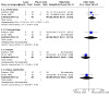
Comparison 3: Laser versus plastic tube, Outcome 2: Adverse effects
3.3. Analysis.

Comparison 3: Laser versus plastic tube, Outcome 3: Technical success of procedure
3.4. Analysis.

Comparison 3: Laser versus plastic tube, Outcome 4: Procedure mortality
3.5. Analysis.

Comparison 3: Laser versus plastic tube, Outcome 5: Dysphagia improvement
3.6. Analysis.

Comparison 3: Laser versus plastic tube, Outcome 6: All adverse effects
Comparison 4. Laser versus laser plus brachytherapy.
| Outcome or subgroup title | No. of studies | No. of participants | Statistical method | Effect size |
|---|---|---|---|---|
| 4.1 Recurrent dysphagia | 3 | 87 | Odds Ratio (M‐H, Fixed, 95% CI) | 0.22 [0.06, 0.87] |
| 4.2 Adverse effects | 4 | Odds Ratio (M‐H, Fixed, 95% CI) | Subtotals only | |
| 4.2.1 Perforation | 3 | 87 | Odds Ratio (M‐H, Fixed, 95% CI) | 2.89 [0.28, 29.29] |
| 4.2.2 Haemorrhage | 3 | 87 | Odds Ratio (M‐H, Fixed, 95% CI) | 2.78 [0.10, 74.70] |
| 4.2.3 Fistula | 4 | 124 | Odds Ratio (M‐H, Fixed, 95% CI) | 1.10 [0.30, 3.97] |
| 4.2.4 Sepsis | 2 | 48 | Odds Ratio (M‐H, Fixed, 95% CI) | Not estimable |
| 4.2.5 Bolus obstruction | 2 | 48 | Odds Ratio (M‐H, Fixed, 95% CI) | Not estimable |
| 4.2.6 Oesophagitis | 3 | 87 | Odds Ratio (M‐H, Fixed, 95% CI) | 0.34 [0.06, 1.85] |
| 4.2.7 All adverse effects | 4 | 124 | Odds Ratio (M‐H, Fixed, 95% CI) | 0.74 [0.31, 1.77] |
| 4.3 30‐day mortality | 1 | Odds Ratio (M‐H, Fixed, 95% CI) | Subtotals only | |
| 4.4 Technical success of procedure | 3 | 87 | Odds Ratio (M‐H, Fixed, 95% CI) | 15.35 [0.73, 321.58] |
| 4.5 Procedure Mortality | 4 | 124 | Odds Ratio (M‐H, Fixed, 95% CI) | 5.00 [0.22, 115.05] |
4.2. Analysis.
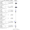
Comparison 4: Laser versus laser plus brachytherapy, Outcome 2: Adverse effects
4.3. Analysis.

Comparison 4: Laser versus laser plus brachytherapy, Outcome 3: 30‐day mortality
4.5. Analysis.

Comparison 4: Laser versus laser plus brachytherapy, Outcome 5: Procedure Mortality
Comparison 5. Laser versus photodynamic therapy (PDT).
| Outcome or subgroup title | No. of studies | No. of participants | Statistical method | Effect size |
|---|---|---|---|---|
| 5.1 Dysphagia improvement (2‐point grade or more) | 2 | 278 | Odds Ratio (M‐H, Random, 95% CI) | 0.97 [0.51, 1.86] |
| 5.2 Adverse effects | 2 | Odds Ratio (M‐H, Fixed, 95% CI) | Subtotals only | |
| 5.2.1 Fever | 2 | 278 | Odds Ratio (M‐H, Fixed, 95% CI) | 0.29 [0.12, 0.70] |
| 5.2.2 Perforation | 2 | 278 | Odds Ratio (M‐H, Fixed, 95% CI) | 5.55 [1.18, 26.20] |
| 5.2.3 Photosensitivity | 2 | 278 | Odds Ratio (M‐H, Fixed, 95% CI) | 0.03 [0.00, 0.24] |
| 5.2.4 All adverse effects | 2 | 278 | Odds Ratio (M‐H, Fixed, 95% CI) | 0.60 [0.33, 1.07] |
Comparison 6. Covered Ultraflex SEMS versus covered Wallstent.
| Outcome or subgroup title | No. of studies | No. of participants | Statistical method | Effect size |
|---|---|---|---|---|
| 6.1 Dysphagia improvement | 2 | 120 | Mean Difference (IV, Fixed, 95% CI) | 0.15 [‐0.04, 0.33] |
| 6.2 Persistent or recurrent dysphagia | 2 | 120 | Odds Ratio (M‐H, Fixed, 95% CI) | 1.27 [0.49, 3.31] |
| 6.3 Technical success | 2 | 120 | Odds Ratio (M‐H, Fixed, 95% CI) | 3.00 [0.12, 76.31] |
| 6.4 30‐day mortality | 2 | 120 | Odds Ratio (M‐H, Fixed, 95% CI) | 1.18 [0.44, 3.18] |
| 6.5 All adverse effects | 2 | 120 | Odds Ratio (M‐H, Fixed, 95% CI) | 0.61 [0.27, 1.38] |
| 6.6 Adverse effects | 2 | Odds Ratio (M‐H, Fixed, 95% CI) | Subtotals only | |
| 6.6.1 Perforation | 2 | 120 | Odds Ratio (M‐H, Fixed, 95% CI) | 1.29 [0.22, 7.58] |
| 6.6.2 Haemorrhage | 2 | 120 | Odds Ratio (M‐H, Fixed, 95% CI) | 1.41 [0.37, 5.33] |
| 6.6.3 Migration | 2 | 120 | Odds Ratio (M‐H, Fixed, 95% CI) | 1.93 [0.54, 6.87] |
| 6.6.4 Tumour overgrowth | 2 | 120 | Odds Ratio (M‐H, Fixed, 95% CI) | 0.27 [0.05, 1.41] |
| 6.6.5 Reflux | 2 | 120 | Odds Ratio (M‐H, Fixed, 95% CI) | 0.61 [0.13, 2.92] |
| 6.6.6 Bolus obstruction | 2 | 120 | Odds Ratio (M‐H, Fixed, 95% CI) | 0.62 [0.10, 4.00] |
| 6.7 Procedure related mortality | 2 | 120 | Odds Ratio (M‐H, Fixed, 95% CI) | 0.97 [0.06, 16.17] |
6.4. Analysis.

Comparison 6: Covered Ultraflex SEMS versus covered Wallstent, Outcome 4: 30‐day mortality
6.5. Analysis.

Comparison 6: Covered Ultraflex SEMS versus covered Wallstent, Outcome 5: All adverse effects
6.6. Analysis.

Comparison 6: Covered Ultraflex SEMS versus covered Wallstent, Outcome 6: Adverse effects
6.7. Analysis.

Comparison 6: Covered Ultraflex SEMS versus covered Wallstent, Outcome 7: Procedure related mortality
Comparison 7. SEMS versus plastic tube (degree of concealment).
| Outcome or subgroup title | No. of studies | No. of participants | Statistical method | Effect size |
|---|---|---|---|---|
| 7.1 Persistent or recurrent dysphagia (analysis by concealment of allocation) | 7 | Odds Ratio (M‐H, Random, 95% CI) | Subtotals only | |
| 7.1.1 Concealment of allocation A | 4 | 323 | Odds Ratio (M‐H, Random, 95% CI) | 0.49 [0.19, 1.28] |
| 7.1.2 Concealment of allocation non‐A | 3 | 110 | Odds Ratio (M‐H, Random, 95% CI) | 0.29 [0.07, 1.21] |
| 7.2 Technical success (analysis by concealment of allocation) | 7 | Odds Ratio (M‐H, Fixed, 95% CI) | Subtotals only | |
| 7.2.1 Concealment of allocation A | 4 | 323 | Odds Ratio (M‐H, Fixed, 95% CI) | 1.26 [0.33, 4.85] |
| 7.2.2 Concealment of allocation non‐A | 3 | 110 | Odds Ratio (M‐H, Fixed, 95% CI) | 4.90 [1.03, 23.24] |
| 7.3 Procedure mortality (analysis by concealment of allocation) | 7 | Odds Ratio (M‐H, Fixed, 95% CI) | Subtotals only | |
| 7.3.1 Concealment of allocation A | 4 | 323 | Odds Ratio (M‐H, Fixed, 95% CI) | 0.41 [0.17, 1.02] |
| 7.3.2 Concealment of allocation non‐A | 3 | 110 | Odds Ratio (M‐H, Fixed, 95% CI) | 0.13 [0.01, 2.66] |
| 7.4 30‐day mortality (analysis by concealment of allocation) | 4 | Odds Ratio (M‐H, Fixed, 95% CI) | Subtotals only | |
| 7.4.1 Concealment of allocation A | 3 | 273 | Odds Ratio (M‐H, Fixed, 95% CI) | 0.71 [0.38, 1.30] |
| 7.4.2 Concealment of allocation non‐A | 1 | 31 | Odds Ratio (M‐H, Fixed, 95% CI) | 0.50 [0.12, 2.14] |
| 7.5 All major side effects (analysis by concealment of allocation) | 7 | Odds Ratio (M‐H, Fixed, 95% CI) | Subtotals only | |
| 7.5.1 Concealment of allocation A | 4 | 303 | Odds Ratio (M‐H, Fixed, 95% CI) | 0.23 [0.13, 0.39] |
| 7.5.2 Concealment of allocation non‐A | 3 | 110 | Odds Ratio (M‐H, Fixed, 95% CI) | 0.25 [0.10, 0.65] |
| 7.6 Adverse effects (analysis by concealment of allocation A) | 4 | Odds Ratio (M‐H, Fixed, 95% CI) | Subtotals only | |
| 7.6.1 Perforation (concealment of allocation A) | 4 | 322 | Odds Ratio (M‐H, Fixed, 95% CI) | 0.26 [0.07, 0.95] |
| 7.6.2 Fistula (concealment of allocation A) | 3 | 167 | Odds Ratio (M‐H, Fixed, 95% CI) | 1.80 [0.52, 6.28] |
| 7.6.3 Haemorrhage (concealment of allocation A) | 4 | 323 | Odds Ratio (M‐H, Fixed, 95% CI) | 0.88 [0.47, 1.64] |
| 7.6.4 Chest pain (concealment of allocation A) | 3 | 286 | Odds Ratio (M‐H, Fixed, 95% CI) | 1.35 [0.76, 2.41] |
| 7.6.5 Migration (concealment of allocation A) | 4 | 327 | Odds Ratio (M‐H, Fixed, 95% CI) | 0.28 [0.14, 0.57] |
| 7.6.6 Tumour ingrowth (concealment of allocation A) | 3 | 167 | Odds Ratio (M‐H, Fixed, 95% CI) | 3.30 [0.63, 17.32] |
| 7.6.7 Tumour overgrowth (concealment of allocation A) | 4 | 323 | Odds Ratio (M‐H, Fixed, 95% CI) | 1.07 [0.54, 2.11] |
| 7.6.8 Bolus obstruction (concealment of allocation A) | 4 | 328 | Odds Ratio (M‐H, Fixed, 95% CI) | 0.32 [0.14, 0.74] |
| 7.6.9 Stent malfunction (concealment of allocation A) | 4 | 328 | Odds Ratio (M‐H, Fixed, 95% CI) | 0.35 [0.13, 1.00] |
| 7.7 Adverse effects (analysis by concealment of allocation: non‐A) | 3 | Odds Ratio (M‐H, Fixed, 95% CI) | Subtotals only | |
| 7.7.1 Perforation (concealment of allocation non‐A) | 3 | 110 | Odds Ratio (M‐H, Fixed, 95% CI) | 0.15 [0.02, 1.31] |
| 7.7.2 Fistula (concealment of allocation non‐A) | 3 | 110 | Odds Ratio (M‐H, Fixed, 95% CI) | Not estimable |
| 7.7.3 Haemorrhage (concealment of allocation non‐A) | 3 | 110 | Odds Ratio (M‐H, Fixed, 95% CI) | 0.18 [0.01, 4.01] |
| 7.7.4 Migration (concealment of allocation non‐A) | 3 | 110 | Odds Ratio (M‐H, Fixed, 95% CI) | 0.21 [0.05, 0.87] |
| 7.7.5 Tumour ingrowth (concealment of allocation non‐A) | 3 | 110 | Odds Ratio (M‐H, Fixed, 95% CI) | 5.86 [0.26, 130.36] |
| 7.7.6 Tumour overgrowth (concealment of allocation non‐A) | 3 | 110 | Odds Ratio (M‐H, Fixed, 95% CI) | 0.64 [0.16, 2.59] |
| 7.7.7 Bolus obstruction (concealment of allocation non‐A) | 3 | 110 | Odds Ratio (M‐H, Fixed, 95% CI) | 0.73 [0.22, 2.40] |
| 7.7.8 Stent malfunction (concealment of allocation non‐A) | 3 | 110 | Odds Ratio (M‐H, Fixed, 95% CI) | Not estimable |
7.1. Analysis.

Comparison 7: SEMS versus plastic tube (degree of concealment), Outcome 1: Persistent or recurrent dysphagia (analysis by concealment of allocation)
7.2. Analysis.
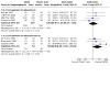
Comparison 7: SEMS versus plastic tube (degree of concealment), Outcome 2: Technical success (analysis by concealment of allocation)
7.3. Analysis.

Comparison 7: SEMS versus plastic tube (degree of concealment), Outcome 3: Procedure mortality (analysis by concealment of allocation)
7.4. Analysis.

Comparison 7: SEMS versus plastic tube (degree of concealment), Outcome 4: 30‐day mortality (analysis by concealment of allocation)
7.5. Analysis.

Comparison 7: SEMS versus plastic tube (degree of concealment), Outcome 5: All major side effects (analysis by concealment of allocation)
7.6. Analysis.

Comparison 7: SEMS versus plastic tube (degree of concealment), Outcome 6: Adverse effects (analysis by concealment of allocation A)
7.7. Analysis.

Comparison 7: SEMS versus plastic tube (degree of concealment), Outcome 7: Adverse effects (analysis by concealment of allocation: non‐A)
Comparison 8. Anti‐reflux versus standard open stent.
| Outcome or subgroup title | No. of studies | No. of participants | Statistical method | Effect size |
|---|---|---|---|---|
| 8.1 Dysphagia improvement | 2 | 106 | Mean Difference (IV, Fixed, 95% CI) | 12.52 [2.14, 22.90] |
| 8.2 Quality of life | 2 | 106 | Std. Mean Difference (IV, Fixed, 95% CI) | ‐0.04 [‐0.42, 0.35] |
| 8.3 Reflux score | 2 | 106 | Std. Mean Difference (IV, Random, 95% CI) | 0.36 [‐0.02, 0.75] |
| 8.4 Dyspnea score | 2 | 106 | Std. Mean Difference (IV, Fixed, 95% CI) | ‐0.62 [‐1.02, ‐0.23] |
| 8.5 All adverse effects | 2 | 106 | Odds Ratio (M‐H, Fixed, 95% CI) | 0.86 [0.38, 1.94] |
| 8.6 Adverse effects | 2 | 530 | Odds Ratio (M‐H, Fixed, 95% CI) | 0.84 [0.42, 1.67] |
| 8.6.1 Stent migration | 2 | 106 | Odds Ratio (M‐H, Fixed, 95% CI) | 0.68 [0.19, 2.50] |
| 8.6.2 Stent occlusion | 2 | 106 | Odds Ratio (M‐H, Fixed, 95% CI) | 1.10 [0.35, 3.49] |
| 8.6.3 Bleeding | 2 | 106 | Odds Ratio (M‐H, Fixed, 95% CI) | 1.93 [0.31, 11.98] |
| 8.6.4 Esophageal perforation | 2 | 106 | Odds Ratio (M‐H, Fixed, 95% CI) | 0.40 [0.04, 3.94] |
| 8.6.5 Gastric perforation | 2 | 106 | Odds Ratio (M‐H, Fixed, 95% CI) | 0.40 [0.04, 3.94] |
Comparison 9. Brachytherapy versus brachytherapy plus radiotherapy.
| Outcome or subgroup title | No. of studies | No. of participants | Statistical method | Effect size |
|---|---|---|---|---|
| 9.1 Adverse effects | 2 | 554 | Odds Ratio (M‐H, Random, 95% CI) | 1.17 [0.44, 3.15] |
| 9.1.1 Stricture | 2 | 277 | Odds Ratio (M‐H, Random, 95% CI) | 1.43 [0.16, 12.85] |
| 9.1.2 Fistula | 2 | 277 | Odds Ratio (M‐H, Random, 95% CI) | 1.09 [0.24, 4.89] |
Characteristics of studies
Characteristics of included studies [ordered by study ID]
Adam 1997.
| Study characteristics | ||
| Methods | RCT | |
| Participants | UK, 60 patients with squamous and adenocarcinoma | |
| Interventions | Covered SEMS (Wall) vs Strecker uncovered SEMS versus laser | |
| Outcomes | Median improvement at 1 month, recurrent dysphagia n (%), intervention for recurrence n (%), adverse effects | |
| Notes | — | |
| Risk of bias | ||
| Bias | Authors' judgement | Support for judgement |
| Random sequence generation (selection bias) | Low risk | The patients were randomly assigned to undergo placement of a plastic‐covered metallic stent, placement of an uncovered metallic stent or laser therapy, according to a computer‐generated random‐number chart |
Alderson 1990.
| Study characteristics | ||
| Methods | RCT | |
| Participants | UK, 40 patients with adeno and squamous carcinoma of middle and lower oesophagus | |
| Interventions | Laser versus plastic tube | |
| Outcomes | Improvement of dysphagia n (%), adverse effects | |
| Notes | — | |
| Risk of bias | ||
| Bias | Authors' judgement | Support for judgement |
| Random sequence generation (selection bias) | High risk | Randomisation was based on tumour location |
Amdal 2013.
| Study characteristics | ||
| Methods | RCT | |
| Participants | Norway, 41 patients | |
| Interventions | SEMS and brachytherapy versus brachytherapy | |
| Outcomes | dysphagia, pain complain | |
| Notes | ||
| Risk of bias | ||
| Bias | Authors' judgement | Support for judgement |
| Random sequence generation (selection bias) | Low risk | The patients were randomised by our Clinical Trials Unit using computer‐based real time permuted block randomisation |
| Allocation concealment (selection bias) | Low risk | The patients were randomised by our Clinical Trials Unit using computer‐based real time permuted block randomisation |
| Blinding of participants and personnel (performance bias) All outcomes | High risk | Blinding was impossible in the nature of interventions |
Angelini 1991.
| Study characteristics | ||
| Methods | RCT | |
| Participants | Italy, 34 patients with squamous and adenocarcinoma | |
| Interventions | Laser versus polidocanol injection | |
| Outcomes | 1‐point grade improvement at 1 month, recurrent dysphagia n (%), time to recurrence, interventions for recurrence n (%), survival, adverse effects | |
| Notes | — | |
| Risk of bias | ||
| Bias | Authors' judgement | Support for judgement |
| Random sequence generation (selection bias) | Low risk | 37 consecutive patients referred to our endoscopy service have been allocated by the random numbers table of Fisher and Yates |
Anghorn 1983.
| Study characteristics | ||
| Methods | RCT | |
| Participants | Africa, 106 patients with adeno and squamous carcinoma | |
| Interventions | Plastic stent versus gastric bypass surgery | |
| Outcomes | Improvement in dysphagia n (%), mortality, morbidity and complications n (%) | |
| Notes | — | |
| Risk of bias | ||
| Bias | Authors' judgement | Support for judgement |
| Random sequence generation (selection bias) | Unclear risk | A total of 106 patients admitted were prospectively randomized for palliative treatment |
Barr 1990.
| Study characteristics | ||
| Methods | RCT | |
| Participants | UK, 40 patients with adeno and squamous carcinoma | |
| Interventions | Laser versus laser plus plastic tube (AT) | |
| Outcomes | Best mean dysphagia grade and mean grade, recurrent dysphagia n (%) mean survival in weeks adverse effects, LASA quality of life | |
| Notes | ||
| Risk of bias | ||
| Bias | Authors' judgement | Support for judgement |
| Random sequence generation (selection bias) | Low risk | Patients were randomised on referral to receive laser therapy only or initial laser therapy followed by endoscopic intubation using sealed envelopes |
| Allocation concealment (selection bias) | Low risk | Patients were randomised on referral to receive laser therapy only or initial laser therapy followed by endoscopic intubation using sealed envelopes |
Bergquist 2005.
| Study characteristics | ||
| Methods | RCT, multicentre | |
| Participants | Sweden, 65 patients with advanced oesophageal or gastro‐oesophageal junction cancers | |
| Interventions | SEMS vs brachytherapy. Iridium source, 3 fractions of 7 Gy | |
| Outcomes | Health related quality of life main outcome; secondary outcomes included dysphagia improvement | |
| Notes | — | |
| Risk of bias | ||
| Bias | Authors' judgement | Support for judgement |
| Random sequence generation (selection bias) | Low risk | To ensure an even distribution of patients between the two treatment groups according to age, sex, grade of dysphagia, tumour histology, tumour site and treatment, a computer‐based randomisation algorithm was used and conducted by The Regional Cancer Register of Göteborg in Sweden |
| Allocation concealment (selection bias) | Low risk | To ensure an even distribution of patients between the two treatment groups according to age, sex, grade of dysphagia, tumour histology, tumour site and treatment, a computer‐based randomization algorithm was used and conducted by The Regional Cancer Register of Göteborg in Sweden |
Canto 2002.
| Study characteristics | ||
| Methods | RCT | |
| Participants | USA, 56 patients with squamous and adenocarcinoma | |
| Interventions | SEMS versus PDT | |
| Outcomes | Mean dysphagia, also dysphagia improvement in 3 weeks n (%), survival, adverse effects, EORTC and SF‐36 at 3 weeks and every 3 months | |
| Notes | Only abstract | |
| Risk of bias | ||
| Bias | Authors' judgement | Support for judgement |
| Random sequence generation (selection bias) | Unclear risk | Randomisation was stratified by CA location and presence of distant metastasis |
Carrazone 1999.
| Study characteristics | ||
| Methods | RCT | |
| Participants | Italy, 47 patients fungating adeno and squamous carcinoma | |
| Interventions | Laser versus ethanol injection | |
| Outcomes | Return to oral feeding n (%), mean dysphagia free interval, mean number of endoscopies, adverse effects, survival n (%) | |
| Notes | — | |
| Risk of bias | ||
| Bias | Authors' judgement | Support for judgement |
| Random sequence generation (selection bias) | Unclear risk | All patients (44 men and 3 women, mean age 63 years, range 27–88) were randomly assigned to endoscopic Nd:YAG laser therapy (n = 24) or intratumoural alcohol injection (n = 23) (Table I) |
Carter 1992.
| Study characteristics | ||
| Methods | RCT | |
| Participants | UK, 40 patients adeno and squamous carcinoma | |
| Interventions | Plastic tube versus laser | |
| Outcomes | Median best dysphagia and before death, recurrent dysphagia, median survival, adverse effects | |
| Notes | — | |
| Risk of bias | ||
| Bias | Authors' judgement | Support for judgement |
| Random sequence generation (selection bias) | Unclear risk | Forty patients were allocated to treatment by endoscopic intubation or endoscopic laser therapy. Randomisation was based on tumour location |
Dai 2013.
| Study characteristics | ||
| Methods | RCT | |
| Participants | China, 67 patients | |
| Interventions | A conventional stent versus an iodine‐eluting esophageal stent | |
| Outcomes | Dysphagia score, median survival time. Side effects, complications and security assessment | |
| Notes | ||
| Risk of bias | ||
| Bias | Authors' judgement | Support for judgement |
| Random sequence generation (selection bias) | Low risk | Patients were randomly assigned into two groups, those who received a conventional stent (group A; n=36) and those who received an iodine‐eluting esophageal stent (group B; n=31) |
| Allocation concealment (selection bias) | Low risk | Patients were randomly assigned into two groups, those who received a conventional stent (group A; n=36) and those who received an iodine‐eluting esophageal stent (group B; n=31) |
Dallal 2001.
| Study characteristics | ||
| Methods | RCT | |
| Participants | UK, 65 patients squamous and adenocarcinoma | |
| Interventions | SEMS versus laser or APC or both | |
| Outcomes | Median dysphagia improvement, QOL, survival n (%), adverse effects | |
| Notes | — | |
| Risk of bias | ||
| Bias | Authors' judgement | Support for judgement |
| Random sequence generation (selection bias) | Low risk | Patients were randomised with a sealed envelope technique in blocks of 6; this accounts for the inequality of numbers in each arm of the study |
| Allocation concealment (selection bias) | Low risk | Patients were randomised with a sealed envelope technique in blocks of 6; this accounts for the inequality of numbers in each arm of the study |
| Blinding of participants and personnel (performance bias) All outcomes | Unclear risk | Blinding was impossible in the nature of interventions |
De Palma 1996.
| Study characteristics | ||
| Methods | RCT | |
| Participants | Italy, 39 patients with oesophageal carcinoma | |
| Interventions | SEMS (covered UF) versus WC plastic tubes | |
| Outcomes | Immediate mean dysphagia scores, recurrent dysphagia n (%), interventions for recurrence n (%), survival, adverse effects | |
| Notes | — | |
| Risk of bias | ||
| Bias | Authors' judgement | Support for judgement |
| Random sequence generation (selection bias) | Low risk | The patients were allocated by the random number table of Fisher and Yates to traditional stent (group A, 20 patients) or expandable metal stent (group B, 19 patients) |
| Allocation concealment (selection bias) | Low risk | The patients were allocated by the random number table of Fisher and Yates to traditional stent (group A, 20 patients) or expandable metal stent (group B, 19 patients) |
Fu 2004.
| Study characteristics | ||
| Methods | RCT | |
| Participants | China, 53 patients with squamous and adenocarcinoma | |
| Interventions | SEMS (GT‐Z and UF) versus SEMS with chemoradiotherapy | |
| Outcomes | Median dysphagia scores, adverse effects n (%) | |
| Notes | — | |
| Risk of bias | ||
| Bias | Authors' judgement | Support for judgement |
| Blinding of participants and personnel (performance bias) All outcomes | High risk | Blinding was impossible in the nature of interventions |
Fuchs 1991.
| Study characteristics | ||
| Methods | RCT | |
| Participants | Germany, 47 patients with adeno and squamous carcinoma | |
| Interventions | Laser versus plastic tube | |
| Outcomes | 1 and 2‐grade improvement n (%), improvement in performance status n (%) | |
| Notes | — | |
| Risk of bias | ||
| Bias | Authors' judgement | Support for judgement |
| Random sequence generation (selection bias) | Unclear risk | Of 124 subjects, only 40 met the criteria for randomization. After their randomization, 4 patients initially started on PEP had to be withdrawn from this therapy due to a high risk of tube dislocation and 3 cases in the ELT group were considered to be withdrawal cases since a PEP treatment would have borne a high risk of dislocation |
| Blinding of participants and personnel (performance bias) All outcomes | High risk | Blinding was impossible in the nature of interventions |
Guo 2008.
| Study characteristics | ||
| Methods | RCT | |
| Participants | China, 53 patients | |
| Interventions | MTN‐S stent versus I125 stent | |
| Outcomes | Dysphagia grades, median and mean survival times | |
| Notes | ‐ | |
| Risk of bias | ||
| Bias | Authors' judgement | Support for judgement |
| Random sequence generation (selection bias) | Low risk | Patients were randomly assigned to the irradiation stent group or the control group by using Proc Plan Seed210002 |
| Allocation concealment (selection bias) | Low risk | Patients were randomly assigned to the irradiation stent group or the control group by using Proc Plan Seed210002 |
| Blinding (performance bias and detection bias) All outcomes | Low risk | Except for the interventional radiologists, all patients,the nurse following up patients, and the statistician performing the analyses in our study were blinded to the type of stent used |
| Blinding of participants and personnel (performance bias) All outcomes | Low risk | Except for the interventional radiologists, all patients,the nurse following up patients, and the statistician performing the analyses in our study were blinded to the type of stent used |
| Blinding of outcome assessment (detection bias) All outcomes | Low risk | Except for the interventional radiologists, all patients,the nurse following up patients, and the statistician performing the analyses in our study were blinded to the type of stent used |
Heier 1995.
| Study characteristics | ||
| Methods | RCT | |
| Participants | USA, 42 patients with squamous and adenocarcinoma, previous failed therapy, refusal of surgery | |
| Interventions | PDT versus laser | |
| Outcomes | Mean oesophageal grade and performance status, mean time to recurrence, survival, adverse effects | |
| Notes | — | |
| Risk of bias | ||
| Bias | Authors' judgement | Support for judgement |
| Random sequence generation (selection bias) | Unclear risk | Randomisation was stratified by tumour length (greater than versus less than 10 cm) and prior therapy |
Homs 2004a.
| Study characteristics | ||
| Methods | RCT | |
| Participants | Netherlands, 209 patients with squamous and adenocarcinoma with dysphagia 2‐4 | |
| Interventions | SEMS (covered UF) versus brachytherapy | |
| Outcomes | 1‐month, 1‐grade improvement in dysphagia n (%), mean difference in dysphagia‐free survival, EORTC, EQ 5D, generic HRQOL, recurrent dysphagia n (%), adverse effects n (%), interventions for recurrence (%), survival median | |
| Notes | Two publications | |
| Risk of bias | ||
| Bias | Authors' judgement | Support for judgement |
| Random sequence generation (selection bias) | Unclear risk | For randomisation, patients were stratified for location of the tumour (oesophagus or oesophagogastric junction) and for administration of chemotherapy before treatment. Randomisation was centrally done by telephone by staff at the trial office of the Department of Oncology, Erasmus MC Rotterdam. |
| Allocation concealment (selection bias) | Low risk | For randomisation, patients were stratified for location of the tumour (oesophagus or oesophagogastric junction) and for administration of chemotherapy before treatment. Randomisation was centrally done by telephone by staff at the trial office of the Department of Oncology, Erasmus MC Rotterdam |
| Blinding of participants and personnel (performance bias) All outcomes | High risk | Blinding was impossible in the nature of interventions |
Homs 2004b.
| Study characteristics | ||
| Methods | RCT | |
| Participants | Netherlands, 209 patients with squamous and adenocarcinoma with dysphagia grade 2 to 4 | |
| Interventions | SEMS (covered UF) versus brachytherapy | |
| Outcomes | 1‐month, 1‐grade improvement in dysphagia n (%), mean difference in dysphagia‐free survival, EORTC, EQ 5D, generic HRQOL, recurrent dysphagia n(%), adverse effects n (%), interventions for recurrence (%), survival median | |
| Notes | Two publications | |
| Risk of bias | ||
| Bias | Authors' judgement | Support for judgement |
| Random sequence generation (selection bias) | Unclear risk | We conducted a randomised trial comparing metal stent placement with single dose brachytherapy for the palliation of oesophageal cancer |
| Blinding of participants and personnel (performance bias) All outcomes | High risk | Blinding was impossible in the nature of interventions |
Homs 2004c.
| Study characteristics | ||
| Methods | RCT | |
| Participants | Netherlands, 30 patients with lower oesophageal or GOJ cancers | |
| Interventions | Covered open SEMS versus anti‐reflux SEMS | |
| Outcomes | EORTC OES‐23 and pH monitoring for reflux were primary outcomes; secondary outcomes included dysphagia improvement, overall survival, complications | |
| Notes | — | |
| Risk of bias | ||
| Bias | Authors' judgement | Support for judgement |
| Random sequence generation (selection bias) | Low risk | Computer‐generated block randomisation lists were prepared with block sizes of 4 and 6 in random order. Randomisation by telephone was centrally performed at the trial office of the Department of Oncology of our medical centre |
| Allocation concealment (selection bias) | Low risk | A ‐ Adequate. Computer‐generated block randomisation lists were prepared with block sizes of 4 and 6 in random order. Randomisation by telephone was centrally performed at the trial office of the Department of Oncology of our medical centre |
| Blinding of participants and personnel (performance bias) All outcomes | Low risk | Randomisation by telephone was centrally performed at the trial office of the Department of Oncology of our medical centre. Patients were blinded as to the type of stent they received |
Horneaux 2001.
| Study characteristics | ||
| Methods | RCT | |
| Participants | Brazil, 40 patients with stage III, IV SCC | |
| Interventions | SEMS (Esophacoil) versus Posthelwaite surgical bypass | |
| Outcomes | Mean dysphagia scores at 30 days and 120 days, mean performance status, procedure related morbidity, mortality n (%), survival | |
| Notes | Portuguese | |
Javed 2012.
| Study characteristics | ||
| Methods | RCT | |
| Participants | India, 84 patients with inoperable esophageal cancer and with high grade dysphagia | |
| Interventions | covered Ultraflex stent versus a combination of stent and EBRT | |
| Outcomes | Dysphagia scores; Overall median survival, the incidence of complications | |
| Notes | ‐ | |
| Risk of bias | ||
| Bias | Authors' judgement | Support for judgement |
| Random sequence generation (selection bias) | Low risk | Patients included for the study were randomised (with the help of a computer‐generated random number table) into two groups using a sealed envelope technique |
| Allocation concealment (selection bias) | Low risk | Patients included for the study were randomized (with the help of a computer‐generated random number table) into two groups using a sealed envelope technique |
Kim 2009.
| Study characteristics | ||
| Methods | RCT | |
| Participants | South Korea, 37 consecutive patients with malignant dysphagia due to inoperable oesophageal or gastric cardia cancer | |
| Interventions | Double‐layered Niti‐S stent versus covered metal stents | |
| Outcomes | Dysphagia score; overall complications | |
| Notes | ||
| Risk of bias | ||
| Bias | Authors' judgement | Support for judgement |
| Random sequence generation (selection bias) | Unclear risk | 37 consecutive patients with malignant dysphagia due to inoperable oesophageal or gastric cardia cancer were included in the study. The patients were randomly assigned to treatment with a double‐layered or covered Niti‐S‐stent |
Knyrim 1993.
| Study characteristics | ||
| Methods | RCT | |
| Participants | Germany, 42 patients with adeno and squamous carcinoma | |
| Interventions | Wilson cook plastic tube versus Wallstent uncovered | |
| Outcomes | At least 1 grade improvement at 6 weeks, median dysphagia at 6 weeks, recurrent dysphagia n (%), mean rate of interventions, mean survival, adverse effects | |
| Notes | — | |
| Risk of bias | ||
| Bias | Authors' judgement | Support for judgement |
| Random sequence generation (selection bias) | Low risk | The patients enrolled were randomly assigned to treatment with a plastic prosthesis or a metal stent according to a computer‐generated random number chart |
| Allocation concealment (selection bias) | Low risk | The patients enrolled were randomly assigned to treatment with a plastic prosthesis or a metal stent according to a computer‐generated random number chart |
Konigsrainer 2000.
| Study characteristics | ||
| Methods | RCT | |
| Participants | Austria, 39 patients with squamous and adenocarcinoma | |
| Interventions | SEMS (Wallstent) versus SEMS plus limited laser versus laser plus EBRT | |
| Outcomes | Mean dysphagia, recurrent dysphagia n (%), mean time to recurrence, adverse effects | |
| Notes | — | |
| Risk of bias | ||
| Bias | Authors' judgement | Support for judgement |
| Random sequence generation (selection bias) | Unclear risk | A total of 39 consecutive patients with unresectable oesophageal cancer were randomly allocated to either receive combined laser and radiotherapy group or implantation of an expanding metal stent group |
| Blinding of participants and personnel (performance bias) All outcomes | High risk | Blinding was impossible in the nature of interventions |
Lightdale 1995.
| Study characteristics | ||
| Methods | RCT | |
| Participants | USA, 236 patients with squamous and adenocarcinoma | |
| Interventions | PDT versus laser | |
| Outcomes | Mean change at 1 week and 1 month, objective tumour response at same intervals, mean time to recurrence, median survival, adverse effects | |
| Notes | — | |
| Risk of bias | ||
| Bias | Authors' judgement | Support for judgement |
| Random sequence generation (selection bias) | Low risk | Informed consent was obtained from all patients, who were assigned to one of four strata based on the length of their tumour at the time of randomisation (greater or less than 10 cm) and whether or not they had received prior therapy for their cancer. Within centre and stratum, patients were allocated sequentially to treatment with either PDT or Nd:YAG laser therapy using a computerised randomisation schema with a blocking factor of 4 |
| Allocation concealment (selection bias) | Low risk | Informed consent was obtained from all patients, who were assigned to one of four strata based on the length of their tumour at the time of randomisation (greater or less than 10 cm) and whether or not they had received prior therapy for their cancer. Within centre and stratum, patients were allocated sequentially to treatment with either PDT or Nd:YAG laser therapy using a computerised randomisation schema with a blocking factor of 4 |
Low 1992.
| Study characteristics | ||
| Methods | RCT | |
| Participants | UK, 23 patients with predominant adenocarcinoma | |
| Interventions | Laser versus brachytherapy | |
| Outcomes | Dysphagia improvement n (%) in 2 months and 6 months or death, recurrent dysphagia n (%), interventions n (%) | |
| Notes | — | |
| Risk of bias | ||
| Bias | Authors' judgement | Support for judgement |
| Random sequence generation (selection bias) | Unclear risk | 23 consecutive patients were randomised to receive either brachytherapy or laser therapy |
Mannell 1986.
| Study characteristics | ||
| Methods | RCT | |
| Participants | South Africa, 170 patients with squamous cell carcinoma | |
| Interventions | Plastic stent versus dilatation and bleomycin | |
| Outcomes | Dysphagia score n (%), mean dysphagia‐free survival, mean overall survival, adverse effects | |
| Notes | — | |
| Risk of bias | ||
| Bias | Authors' judgement | Support for judgement |
| Random sequence generation (selection bias) | Unclear risk | Patients with advanced oesophageal cancer who were eligible for this trial were randomly allocated to group 1 or to group 2 |
| Blinding of participants and personnel (performance bias) All outcomes | High risk | Blinding was impossible in the nature of interventions |
Mehta 2008.
| Study characteristics | ||
| Methods | RCT | |
| Participants | India, 62 patients with previously untreated, inoperable, locally advanced carcinoma oesophagus | |
| Interventions | ERT+BT versus ERT | |
| Outcomes | dysphagia scores; complications | |
| Notes | ‐ | |
| Risk of bias | ||
| Bias | Authors' judgement | Support for judgement |
| Random sequence generation (selection bias) | Low risk | Tippet's random‐number table was used for randomisation of patients to the three arms, with at least 20 patients in each arm |
| Allocation concealment (selection bias) | Low risk | Tippet's random‐number table was used for randomisation of patients to the three arms, with at least 20 patients in each arm |
| Blinding of participants and personnel (performance bias) All outcomes | High risk | Blinding was impossible in the nature of interventions |
O'Donnell 2002.
| Study characteristics | ||
| Methods | RCT | |
| Participants | UK, 50 patients with inoperable oesophageal carcinoma | |
| Interventions | Cook plastic tubes versus covered UF and Wallstents | |
| Outcomes | Dysphagia improvement n (%), survival, adverse effects EORTC QLQ‐30, cost | |
| Notes | — | |
| Risk of bias | ||
| Bias | Authors' judgement | Support for judgement |
| Random sequence generation (selection bias) | Low risk | Patients were randomised using a list of computer‐generated numbers concealed in sealed envelopes to receive either a plastic endoprosthesis or metallic stent |
| Allocation concealment (selection bias) | Low risk | Patients were randomised using a list of computer‐generated numbers concealed in sealed envelopes to receive either a plastic endoprosthesis or metallic stent |
Power 2007.
| Study characteristics | ||
| Methods | RCT | |
| Participants | Ireland, 49 consecutive patients with malignant dysphagia | |
| Interventions | anti‐reflux stent versus standard stent | |
| Outcomes | dysphagia scores; adverse effects | |
| Notes | ‐ | |
| Risk of bias | ||
| Bias | Authors' judgement | Support for judgement |
| Random sequence generation (selection bias) | Low risk | Randomisation was by the closed envelope technique and primarily pertained to a cohort size of 40 patients |
| Allocation concealment (selection bias) | Low risk | Randomisation was by the closed envelope technique and primarily pertained to a cohort size of 40 patients |
| Blinding (performance bias and detection bias) All outcomes | Low risk | A research nurse who assessed the patients’ health related quality of life (HRQoL) and follow‐up was blinded as to the allocation of patients to each group |
| Blinding of participants and personnel (performance bias) All outcomes | Low risk | The patients were blinded as to the type of stent they received.A research nurse who assessed the patients’ health related quality of life (HRQOL) and follow‐up was blinded as to the allocation of patients to each group |
Reed 1991.
| Study characteristics | ||
| Methods | RCT | |
| Participants | USA, 27 patients with squamous carcinoma of the mid and lower oesophagus | |
| Interventions | Plastic tube (AT) versus plastic tube plus EBRT versus plastic tube plus laser | |
| Outcomes | Able to eat solids achieved in n (%), mean improvement in dysphagia, performance status, overall survival, recurrence dysphagia n (%) adverse effects | |
| Notes | All but one black | |
| Risk of bias | ||
| Bias | Authors' judgement | Support for judgement |
| Random sequence generation (selection bias) | Unclear risk | Twenty‐seven patients were prospectively randomised to one of three palliative treatment arms |
Ries 1989.
| Study characteristics | ||
| Methods | RCT | |
| Participants | Germany, 37 patients with adeno and squamous carcinoma | |
| Interventions | Laser versus laser plus brachytherapy | |
| Outcomes | Mean dysphagia‐free interval, mean number of endoscopies, mean survival | |
| Notes | — | |
| Risk of bias | ||
| Bias | Authors' judgement | Support for judgement |
| Blinding of participants and personnel (performance bias) All outcomes | High risk | Blinding was impossible in the nature of interventions |
Rosenblatt 2010.
| Study characteristics | ||
| Methods | RCT | |
| Participants | Austria, 219 patients | |
| Interventions | HDRBT+EBRT versus HDRBT | |
| Outcomes | Dysphagia relief experience (DRE); various scores, performance status, weight and adverse events | |
| Notes | ‐ | |
| Risk of bias | ||
| Bias | Authors' judgement | Support for judgement |
| Random sequence generation (selection bias) | Unclear risk | This was a prospective multicentre randomised clinical trial. Six countries (Brazil, China, Croatia, India, South Africa and Sudan) participated in patient accrual, treatment and follow up |
| Blinding of participants and personnel (performance bias) All outcomes | High risk | Therapies were administered in a non‐blinded manner, without placebo |
Roseveare 1998.
| Study characteristics | ||
| Methods | RCT | |
| Participants | UK, 31 patients with squamous and adenocarcinoma | |
| Interventions | Plastic stents (AT) versus SEMS (GT‐Z stents) | |
| Outcomes | Mean dysphagia, nutrition status, survival and adverse effects | |
| Notes | — | |
| Risk of bias | ||
| Bias | Authors' judgement | Support for judgement |
| Random sequence generation (selection bias) | Unclear risk | Randomisation was undertaken at the time of insertion, following endoscopic assessment |
Rupinski 2011.
| Study characteristics | ||
| Methods | RCT | |
| Participants | Poland, 93 patients with malignant dysphagia | |
| Interventions | APC+HDR versus APC+PDT versus APC | |
| Outcomes | dysphagia improvement(n); survival, QOL, treatment‐associated complications, and treatment tolerance | |
| Notes | ‐ | |
| Risk of bias | ||
| Bias | Authors' judgement | Support for judgement |
| Random sequence generation (selection bias) | Low risk | Block randomisation (n = 6) was computer‐generated by independent statistical unit of the Institute of Oncology |
| Allocation concealment (selection bias) | Low risk | Block randomization (n = 6) was computer generated by independent statistical unit of the Institute of Oncology. The staff members of the statistics unit had no further role in the study |
| Blinding (performance bias and detection bias) All outcomes | High risk | The staff members of the statistics unit had no further role in the study. Because of the specificity of the interventions used, the study was open label |
| Blinding of participants and personnel (performance bias) All outcomes | High risk | Because of the specificity of the interventions used, the study was open label |
| Blinding of outcome assessment (detection bias) All outcomes | High risk | Because of the specificity of the interventions used, the study was open label |
Sabharwal 2003.
| Study characteristics | ||
| Methods | RCT | |
| Participants | UK, 53 patients with lower third oesophageal carcinoma | |
| Interventions | SEMS (Wallstent) vs SEMS (Ultraflex) | |
| Outcomes | Mean dysphagia at days 1 and 30, recurrent dysphagia n (%), adverse effects n (%) | |
| Notes | — | |
| Risk of bias | ||
| Bias | Authors' judgement | Support for judgement |
| Random sequence generation (selection bias) | Low risk | Patients were randomised into two groups to receive either the Flamingo Wallstent or the Ultraflex stent. This was done by randomly selecting sealed envelopes (100) with the label Ultraflex (50) or Flamingo (50) enclosed inside |
| Allocation concealment (selection bias) | Low risk | Patients were randomised into two groups to receive either the Flamingo Wallstent or the Ultraflex stent. This was done by randomly selecting sealed envelopes (100) with the label Ultraflex (50) or Flamingo (50) enclosed inside |
Sabharwal 2008.
| Study characteristics | ||
| Methods | RCT | |
| Participants | UK, 49 patients with dysphagia due to inoperable carcinoma in the lower third of the oesophagus | |
| Interventions | Anti‐reflux stent versus covered standard open stent | |
| Outcomes | Dysphagia score | |
| Notes | ‐ | |
| Risk of bias | ||
| Bias | Authors' judgement | Support for judgement |
| Random sequence generation (selection bias) | Low risk | This was done by randomly selecting sealed envelopes with the label Ultraflex or FerX Ella enclosed inside |
| Allocation concealment (selection bias) | Low risk | This was done by randomly selecting sealed envelopes with the label Ultraflex or FerX Ella enclosed inside |
Sander 1991.
| Study characteristics | ||
| Methods | RCT | |
| Participants | Germany, 43 patients with adeno and squamous carcinoma | |
| Interventions | Laser versus laser plus brachytherapy | |
| Outcomes | Mean time to recurrence, mean interventions for recurrent dysphagia and mean survival | |
| Notes | — | |
| Risk of bias | ||
| Bias | Authors' judgement | Support for judgement |
| Random sequence generation (selection bias) | Unclear risk | Patients who met the pre‐conditions were randomised to one of the two treatment groups laser and laser plus afterloading |
| Allocation concealment (selection bias) | Unclear risk | Patients who met the pre‐conditions were randomised to one of the two treatment groups laser and laser plus afterloading. B ‐ Unclear |
| Blinding of participants and personnel (performance bias) All outcomes | High risk | Blinding was impossible in the nature of interventions |
Sanyika 1999.
| Study characteristics | ||
| Methods | RCT | |
| Participants | South Africa, 40 patients with SCC | |
| Interventions | Procter Livingstone tubes versus SEMS (Wallstents) | |
| Outcomes | At least 1 grade dysphagia improvement at 1 month and 2 months, intervention for recurrent dysphagia n (%), adverse effects, survival at 3 months | |
| Notes | — | |
| Risk of bias | ||
| Bias | Authors' judgement | Support for judgement |
| Random sequence generation (selection bias) | Unclear risk | Patients with dysphagia from oesophageal carcinoma were randomised (simple random sampling without replacement) on admission to hospital into two groups |
Sargeant 1997.
| Study characteristics | ||
| Methods | RCT | |
| Participants | UK, 67 patients with adeno and squamous carcinoma | |
| Interventions | Laser versus laser plus EBRT | |
| Outcomes | Mean dysphagia‐free interval, mean time to further treatments, adeno and squamous results separately, adverse effects, survival | |
| Notes | — | |
| Risk of bias | ||
| Bias | Authors' judgement | Support for judgement |
| Random sequence generation (selection bias) | Low risk | Patients were stratified according to histology (squamous cell cancer or adenocarcinoma) before randomisation by sealed envelopes |
| Allocation concealment (selection bias) | Low risk | Patients were stratified according to histology (squamous cell cancer or adenocarcinoma) before randomisation by sealed envelopes |
| Blinding of participants and personnel (performance bias) All outcomes | High risk | Blinding was impossible in the nature of interventions |
Shenfine 2009.
| Study characteristics | ||
| Methods | RCT | |
| Participants | UK, 217 patients with inoperable squamous and adenocarcinoma | |
| Interventions | SEMS (18 mm) versus SEMS (24 mm) vs plastic tube (AT), non‐stent therapies | |
| Outcomes | Mean dysphagia at 6 weeks, mean survival at 6 weeks, QOL, QALY, adverse effects | |
| Notes | ||
| Risk of bias | ||
| Bias | Authors' judgement | Support for judgement |
| Random sequence generation (selection bias) | Low risk | A total of 215 patients were referred for centralised, independent, computer‐generated block randomisation by non‐medical research staff |
| Allocation concealment (selection bias) | Low risk | A total of 215 patients were referred for centralised, independent, computer‐generated block randomisation by non‐medical research staff |
| Blinding (performance bias and detection bias) All outcomes | Low risk | Research staff and patients were blinded to the received stent type for the three stent treatment arms |
| Blinding of participants and personnel (performance bias) All outcomes | Low risk | Research staff and patients were blinded to the received stent type for the three stent treatment arms |
| Blinding of outcome assessment (detection bias) All outcomes | Low risk | Research staff and patients were blinded to the received stent type for the three stent treatment arms |
Shim 2005.
| Study characteristics | ||
| Methods | RCT | |
| Participants | Korea, 36 patients with lower oesophageal and cardiac carcinoma | |
| Interventions | SEMS (UF) versus SEMS (DO stent) versus SEMS (S‐Stent) | |
| Outcomes | Mean dysphagia, reflux symptoms n (%), adverse effects, % total pH < 4, mean De Meester score | |
| Notes | — | |
| Risk of bias | ||
| Bias | Authors' judgement | Support for judgement |
| Random sequence generation (selection bias) | Unclear risk | 36 consecutive patients with dysphagia caused by inoperable carcinoma of the oesophago‐gastric junction were randomly assigned to undergo insertion of either a newly designed anti‐reflux stent, a Dostent, or a standard open stent |
Siersema 1998.
| Study characteristics | ||
| Methods | RCT | |
| Participants | Netherlands, 75 patients with adeno and squamous carcinoma | |
| Interventions | Celestin tubes vs SEMS | |
| Outcomes | Mean dysphagia at 4 weeks, hospital stay, recurrent dysphagia n (%), overall survival, adverse effects particular relation to prior RCT | |
| Notes | — | |
| Risk of bias | ||
| Bias | Authors' judgement | Support for judgement |
| Random sequence generation (selection bias) | Low risk | All patients who fulfilled the inclusion criteria were assigned to treatment with a latex prosthesis or a coated self‐expanding metal stent according to a computer‐generated allocation performed by the Trialbureau of the University Hospital Rotterdam |
| Allocation concealment (selection bias) | Low risk | All patients who fulfilled the inclusion criteria were assigned to treatment with a latex prosthesis or a coated self‐expanding metal stent according to a computer‐generated allocation performed by the Trialbureau of the University Hospital Rotterdam |
Siersema 2001.
| Study characteristics | ||
| Methods | RCT | |
| Participants | Netherlands, 100 patients with squamous and adenocarcinoma | |
| Interventions | Comparison between 3 different SEMS (UF) versus Wallstent versus GT‐Z stents | |
| Outcomes | Mean dysphagia scores at 4 weeks, recurrent dysphagia n (%), survival, performance status at 4 weeks, adverse effects | |
| Notes | — | |
| Risk of bias | ||
| Bias | Authors' judgement | Support for judgement |
| Random sequence generation (selection bias) | Unclear risk | 100 consecutive patients with dysphagia caused by inoperable carcinoma of the oesophagus or gastric cardia, or recurrent dysphagia after prior radiation for esophageal cancer with curative or palliative intent, were randomized to undergo stent insertion of either a partially covered Ultraflex stent, a partially covered Flamingo Wallstent, or a covered Gianturco‐Z stent |
Spencer 2002.
| Study characteristics | ||
| Methods | RCT | |
| Participants | UK, 22 patients with adenocarcinoma of oesophagus and cardia | |
| Interventions | Laser versus laser with brachytherapy | |
| Outcomes | Median dysphagia scores at 2, 4, 6, 10 weeks, median time to recurrent dysphagia, survival, LASA QOL | |
| Notes | — | |
| Risk of bias | ||
| Bias | Authors' judgement | Support for judgement |
| Random sequence generation (selection bias) | Unclear risk | Patients able to eat a soft diet after laser re‐canalisation were randomised to no further therapy or a single treatment with brachytherapy (10 Gy) |
| Blinding of participants and personnel (performance bias) All outcomes | High risk | Blinding was impossible in the nature of interventions |
Sur 2004.
| Study characteristics | ||
| Methods | RCT | |
| Participants | South Africa, 60 patients with inoperable, non‐metastatic squamous carcinoma | |
| Interventions | Brachytherapy, 16 Gy in 2 fractions over 3 days compared to brachytherapy followed by 30 Gy EBRT over 2 weeks | |
| Outcomes | Dysphagia‐free survival at 6 months, median overall survival in months, fistula and stricture rates | |
| Notes | — | |
| Risk of bias | ||
| Bias | Authors' judgement | Support for judgement |
| Random sequence generation (selection bias) | Unclear risk | Sixty patients with advanced inoperable oesophageal cancer were entered into a randomised prospective pilot study from July 2000 to December 2000 |
| Allocation concealment (selection bias) | Unclear risk | B ‐ Unclear |
Tan 1998.
| Study characteristics | ||
| Methods | RCT | |
| Participants | UK, 26 patients with adeno and squamous carcinoma | |
| Interventions | Laser versus laser plus brachytherapy | |
| Outcomes | Mean improvement at 2 weeks, mean dysphagia‐free interval, no. of endoscopies, mean interval between further intervention, recurrent dysphagia n (%), interventions for recurrent dysphagia n (%) | |
| Notes | — | |
| Risk of bias | ||
| Bias | Authors' judgement | Support for judgement |
| Random sequence generation (selection bias) | Unclear risk | We report here the interim results of an ongoing prospective, randomised trial designed to establish whether brachytherapy following laser recanalisation of oesophageal cancer offers any advantages over laser therapy alone |
| Blinding of participants and personnel (performance bias) All outcomes | High risk | Blinding was impossible in the nature of interventions |
Turrisi 2002.
| Study characteristics | ||
| Methods | RCT | |
| Participants | USA, 32 patients with locally advanced non‐resectable carcinoma | |
| Interventions | SEMS (UF) vs EBRT, 20 Gy in 5 fractions | |
| Outcomes | Median dysphagia‐free interval, median survival, mortality n (%), recurrent dysphagia n (%), interventions for recurrence. Quality of life assessment using EORTC QLQ‐30 and DDQ‐15 Plus questionnaires | |
| Notes | Abstract only. Early completion of the trial due to slow accrual of patients | |
| Risk of bias | ||
| Bias | Authors' judgement | Support for judgement |
| Random sequence generation (selection bias) | Unclear risk | The primary analysis measured time to first occurrence of recurrent dysphagia, or failure of the randomly allocated intervention (S versus RT: 20 Gy 5 Fx–repeat in 3 weeks, total dose 40Gy in 10 fractions) leading to crossover, or death |
| Blinding of participants and personnel (performance bias) All outcomes | High risk | Blinding was impossible in the nature of interventions |
Vakil 2001.
| Study characteristics | ||
| Methods | RCT | |
| Participants | USA, 62 patients with GOJ adenocarcinoma | |
| Interventions | Covered versus uncovered SEMS | |
| Outcomes | Mean dysphagia scores at 1, 3, 6 months, mean performance status 1 to 6 months, interventions for recurrent dysphagia, adverse effects | |
| Notes | — | |
| Risk of bias | ||
| Bias | Authors' judgement | Support for judgement |
| Random sequence generation (selection bias) | Low risk | Randomisation was performed in blocks of four using opaque, sealed envelopes (randomisation with concealed allocation) |
| Allocation concealment (selection bias) | Low risk | Randomisation was performed in blocks of four using opaque, sealed envelopes (randomisation with concealed allocation) |
| Blinding (performance bias and detection bias) All outcomes | High risk | Blinding was not maintained during the follow‐up period |
Verschuur 2008.
| Study characteristics | ||
| Methods | RCT | |
| Participants | Netherlands, 125 patients with dysphagia from inoperable carcinoma of the esophagus or gastric cardia | |
| Interventions | Ultrafelx stent versus Polyflex Stent versus Niti‐S stent | |
| Outcomes | Dysphagia score; adverse effects | |
| Notes | ‐ | |
| Risk of bias | ||
| Bias | Authors' judgement | Support for judgement |
| Random sequence generation (selection bias) | Low risk | Randomisation was performed by the Trial Office of the Department of Oncology, Erasmus MC Rotterdam, using a computer‐generated allocation protocol |
| Allocation concealment (selection bias) | Low risk | Randomisation was performed by the Trial Office of the Department of Oncology, Erasmus MC Rotterdam, using a computer‐generated allocation protocol |
Wenger 2006.
| Study characteristics | ||
| Methods | RCT | |
| Participants | Sweden, 41 patients with lower oesophageal or GOJ carcinoma | |
| Interventions | Covered open SEMS versus anti‐reflux SEMS | |
| Outcomes | HRQOL using EORTC QLQ‐30 and EORTC QLQ‐OES 18 main outcome measure. Secondary outcomes included dysphagia improvement, overall survival and complication rates | |
| Notes | — | |
| Risk of bias | ||
| Bias | Authors' judgement | Support for judgement |
| Random sequence generation (selection bias) | Unclear risk | The randomisation process was conducted at the Regional Oncological Center at Karolinska University Hospital (i.e., separately from all participating hospital departments and the study secretariat) The patients were kept blinded to the type of stent inserted |
| Allocation concealment (selection bias) | Unclear risk | The randomisation process was conducted at the Regional Oncological Center at Karolinska University Hospital (i.e., separately from all participating hospital departments and the study secretariat) The patients were kept blinded to the type of stent inserted |
| Blinding of participants and personnel (performance bias) All outcomes | Low risk | The patients were kept blinded to the type of stent inserted |
Wenger 2010.
| Study characteristics | ||
| Methods | RCT | |
| Participants | Sweden, 65 patients with an inoperable cancer of the distal esophagus or cardia, and dysphagia of at least grade 2 | |
| Interventions | Polyflex versus SEMS | |
| Outcomes | HRQOL, dysphagia improvement, overall survival and complication rates | |
| Notes | ||
| Risk of bias | ||
| Bias | Authors' judgement | Support for judgement |
| Random sequence generation (selection bias) | Low risk | The randomisation process was conducted, via telephone, from an overall list for all centres, with sealed envelopes declaring which stent was to be used, by the Regional Oncological Center at Karolinska University Hospital |
| Allocation concealment (selection bias) | Low risk | The randomisation process was conducted, via telephone, from an overall list for all centres, with sealed envelopes declaring which stent was to be used, by the Regional Oncological Center at Karolinska University Hospital |
| Blinding of participants and personnel (performance bias) All outcomes | Low risk | The patients were kept unaware of the type of stent inserted |
APC = argon plasma coagulation AT = Atkinson tubes EBRT = external beam radiotherapy EORTC = European Organisation for Research and Treatment of Cancer GOJ = gastro‐oesophageal junction GT‐Z = Gianturco Z‐stent HRQOL = health related quality of life HTA = health technology assessment LASA = linear analogue self‐assessment PDT = photodynamic therapy QALY = quality‐adjusted life year QOL = quality of life RCT = randomised controlled trial SCC = squamous cell carcinoma SEMS = self‐expanding metallic stent UF = Ultraflex vs = versus WC = Wilson Cook
Characteristics of excluded studies [ordered by study ID]
| Study | Reason for exclusion |
|---|---|
| Airoldi 2003 | Survival and response rates in recurrent tumour only; dysphagia improvement not the outcome |
| Alberts 1984 | Survival was the primary outcome; dysphagia improvement was not an outcome |
| Alfke 1996 | RCT. Part of the included publication Knyrim 1993, hence not included separately |
| Anand 1998 | RCT on patients fit for intensive chemotherapy and radiation with curative intent. Majority of the patients were deemed resectable at entry into the study, hence does not meet this review's inclusion criteria of palliative intent in unresectable patients |
| Anderson 1984 | RCT with curative intent. Main outcome survival. No details available on dysphagia improvement |
| Aoki 2001 | Not a randomised controlled trial |
| Ashit M 2009 | RCT. Published in abstract form only. Complete details not available to extract data |
| Barone 1993 | Case control study; not a randomised controlled trial |
| Bergquist 2012 | No comparisons between different modalities |
| Bethge 1997 | Non‐randomised prospective study of SEMS in cancer near the upper oesophagus; no comparison between different modalities |
| Boeckel 2010 | Not a randomised controlled trial |
| Boudet 1994 | Survival was the primary outcome; dysphagia improvement was not the primary outcome |
| Brell 2009 | Not a randomised controlled trial |
| Chou 2010 | Not a randomised controlled trial |
| Conigliaro 2007 | Not a randomised controlled trial |
| Cwikiel 1996 | Non‐randomised study. Prospective evaluation of stent treatment and comparison to a retrospective cohort of patients treated with other modalities, hence did not meet the inclusion criteria |
| Dimofte 2004 | Not a randomised controlled trial; dysphagia not the primary outcome |
| Ellis 1977 | Not a randomised controlled trial |
| Fakhrian 2012 | Not a randomised controlled trial |
| Fritz 2003 | Study with curative treatment intent; survival and remission rates were the main outcomes |
| Han 2004 | Unable to obtain full article or data from the authors. Available details inadequate to assess quality and extract data |
| Hatlevoll 1992 | RCT with curative intent; dysphagia was not the primary outcome |
| Hishikawa 1991 | RCT comparing the effectiveness of adjuvant chemotherapy prior to external beam radiotherapy. Dysphagia not the primary outcome. Survival and tumour response were the primary outcomes |
| Homs 2004d | Prospective follow‐up study of patients having SEMS insertion; not a randomised controlled trial |
| Jensen 1988 | Not a randomised controlled trial |
| Kharadi 1997 | RCT comparing external beam radiotherapy and dilatation with or without intubation in patients with inoperable squamous carcinoma. Main outcome was overall survival. Secondary outcomes included dysphagia improvement, ECOG performance status and other QOL indices |
| Kolaric 1976 | Dysphagia not the main outcome; tumour remission was the primary outcome |
| Kolaric 1980 | RCT comparing chemotherapy with chemoradiotherapy; tumour response was the primary outcome and dysphagia improvement was not an outcome |
| Kostopoulos 2003 | Not a randomised controlled trial |
| Kozarek 1995 | Multicentre prospective follow‐up study of SEMS; not a randomised controlled trial |
| Laasch 2002 | Not a randomised controlled trial |
| Leary 2009 | Not a randomised controlled trial |
| Loizou 1991 | Not a randomised controlled trial |
| Loizou 1992 | Quality of life assessment study from a prospective non‐randomised trial |
| Manomaipiboon 2001 | Non‐randomised comparative study of surgery and stent insertion |
| May 1996 | Not a randomised controlled trial |
| Nakashima 2012 | Case report |
| Naveau 1989 | RCT comparing 2 different techniques of laser treatment. No comparison of different modalities. Included patients with rectal tumours |
| O'Rourke 1992 | Not a randomised controlled trial. Combined quality of palliation including swallowing ability, Karnofsky performance, severity of pain the main outcome |
| Osaka 2011 | Not a randomised controlled trial |
| Osugi 2002 | Non‐randomised case control study; 24‐hour pH measurement after the stent placement was the main outcome |
| Paolucci 1990 | Not a randomised controlled trial |
| Qiu 2013 | Not a randomised controlled trial |
| Radford 1989 | RCT on different types laser treatment; not comparison of different modality, hence did not meet the inclusion criteria |
| Resbeut 1985 | Not a randomised controlled trial. Tumour response rate and survival were the main outcomes. Dysphagia improvement was not an outcome |
| Rolachon 1998 | Not a randomised controlled trial |
| Roussel 1989 | Dysphagia not the primary outcome; survival, performance status, adverse effects and weight loss were the outcomes |
| Rueth 2012 | Not a randomised controlled trial |
| Rupinski 2000 | RCT. Published in abstract form only. Complete details not available to extract data |
| Schmassmann 1997 | Not a randomised controlled trial |
| Schmid 1993 | Improvement in dysphagia was not an outcome, hence did not meet the inclusion criteria for this review |
| Schumacher 1998 | Not a randomised controlled trial |
| Sculpher 1995 | Cost‐effectiveness model only; not a randomised controlled study |
| Shaheen 2013 | Dysphagia improvement not the primary outcome |
| Shenfine 2005 | Full text (Shenfine 2009) published |
| Shin 2005 | Not a randomised controlled trial; historic controls with permanent stent placement compared to patients with temporary stent placement during concurrent radiotherapy |
| Siddiqui 2012 | Not a randomised controlled trial |
| Siersema 2000 | Prospective non‐randomised comparison between large and small diameter metal stents |
| Sur 1998a | RCT. Brachytherapy dose optimisation study comparing 18 Gy in 3 fractions, 16 Gy in 2 fractions and 12 Gy in 2 fractions. Not a comparison between 2 modalities, hence did not meet the inclusion criteria for this review |
| Sur 1999 | RCT of brachytherapy with and without chemosensitisation with single continuous 5‐day infusion of 5‐FU. No comparison between 2 different modalities, hence did not meet the inclusion criteria of this review |
| Sur 2002 | RCT of brachytherapy alone in different doses (18 Gy in 3 fractions and 16 Gy in 2 fractions). No comparison between different modalities |
| Teubbutt 2002 | Tumour response, survival, toxicity and quality of life were the primary outcomes |
| Tomblyn 2012 | Not a randomised controlled trial |
| Tranberg 1995 | Not a randomised controlled trial |
| Turkyilmaz 2010 | Not a randomised controlled trial |
| Uitdehaag 2009 | RCT of stent insertion alone in different stent delivery systems. No comparison between different modalities |
| Van 2012 | Dysphagia not the primary outcome; re‐intervention rate, technical success, stent dysfunction, complications, and survival were the outcomes |
| Wenger 2005 | Cost and economic evaluation of the included study Bergquist 2005 |
ECOG = Eastern Cooperative Oncology Group QOL = quality of life RCT = randomised controlled trial SEMS = self‐expanding metallic stent
Differences between protocol and review
During the protocol stage of the review, we planned for two authors to independently assess the studies for quality, methodology and data collection. However, due to unforeseen circumstances, this was not possible.
Post hoc subgroup analysis was performed for the primary outcome of dysphagia improvement by excluding the group with 24 mm diameter SEMS (Analysis 1.2).
1.2. Analysis.

Comparison 1: SEMS versus plastic tube (main analysis), Outcome 2: Subgroup analysis dysphagia improvement
Contributions of authors
This review was written by Yingxue Dai and Shujuan Yang.
The database searches were primarily done by the Cochrane UGPD Review Group, and other searches were done by Yingxue Dai and Shujuan Yang.
Study selection was by Yingxue Dai, Yao Xie and Shujuan Yang.
Assessment of methodological quality was by Yingxue Dai, Yao Xie, Xu‐Dong Liu, Xiongfei Pan,Chaoying L, Jianxin Zhang, Xinyue Chen and Jing Zhou.
Data extraction was by Yingxue Dai and Shujuan Yang.
Sources of support
Internal sources
Sichuan University, China
External sources
No sources of support provided
Declarations of interest
None
Edited (no change to conclusions)
References
References to studies included in this review
Adam 1997 {published data only}
- Adam A, Ellul J, Watkinson AF, Tan BS, Morgan RA, Saunders MP, et al. Palliation of inoperable esophageal carcinoma: a prospective randomized trial of laser therapy and stent placement. Radiology 1997;202(2):344-8. [PMID: ] [DOI] [PubMed] [Google Scholar]
Alderson 1990 {published data only}
- Alderson D, Wright PD. Laser recanalization versus endoscopic intubation in the palliation of malignant dysphagia. British Journal of Surgery 1990;77(10):1151-3. [PMID: ] [DOI] [PubMed] [Google Scholar]
Amdal 2013 {published data only}
- Amdal CD, Jacobsen AB, Sandstad B, Warloe T, Bjordal K, et al. Palliative brachytherapy with or without primary stent placement in patients with oesophageal cancer, a randomised phase III trial. Radiotherapy and Oncology 2013;107(3):428-33. [PMID: ] [DOI] [PubMed] [Google Scholar]
Angelini 1991 {published data only}
- Angelini G, Pasini AF, Ederle A, Castanini A, Talamini G, Bulighin G. Nd:YAG laser versus polidocanol injection for palliation of esophageal malignancy: a prospective, randomized study. Gastrointestinal Endoscopy 1991;37(6):607-10. [PMID: ] [DOI] [PubMed] [Google Scholar]
Anghorn 1983 {published data only}
- Anghorn IB, Hafejee AA. Pulsion intubation v. retrosternal gastric bypass for palliation of unresectable carcinoma of the upper thoracic oesophagus. British Journal of Surgery 1983;70(6):335-8. [PMID: ] [DOI] [PubMed] [Google Scholar]
Barr 1990 {published data only}
- Barr H, Krasner N, Raouf A, Walker RJ. Prospective randomised trial of laser therapy only and laser therapy followed by endoscopic intubation for the palliation of malignant dysphagia. Gut 1990;31(3):252-8. [PMID: ] [DOI] [PMC free article] [PubMed] [Google Scholar]
Bergquist 2005 {published data only}
- Bergquist H, Wenger U, Johnsson E, Nyman J, Ejnell H, Hammerlid E, et al. Stent insertion or endoluminal brachytherapy as palliation of patients with advanced cancer of the esophagus and gastroesophageal junction. Results of a randomized, controlled clinical trial. Diseases of the Esophagus 2005;18(3):131-9. [PMID: ] [DOI] [PubMed] [Google Scholar]
Canto 2002 {unpublished data only}
- Canto MI, Smith C, McClelland L, Kantsevoy S, Heath E, Zahurak M, et al. Randomized trial of PDT vs stent for palliation of malignant dysphagia: cost effectiveness and quality of life. Gastrointestinal Endoscopy 2002.
Carrazone 1999 {published data only}
- Carrazzone A, Bonavina L, Segalin A, Ceriani C, Peracchia A. Endoscopic palliation of oesophageal cancer: results of a prospective comparison of ND:YAG laser and ethanol injection. European Journal of Surgery 1999;165(4):351-6. [PMID: ] [DOI] [PubMed] [Google Scholar]
Carter 1992 {published data only}
- Carter R, Smith JS, Anderson JR. Laser recanalization versus endoscopic intubation in the palliation of malignant dysphagia: a randomized prospective study. British Journal of Surgery 1992;79(11):1167-70. [PMID: ] [DOI] [PubMed] [Google Scholar]
Dai 2013 {published data only}
- Dai Z, Zhou D, Hu J, Zhang L, Lin Y, Zhang J, et al. Clinical application of iodine‑eluting stent in patients with advanced esophageal cancer. Oncology Letters 2013;6(3):713-8. [PMID: ] [DOI] [PMC free article] [PubMed] [Google Scholar]
Dallal 2001 {published data only}
- Dallal HJ, Smith GD, Grieve DC, Ghosh S, Penman ID, Palmer KR. A randomized trial of thermal ablative therapy versus expandable metal stents in the palliative treatment of patients with esophageal carcinoma. Gastrointestinal Endoscopy 2001;54(5):549-57. [PMID: ] [DOI] [PubMed] [Google Scholar]
De Palma 1996 {published data only}
- De Palma GD, di Matteo E, Romano G, Fimmano A, Rondinone G, Catanzano C. Plastic prosthesis versus expandable metal stents for palliation of inoperable esophageal thoracic carcinoma; a controlled prospective study. Gastrointestinal Endoscopy 1996;43(5):478-82. [PMID: ] [DOI] [PubMed] [Google Scholar]
Fu 2004 {published data only}
- Fu JH, Rong TH, Li XD, Yu H, Ma GW, Min HQ. Treatment of unresectable esophageal carcinoma by stenting with or without radiochemotherapy. Zhonghua Zhong Liu Za Zhi [Chinese Journal of Oncology] 2004;26(2):109-11. [PMID: ] [PubMed] [Google Scholar]
Fuchs 1991 {published data only}
- Fuchs KH, Freys SM, Schaube H, Eckstein AK, Selch A, Hamelmann H. Randomized comparison of endoscopic palliation of malignant esophageal stenoses. Surgical Endoscopy 1991;5(2):63-7. [PMID: ] [DOI] [PubMed] [Google Scholar]
Guo 2008 {published and unpublished data}
- Guo JH, Teng GJ, Zhu GY, He SC, Fang W, Deng G, et al. Self expandable esophageal stent loaded with 125I seeds: Initial experience in patients with advanced esophageal cancer. Radiology 2008;247(2):574-81. [PMID: ] [DOI] [PubMed] [Google Scholar]
Heier 1995 {published data only}
- Heier SK, Rothman KA, Heier LM, Rosenthal WS. Photodynamic therapy for obstructing esophageal cancer: light dosimetry and randomized comparison with ND:YAG laser therapy. Gastroenterology 1995;109(1):63-72. [PMID: ] [DOI] [PubMed] [Google Scholar]
Homs 2004a {published data only}
- Homs MYV, Steyerberg EW, Eijkenboom WMH, Tilanus HW, Stolpers LJA, Bartelsman JWM, et al. Single dose brachytherapy versus metal stent placement for the palliation of dysphagia from oesophageal cancer: multicentre randomised trial. Lancet 2004;364(9444):1497-504. [PMID: ] [DOI] [PubMed] [Google Scholar]
Homs 2004b {published data only}
- Homs MY, Essink-Bot ML, Borsboom GJJM, Steyerberg EW, Siersema PD, Dutch SIREC Study Group. A prospective comparison between stent placement and single dose brachytherapy. European Journal of Cancer 2004;40(12):1862-71. [PMID: ] [DOI] [PubMed] [Google Scholar]
Homs 2004c {published data only}
- Homs MYV, Wahab PJ, Kuipers EJ, Steyerberg ES, Grool TA, Haringsma J, et al. Esophageal stents with antireflux valve for tumours of the distal esophagus and gastric cardia: a randomized trial. Gastrointestinal Endoscopy 2004;60(5):695-702. [PMID: ] [DOI] [PubMed] [Google Scholar]
Horneaux 2001 {published data only}
- Hourneaux G, Moura E, Sakai P, Cecconello I, Ishioka S. Palliative treatment of advanced esophageal cancer. Comparative study: auto-expandable metal stent and isoperistaltic esophagogastric bypass [Tratamiento paliativo del cancer avanzado de esofago. Estudio comparativo: protesis metalica autoexpansible y tubo gastrico isoperistaltico]. Acta Gastroenterologica Latinoamericana 2001;31(1):13-22. [11370175] [PubMed] [Google Scholar]
Javed 2012 {published and unpublished data}
- Javed A, Pal S, Dash NR, Ahuja V, Mohanti BK, Vishnubhatla S, et al. Palliative stenting with or without radiotherapy for inoperable esophageal carcinoma: a randomized trial. Journal of Gastrointestinal Cancer 2012;43(1):63–9. [PMID: ] [DOI] [PubMed] [Google Scholar]
Kim 2009 {published and unpublished data}
- Kim ES, Jeon SW, Park SY, Cho CM, Tak WY, Kweon YO, et al. Comparison of double-layered and covered Niti-S stents for palliation of malignant dysphagia. Journal of Gastroenterology and Hepatology 2009;24(1):114–9. [PMID: ] [DOI] [PubMed] [Google Scholar]
Knyrim 1993 {published data only}
- Knyrim K, Wagner HJ, Bethge N, Keymling M, Vakil N. A controlled trial of an expansile metal stent for palliation of esophageal obstruction due to inoperable cancer. New England Journal of Medicine 1993;329(18):1302-7. [PMID: ] [DOI] [PubMed] [Google Scholar]
Konigsrainer 2000 {published data only}
- Konigsrainer A, Reidmann B, De Vries A, Ofner D, Spechtenhauser B, Aigner F, et al. Expandable metal stents versus laser combined with radiotherapy for palliation of unresectable esophageal cancer: a prospective randomized trial. Hepato-Gastroenterology 2000;47(33):724-7. [PMID: ] [PubMed] [Google Scholar]
Lightdale 1995 {published data only}
- Lightdale CJ, Heier SK, Marcon NE, McCaughan JS Jr, Gerdes H, Overholt BF, et al. Photodynamic therapy with porfimer sodium versus thermal ablation therapy with Nd:YAG laser for palliation of esophageal cancer: a multicenter randomized trial. Gastrointestinal Endoscopy 1995;42(6):507-12. [PMID: ] [DOI] [PubMed] [Google Scholar]
Low 1992 {published data only}
- Low DE, Pagliero KM. Prospective randomized clinical trial comparing brachytherapy and laser photoablation for palliation of esophageal cancer. Journal of Thoracic and Cardiovascular Surgery 1992;104(1):173-8. [PMID: ] [PubMed] [Google Scholar]
Mannell 1986 {published data only}
- Mannell A, Becker PJ, Melissas J, Diamantes T. Intubation v. dilatation plus bleomycin in the treatment of advanced oesophageal cancer. The results of a prospective randomized trial. South African Journal of Surgery 1986;24(1):15-9. [PMID: ] [PubMed] [Google Scholar]
Mehta 2008 {published and unpublished data}
- Mehta S, Sharma SC, Kapoor R, Kochhar R, Mehta V. Quality of life assessment with different radiotherapy schedules in palliative management of advanced carcinoma esophagus: a prospective randomized study. Indian Journal of Palliative Care 2008;14(2):90-6. [DOI: 10.4103/0973-1075.45452] [DOI] [Google Scholar]
O'Donnell 2002 {published data only}
- O'Donnell CA, Fullarton GM, Watt E, Lennon K, Murray GD, Moss JG. Randomized clinical trial comparing self-expanding metallic stents with plastic endoprosthesis in the palliation of oesophageal cancer. British Journal of Surgery 2002;89(8):985-92. [PMID: ] [DOI] [PubMed] [Google Scholar]
Power 2007 {published and unpublished data}
- Power C, Byrne PJ, Lim K, Ravi N, Moore J, Fitzgerald T, et al. Superiority of anti-reflux stent compared with conventional stents in the palliative management of patients with cancer of the lower esophagus and esophago-gastric junction results of a randomized clinical trial. Diseases of the Esophagus 2007;20(6):466–70. [PMID: ] [DOI] [PubMed] [Google Scholar]
Reed 1991 {published data only}
- Reed CE, Marsh WH, Carlson LS, Seymore CH, Kratz JM. Prospective randomized trial of palliative treatment of unresectable cancer of the esophagus. Annals of Thoracic Surgery 1991;51(4):552-5. [PMID: ] [DOI] [PubMed] [Google Scholar]
Ries 1989 {published data only}
- Ries G, Topfer M, Hagenmuller F, Sander C, Sander R. Palliative treatment of malignant disease of the esophagus and cardia; laser therapy versus laser plus high dose rate iridium 192 afterloading therapy. A prospective randomized study [Palliativbehandlung maligner Stenosen des Osophagus und der Kardia: Lasertherapie versus Laser + High-dose-rate-Iridium-192-Afterloading-Therapie. Eine prospektive und randomisierte Studie]. Strahlentherapie und Onkologie 1989;165(8):584-6. [PMID: ] [PubMed] [Google Scholar]
Rosenblatt 2010 {published and unpublished data}
- Rosenblatt E, Jones G, Sur RK, Donde B, Salvajoli JV, Ghosh-Laskar S, et al. Adding external beam to intra-luminal brachytherapy improves palliation in obstructive squamous cell oesophageal cancer a prospective multi-centre randomized trial of the International Atomic Energy Agency. Radiotherapy and Oncology 2010;97(3):488–94. [PMID: ] [DOI] [PubMed] [Google Scholar]
Roseveare 1998 {published data only}
- Roseveare CD, Patel P, Simmonds N, Goggin PM, Kimble J, Shepherd HA. Metal stents improve dysphagia, nutrition and survival in malignant oesophageal stenosis: a randomized controlled trial comparing modified Gianturco Z-stents with plastic Atkinson tubes. European Journal of Gastroenterology & Hepatology 1998;10(8):653-7. [PMID: ] [PubMed] [Google Scholar]
Rupinski 2011 {published and unpublished data}
- Rupinski M, Zagorowicz E, Regula J, Fijuth J, Kraszewska E, Polkowski M, et al. Randomized comparison of three palliative regimens including brachytherapy, photodynamic therapy, and APC in patients with malignant dysphagia (CONSORT 1a) (Revised II). American Journal of Gastroenterology 2011;106(9):1612–20. [PMID: ] [DOI] [PubMed] [Google Scholar]
Sabharwal 2003 {published data only}
- Sabharwal T, Hamady MS, Chui S, Atkinson S, Mason R, Adam A. A randomised prospective comparison of the Flamingo Wallstent and Ultraflex stent for palliation of dysphagia associated with lower third oesophageal carcinoma. Gut 2003;52(7):922-6. [PMID: ] [DOI] [PMC free article] [PubMed] [Google Scholar]
Sabharwal 2008 {published and unpublished data}
- Sabharwal T, Gulati MS, Fotiadis N, Dourado R, Botha A, Mason R, et al. Randomised comparison of the FerX Ella antireflux stent and the ultraflex stent proton pump inhibitor: proton pump inhibitor combination for prevention of post-stent reflux in patients with esophageal carcinoma involving the esophago-gastric junction. Journal of Gastroenterology and Hepatology 2008;23(5):723–8. [PMID: ] [DOI] [PubMed] [Google Scholar]
Sander 1991 {published data only}
- Sander R, Hagenmueller F, Sander C, Riess G, Classen M. Laser versus laser plus afterloading with iridium-192 in the palliative treatment of malignant stenosis of the esophagus: a prospective, randomized and controlled study. Gastrointestinal Endoscopy 1991;37(4):433-40. [PMID: ] [DOI] [PubMed] [Google Scholar]
Sanyika 1999 {published data only}
- Sanyika C, Corr P, Haffejee A. Palliative treatment of oesophageal carcinoma-efficacy of plastic versus self-expandable stents. South African Medical Journal 1999;89(6):640-3. [PubMed] [Google Scholar]
Sargeant 1997 {published data only}
- Sargeant IR, Tobias JS, Blackman G, Thorpe S, Glover JR, Bown SG. Radiotherapy enhances laser palliation of malignant dysphagia: a randomised study. Gut 1997;40(3):362-9. [PMID: ] [DOI] [PMC free article] [PubMed] [Google Scholar]
Shenfine 2009 {published and unpublished data}
- Shenfine J, McNamee P, Steen N, Bond J, Griffin SM. A randomized controlled clinical trial of palliative therapies for patients with inoperable esophageal cancer. American Journal of Gastroenterology 2009;104(7):1674–85. [PMID: ] [DOI] [PubMed] [Google Scholar]
Shim 2005 {published data only}
- Shim CS, Jung IS, Cheon YK, Ryu CB, Hong SJ, Kim JO, et al. Management of malignant stricture of the esophagogastric junction with a newly designed self-expanding metal stent with an antireflux mechanism. Endoscopy 2005;37(4):335-9. [PMID: ] [DOI] [PubMed] [Google Scholar]
Siersema 1998 {published data only}
- Siersema PD, Hop WC, Dees J, Tilanus HW, Blackenstein M. Coated self-expanding metal stents versus latex prosthesis for esophagogastric cancer with special reference to prior radiation and chemotherapy: a controlled prospective study. Gastrointestinal Endoscopy 1998;47(2):113-20. [PMID: ] [DOI] [PubMed] [Google Scholar]
Siersema 2001 {published data only}
- Siersema PD, Hop WC, Blackenstein M, Tilburg AJ, Bac DJ, Homs MY, et al. A comparison of 3 types of covered metal stents for the palliation of patients with dysphagia caused by esophagogastric carcinoma: a prospective randomized study. Gastrointestinal Endoscopy 2001;54(2):145-53. [PMID: ] [DOI] [PubMed] [Google Scholar]
Spencer 2002 {published data only}
- Spencer GM, Thorpe SM, Blackman GM, Solano H, Tobias HS, Lovat LB, et al. Laser augmented by brachytherapy versus laser alone in the palliation of adenocarcinoma of the oesophagus and cardia: a randomised study. Gut 2002;50(2):224-7. [PMID: ] [DOI] [PMC free article] [PubMed] [Google Scholar]
Sur 2004 {published data only}
- Sur RK, Donde B, Falkson C, Ahmed SN, Levin V, Nag S, et al. Randomized prospective study comparing high-dose-rate intraluminal brachytherapy (HDRILBT) alone with HDRILBT and external beam radiotherapy in the palliation of advanced esophageal cancer. Brachytherapy 2004;3(4):191-5. [PMID: ] [DOI] [PubMed] [Google Scholar]
Tan 1998 {published data only}
- Tan CC, Freeman JG, Holmes GK, Benghiat A. Laser therapy combine with brachytherapy for the palliation of malignant dysphagia. Singapore Medical Journal 1998;39(5):202-7. [PMID: ] [PubMed] [Google Scholar]
Turrisi 2002 {unpublished data only}
- Turrisi AT, Hawes RH, Palesch Y, Redmond C, Williams T, Reed C, et al. The SORTIE Trial: palliation with stent (s) or radiation therapy (RT) 20 Gy in 5 fractions intervention for esophageal cancer dysphagia: a multicenter trial for T-4, M with or without squamous or adenocarcinoma of the esophagus. A randomized trial relief from dysphagia and quality of life (QOL) analysis. International Journal of Radiation Oncology, Biology, Physics 2002.
Vakil 2001 {published data only}
- Vakil N, Morris AI, Marcon N, Segalin A, Peracchia A, Bethge N, et al. A prospective randomized controlled trial of covered expandable metal stents in the palliation of malignant esophageal obstruction at the gastroesophageal junction. American Journal of Gastroenterology 2001;96(6):1791-6. [PMID: ] [DOI] [PubMed] [Google Scholar]
Verschuur 2008 {published and unpublished data}
- Verschuur EM, Repici A, Kuipers EJ, Steyerberg EW, Siersema PD. New design esophageal stents for the palliation of dysphagia from esophageal or gastric cardia cancer: a randomized trial. American Journal of Gastroenterology 2008;103(2):304–12. [PMID: ] [DOI] [PubMed] [Google Scholar]
Wenger 2006 {published data only}
- Wenger U, Johnsson E, Arnelo U, Lundell L, Lagergren J. An antireflux stent versus conventional stents for palliation of distal esophageal or cardia cancer: a randomized clinical study. Surgical Endoscopy 2006;20(11):1675-80. [PMID: ] [DOI] [PubMed] [Google Scholar]
Wenger 2010 {published and unpublished data}
- Blomberg J, Wenger U, Lagergren J, Arnelo U, Agustsson T, Johnsson E, et al. Antireflux stent versus conventional stent in the palliation of distal esophageal cancer. A randomized, multicenter clinical trial. Scandinavian Journal of Gastroenterology 2010;45(2):208–16. [PMID: ] [DOI] [PubMed] [Google Scholar]
References to studies excluded from this review
Airoldi 2003 {published data only}
- Airoldi M, Cortesina G, Giordano C, Pedani F, Bumma C, Gabriele P. Docetaxel and vinorelbine: an effective regimen in recurrent squamous cell esophageal carcinoma. Medical Oncology 2003;20(1):19-24. [DOI] [PubMed] [Google Scholar]
Alberts 1984 {published data only}
- Alberts S, Anderson JD, Goedhals L, Cronje JD, Doman MJ. Palliative radiation schedules with and without chemotherapy in advanced carcinoma of the esophagus. South African Medical Journal [Africaans] 1984;66(8):289-91. [PubMed] [Google Scholar]
Alfke 1996 {published data only}
- Alfke H, Wagner HJ, Knyrim K, Bethge N, Keymling M. Palliative therapy of dysphagia due to malignant esophageal stenoses and quality of life. Medizinizche Welt [German] 1996;47(2):62-9. [Google Scholar]
Anand 1998 {published data only}
- Anand BS, Saeed ZA, Michaletz PA, Winchester CB, Doherty MA, Liem JH, et al. A randomized comparison of dilatation alone versus dilatation plus laser in patients receiving chemotherapy and external beam radiation for esophageal carcinoma. Digestive Diseases & Sciences 1998;43(10):2255-60. [DOI] [PubMed] [Google Scholar]
Anderson 1984 {published data only}
- Anderson AP, Berdal P, Edsmyr S, Hatlevoll R, Nygaard K, Ottosen P, et al. Irradiation, chemotherapy and surgery in esophageal cancer: a randomized clinical study. Radiotherapy and Oncology 1984;2:179-88. [DOI] [PubMed] [Google Scholar]
Aoki 2001 {published data only}
- Aoki T, Osaka Y, Takagi Y, Okada R, Shinohara M, Tsuchida A, et al. Comparative study of self-expandable metallic stent and bypass surgery for inoperable esophageal cancer. Diseases of the Esophagus 2001;14(3-4):208-11. [DOI] [PubMed] [Google Scholar]
Ashit M 2009 {published data only}
- Shah AM, Grimley CE, Brown I, Grieve R, Nwokolo CU, Loft DE. A prospective randomised controlled trial of ethanol-induced tumour necrosis (Etn) with or without adjuvant external beam radiotherapy in the palliation of malignant dysphagia. Gastroenterology 2009;136(5):A-455. [Google Scholar]
Barone 1993 {published data only}
- Barone D, Coccia G, Marziano C, Tricerri R, Bonelli L, Tomassini E, et al. Intraluminal high dose rate brachytherapy combined with external radiotherapy in the treatment of oesophageal cancer. Panminerva Medica 1993;35(2):86-92. [PubMed] [Google Scholar]
Bergquist 2012 {published data only}
- Bergquist H, Johnsson E, Nyman J, Rylander H, Hammerlid E, Friesland S, et al. Combined stent insertion and single high-dose brachytherapy in patients with advanced esophageal cancer - results of a prospective safety study. Diseases of the Esophagus 2012;25(5):410-5. [DOI] [PubMed] [Google Scholar]
Bethge 1997 {published data only}
- Bethge N, Sommer A, Vakil N. A prospective trial of self-expanding metal stents in the palliation of malignant esophageal strictures near the upper oesophageal sphincter. Gastrointestinal Endoscopy 1997;45(3):300-3. [DOI] [PubMed] [Google Scholar]
Boeckel 2010 {published data only}
- Boeckel PG, Repici A, Vleggaar FP, Solito B, Rando G, Cortelezzi C, et al. A new metal stent with a controlled-release system for palliation of malignant dysphagia: a prospective, multicenter study. Gastrointestinal Endoscopy 2010;71(3):455-60. [DOI] [PubMed] [Google Scholar]
Boudet 1994 {published data only}
- Boudet MJ, Pouliquen X, Hay JM, Levard H, Langloois-Zantain O, Dubois G, et al. Chemotherapy with 5 flurouracil and cisplatin in the palliative treatment esophageal cancer. A controlled multicenter study of 156 patients. Gastroenterologie Clinique et Biologique [French] 1994.
Brell 2009 {published data only}
- Brell JM, Krishnamurthi SS, Javle M, Saltzman J, Wollner I, Pelley R, et al. A multi-center phase II study of oxaliplatin, irinotecan, and capecitabine in advanced gastric/gastroesophageal junction carcinoma. Cancer Chemotherapy and Pharmacology 2009;63(5):851-7. [DOI] [PMC free article] [PubMed] [Google Scholar]
Chou 2010 {published data only}
- Chou SH, Li HP, Lee JY, Huang MF, Lee CH, Lee KW. Radical resection or chemoradiotherapy for cervical esophageal cancer? World Journal of Surgery 2010;34(8):1832-9. [DOI] [PubMed] [Google Scholar]
Conigliaro 2007 {published data only}
- Conigliaro R, Battaglia G, Repici A, De Pretis G, Ghezzo L, Bittinger M, et al. Polyflex stents for malignant oesophageal and oesophagogastric stricture: a prospective, multicentric study. European Journal of Gastroenterology & Hepatology 2007;19(3):195-203. [DOI] [PubMed] [Google Scholar]
Cwikiel 1996 {published data only}
- Cwikiel M, Cwikiel W, Albertsson M. Palliation of dysphagia in patients with malignant esophageal strictures. Comparison of results of radiotherapy, chemotherapy and esophageal stent treatment. Acta Oncologica 1996;35(1):75-9. [DOI] [PubMed] [Google Scholar]
Dimofte 2004 {published data only}
- Dimofte G, Crumpei F, Triffina L, Nicolescu S, Leanca D. Cost effectiveness of endoscopically placed stents in the palliation of locally advanced esophageal carcinoma. Romanian Journal of Gastroenterology 2004;13(1):17-22. [PubMed] [Google Scholar]
Ellis 1977 {published data only}
- Ellis FH Jr, Salzman FA. Carcinoma of the esophagus surgery versus radiotherapy. Postgraduate Medicine 1977;61(2):167-74. [DOI] [PubMed] [Google Scholar]
Fakhrian 2012 {published data only}
- Fakhrian K, Heilmann J, Schuster T, Thamm R, Reuschel W, Molls M, Geinitz H. Primary radiotherapy with or without chemotherapy in non-metastatic esophageal squamous cell carcinoma: a retrospective study. Diseases of the Esophagus 2012;25(3):256-62. [DOI] [PubMed] [Google Scholar]
Fritz 2003 {published data only}
- Fritz P, Stoll P, Wannenmacher M, Zierhut D. Concurrent chemotherapy and radiation therapy for unresectable locally advanced carcinoma of the esophagus. Phase II study and clinical review on literature. Strahlentherapie und Onkologie 2003;179(5):328-36. [DOI] [PubMed] [Google Scholar]
Han 2004 {published data only}
- Han YT, Peng L, Fang Q, Li Q. Value of radiotherapy and chemotherapy after SEMS implantation operation in patients with malignant esophageal stricture. Aizheng [Chinese] 2004;23(6):682-4. [PubMed] [Google Scholar]
Hatlevoll 1992 {published data only}
- Hatlevoll R, Hagen S, Hansen HS, Hultborn R, Jakobsen A, Mantyla M, et al. Bleomycin/cisplatin as neoadjuvant chemotherapy before radical radiotherapy in localized, inoperable carcinoma of the esophagus. Radiotherapy and Oncology 1992;24:114-6. [DOI] [PubMed] [Google Scholar]
Hishikawa 1991 {published data only}
- Hishikawa Y, Miura T, Yoshimura H, Ono K, Takahashi M, Nakajima T, et al. A randomized prospective study of adjuvant chemotherapy after radiotherapy in unresectable esophageal carcinoma. Diseases of the Esophagus 1991;4(2):85-90. [Google Scholar]
Homs 2004d {published data only}
- Homs MYV, Steyerberg EW, Kuipers EJ, Gaast A, Haringsma J, Bnalkenstein M, et al. Causes and treatment of recurrent dysphagia after self expanding metal stent placement for palliation of esophageal carcinoma. Endoscopy 2004;36(10):880-6. [DOI] [PubMed] [Google Scholar]
Jensen 1988 {published data only}
- Jensen DM, Machicado G, Randall G, Tung LA, English-Zych S. Comparison of low-power YAG laser and BICAP tumor probe for palliation of esophageal cancer strictures. Gastroenterology 1988;94(6):1263-70. [DOI] [PubMed] [Google Scholar]
Kharadi 1997 {published data only}
- Kharadi MY, Qadir A, Khan FA, Khuroo MS. Comparative evaluation of therapeutic approaches in stage III and IV squamous cell carcinoma of the thoracic esophagus with conventional radiotherapy and endoscopic treatment in combination and endoscopic treatment alone: a randomized prospective trial. International Journal of Radiation Oncology, Biology, Physics 1997;39(2):309-20. [DOI] [PubMed] [Google Scholar]
Kolaric 1976 {published data only}
- Kolaric K, Maricic Z, Dujmovic I, Roth A. Therapy of advanced esophageal cancer with bleomycin, irradiation and combination of bleomycin with irradiation. Tumori 1976;62(3):255-62. [DOI] [PubMed] [Google Scholar]
Kolaric 1980 {published data only}
- Kolaric K, Maricic Z, Roth A, Dujmovic I. Combination of bleomycin and adriamycin with and without radiation in the treatment of inoperable esophageal cancer: a randomized study. Cancer 1980;28:27-30. [DOI] [PubMed] [Google Scholar]
Kostopoulos 2003 {published data only}
- Kostopoulos PP, Zissis MI, Polydrou AA, Premchand PP, Hendickse MT, Shorrock CJ, et al. Are metal stents effective for palliation of malignant dysphagia and fistulas? Digestive and Liver Disease 2003;35(4):275-82. [DOI] [PubMed] [Google Scholar]
Kozarek 1995 {published data only}
- Kozarek RA, Ritz S, Brugge WR, Schapiro RH, Waxman I, Boyce WH, et al. Prospective multi center trial of esophageal z-stent placement for malignant dysphagia and tracheoesophageal fistula. Gastrointestinal Endoscopy 1996;44:562-7. [DOI] [PubMed] [Google Scholar]
Laasch 2002 {published data only}
- Laasch HU, Marriott A, Wilbraham L, Tunnah S, England RE, Martin DF. Effectiveness of open versus antireflux stents for palliation of distal esophageal carcinoma and prevention of symptomatic gastroesophageal reflux. Radiology 2002;225(2):359-65. [DOI] [PubMed] [Google Scholar]
Leary 2009 {published data only}
- Leary A, Assersohn L, Cunningham D, Norman AR, Chong G, Brown G, et al. A phase II trial evaluating capecitabine and irinotecan as second line treatment in patients with oesophago-gastric cancer who have progressed on, or within 3 months of platinum-based chemotherapy. Cancer Chemotherapy and Pharmacology 2009;64(3):455-62. [DOI] [PubMed] [Google Scholar]
Loizou 1991 {published data only}
- Loizou LA, Grigg D, Arkinson M, Robertson C, Bown SG. A prospective comparison of laser therapy and intubation in endoscopic palliation for malignant dysphagia. Gastroenterology 1991;100(5 pt 1):1303-10. [PubMed] [Google Scholar]
Loizou 1992 {published data only}
- Loizou LA, Rampton D, Atkinson M, Robertson C, Bown SG. A prospective assessment of quality of life after endoscopic intubation and laser therapy for malignant dysphagia. Cancer 1992;70(2):386-91. [DOI] [PubMed] [Google Scholar]
Manomaipiboon 2001 {published data only}
- Manamaipiboon A, Wattanasirichaigoon S, Srisaovajate S, Thanapongsathorn W, Nggitphaiboon W, Sanpayup Y. Palliative esophageal stent or surgery in advanced esophageal malignancy. Journal of the Medical Association of Thailand 2001;84(10):1443-8. [PubMed] [Google Scholar]
May 1996 {published data only}
- May A, Hahn EG, Ell C. Self-expanding metal stents for palliation of malignant obstruction in the upper gastrointestinal tract. Comparative assessment of three stent types implemented in 96 cases. Journal of Clinical Gastroenterology 1996;22(4):261-6. [DOI] [PubMed] [Google Scholar]
Nakashima 2012 {published data only}
- Nakashima S, Hatanaka N, Yoshikawa Y, Tominaga H, Sunada S, Shimizu Y, et al. Long-term survival of a case of advanced cancer of the esophagogastric junction with complete response to low-dose 5-fluorouracil and cisplatin chemotherapy. Gan to Kagaku Ryoho. Cancer & Chemotherapy 2012;39(10):1559-61. [PMID: 23064072] [PubMed] [Google Scholar]
Naveau 1989 {published data only}
- Naveau S, Poynard T, Zourabichivilli O, Poitrine A, Chaput JC. A randomised study of a coaxial fiber versus a naked fiber for endoscopic Nd:YAG laser therapy of esophageal and rectal tumours. Gastrointestinal Endoscopy 1989;35(3):201-6. [DOI] [PubMed] [Google Scholar]
O'Rourke 1992 {published data only}
- O'Rourke IC, McNeil RJ, Walker PJ, Bull CA. Objective evaluation of the quality of palliation in patients with oesophageal cancer comparing surgery, radiotherapy and intubation. Australian & New Zealand Journal of Surgery 1992;62(12):922-30. [DOI] [PubMed] [Google Scholar]
Osaka 2011 {published data only}
- Osaka Y, Shinohara M, Hoshino S, Ogata T, Takagi Y, Tsuchida A, et al. Phase II study of combined chemotherapy with docetaxel, CDDP and 5-FU for highly advanced esophageal cancer. Anticancer Research 2011;31(2):633-8. [PubMed] [Google Scholar]
Osugi 2002 {published data only}
- Osugi H, Lee S, Higahino M, Tokuhara T, Kaseno S, Takada N, et al. Usefulness of self-expandable metallic stent with an anti-reflux mechanism as a palliation for malignant strictures at the gastroesophageal junction. Surgical Endoscopy 2002;16(10):1478-82. [DOI] [PubMed] [Google Scholar]
Paolucci 1990 {published data only}
- Paolucci V, Henne T, Schmidt-Matthiesen A. Endoscopic palliative tube and laser therapy in advanced cancer of the oesophagus and cardia. Chirurg [German] 1990;61(1):43-8. [PubMed] [Google Scholar]
Qiu 2013 {published data only}
- Qiu G, Tao Y, Du X, Sun A, Yu J, Ruan R, et al. The impact of prior radiotherapy on fatal complications after self-expandable metallic stents (SEMS) for malignant dysphagia due to esophageal carcinoma. Diseases of the Esophagus 2013;26(2):175-81. [DOI] [PubMed] [Google Scholar]
Radford 1989 {published data only}
- Radford CM, Ahlquist DA, Gostout CJ, Viggiano TR, Balm RK, Zinmeister AR. Prospective comparison of contact with non contact Nd:YAG laser therapy for palliation of esophageal carcinoma. Gastrointestinal Endoscopy 1989;35(5):394-7. [DOI] [PubMed] [Google Scholar]
Resbeut 1985 {published data only}
- Resbeut M, Le Prise-Fleury E, Ben-Hassel M, Goudier MJ, Morice-Rouxel MF, Douillard JY, et al. Squamous cell carcinoma of the esophagus. Treatment by combined vincristine-methotrexate plus folinic acid rescue and cisplatin before radiotherapy. Cancer 1985;56(6):1246-50. [DOI] [PubMed] [Google Scholar]
Rolachon 1998 {published data only}
- Rolachon A, Papillon E, Leroy V, Gueddah N, Fournet J. Are self-expanding metallic esophageal prostheses an effective treatment for malignant stenosis of the esophagus? A prospective study of 32 cases. Gastroenterologie Clinique et Biologique [French] 1998;22(1):19-24. [PubMed] [Google Scholar]
Roussel 1989 {published data only}
- Roussel A, Bleiberg H, Dalesio O, Jacob JH, Haegele P, Jung GM, et al. Palliative therapy of inoperable oesophageal carcinoma with radiotherapy and methotrexate: final results of a controlled clinical trial. International Journal of Radiation Oncology, Biology, Physics 1989;16(1):67-72. [DOI] [PubMed] [Google Scholar]
Rueth 2012 {published data only}
- Rueth NM, Shaw D, D'Cunha J, Cho C, Maddaus MA, Andrade RS. Esophageal stenting and radiotherapy: a multimodal approach for the palliation of symptomatic malignant dysphagia. Annals of Surgical Oncology 2012;19(13):4223-8. [DOI] [PubMed] [Google Scholar]
Rupinski 2000 {unpublished data only}
- Rupinski M, Regula J, Fijuth J, Polkowski M, Wronska E, Butruk E. Argon plasma coagulation plus photodynamic therapy vs argon plasma coagulation plus brachytherapy in palliation of malignant dysphagia: a prospective and randomized study. Gastroenterology 2000;118 (4 part 2):P288.4911. [Google Scholar]
Schmassmann 1997 {published data only}
- Schmassmann A, Meyenberger C, Knuchel J, Binek J, Lammer F, Kleiner B, et al. Self-expanding metal stents in malignant esophageal obstruction: a comparison between two stent types. American Journal of Gastroenterology 1997;92(3):400-6. [PubMed] [Google Scholar]
Schmid 1993 {published data only}
- Schmid EU, Alberts AS, Greff F, Terblanche AP, Scholeman L, Burger W, et al. The value of radiotherapy or chemotherapy after intubation for advanced esophageal carcinoma - a prospective randomized trial. Radiotherapy & Oncology 1993;28(1):27-30. [DOI] [PubMed] [Google Scholar]
Schumacher 1998 {published data only}
- Schumacher B, Lubke H, Frieling T, Haussinger D, Niederau C. Palliative treatment of malignant esophageal stenosis: experience with plastic versus metal stents. Hepato-Gastroenterology 1998;45(21):755-60. [PubMed] [Google Scholar]
Sculpher 1995 {published data only}
- Sculpher MH, Sargeant IR, Loizou LA, Thorpe SM, Spencer GM, Bown SG. A cost analysis of ND:YAG laser ablation versus endoscopic intubation for the palliation of malignant dysphagia. European Journal of Cancer 1995;31(10):1640-6. [DOI] [PubMed] [Google Scholar]
Shaheen 2013 {published data only}
- Shaheen NJ, Denison H, Björck K, Karlsson M, Silberg DG. Efficacy and safety of lesogaberan in gastro-oesophageal reflux disease: a randomised controlled trial. Gut 2012;62(9):1248-55. [DOI] [PubMed] [Google Scholar]
Shenfine 2005 {published data only}
- Shenfine J, McNamee P, Steen N, Bond J, Griffin SM. A pragmatic randomised controlled trial of the cost-effectiveness of palliative therapies for patients with inoperable oesophageal cancer. Health Technology Assessment 2005;9(5):1-121. [DOI] [PubMed] [Google Scholar]
Shin 2005 {published data only}
- Shin JH, Song HY, Kim JK, Kim SB, Lee GH, Park SI, et al. Comparison of temporary and permanent stent placement with concurrent radiation therapy in patients with esophageal carcinoma. Journal of Vascular and Interventional Radiology 2005;16(1):67-74. [DOI] [PubMed] [Google Scholar]
Siddiqui 2012 {published data only}
- Siddiqui AA, Sarkar A, Beltz S, Lewis J, Loren D, Kowalski T, et al. Placement of fully covered self-expandable metal stents in patients with locally advanced esophageal cancer before neoadjuvant therapy. Gastrointestinal Endoscopy 2012;76(1):44-51. [DOI] [PubMed] [Google Scholar]
Siersema 2000 {published data only}
- Siersema PD, Hop WC, Blankenstein M, Dees J. A new design metal stent (Flamingo stent) for palliation of malignant dysphagia: a prospective study. Gastrointestinal Endoscopy 2000;51(2):139-45. [DOI] [PubMed] [Google Scholar]
Sur 1998a {published data only}
- Sur RK, Donde B, Levin VC, Mannell A. Fractionated high dose rate intraluminal brachytherapy in palliation of advanced esophageal cancer. International Journal of Radiation Oncology, Biology, Physics 1998;40(2):447-53. [DOI] [PubMed] [Google Scholar]
Sur 1999 {published data only}
- Sur RK, Donde B, Levin V, Mannell A, Merwe DV. Chemosensitization and brachytherapy in palliation of advanced esophageal cancer. Journal of Brachytherapy International 1999;15(3-4):177-87. [Google Scholar]
Sur 2002 {published data only}
- Sur RK, Levin CV, Donde B, Sharma V, Miszczyk L, Nag S. Prospective randomized trial of HDR brachytherapy as a sole modality in palliation of advanced esophageal carcinoma - an International Atomic Energy Agency study. International Journal of Radiation Oncology, Biology, Physics 2002;53(1):127-33. [DOI] [PubMed] [Google Scholar]
Teubbutt 2002 {published data only}
- Tebbutt NC, Norman A, Cunningham D, Iveson T, Seymour M, Hickish T, et al. A multicentre randomised phase III trial comparing protracted venous infusion (PVI) 5-flurouracil (5-FU) with PVI 5-FU plus mitomycin C in patients with inoperable oesophago-gastric cancer. Annals of Oncology 2002;13(10):1568-75. [DOI] [PubMed] [Google Scholar]
Tomblyn 2012 {published data only}
- Tomblyn MB, Goldman BH, Thomas CR Jr, Benedetti JK, Lenz HJ, Mehta V, et al. Cetuximab plus cisplatin, irinotecan, and thoracic radiotherapy as definitive treatment for locally advanced, unresectable esophageal cancer: a phase-II study of the SWOG (S0414). Journal of Thoracic Oncology 2012;7(5):906-12. [DOI] [PMC free article] [PubMed] [Google Scholar]
Tranberg 1995 {published data only}
- Tranberg KG, Stael von Holstein C, Ivancev K, Cwikiel W, Lunderquist A. The YAG laser and wallstent endoprosthesis for palliation of cancer in the esophagus or gastric cardia. Hepato-Gastroenterology 1995;42(2):139-44. [PubMed] [Google Scholar]
Turkyilmaz 2010 {published data only}
- Turkyilmaz A, Eroglu A, Aydin Y, Kurt A, Bilen Y, Karaoglanoglu N. Complications of metallic stent placement in malignant esophageal stricture and their management. Surgical Laparoscopy, Endoscopy and Percutaneous Techniques 2010;20(1):10-5. [DOI] [PubMed] [Google Scholar]
Uitdehaag 2009 {published data only}
- Uitdehaag MJ, van Hooft JE, Verschuur EM, Repici A, Steyerberg EW, Fockens P, et al. A fully-covered stent (Alimaxx-E) for the palliation of malignant dysphagia: a prospective follow-up study. Gastrointestinal Endoscopy 2009;70(6):1082-9. [DOI] [PubMed] [Google Scholar]
Van 2012 {published data only}
- Heel NC, Haringsma J, Boot H, Cats A, Vanhoutvin SA, Kuipers EJ. Comparison of 2 expandable stents for malignant esophageal disease: a randomized controlled trial. Gastrointestinal Endoscopy 2012;76(1):52-8. [DOI] [PubMed] [Google Scholar]
Wenger 2005 {published data only}
- Wenger U, Johnsson E, Bergquist H, Nyman J, Ejnell H, Lagergren J, et al. Health economic evaluation of stent or endoluminal brachytherapy as a palliative strategy in patients with incurable cancer of the oesophagus or gastro-oesophageal junction: results of a randomized clinical trial. European Journal of Gastroenterology and Hepatology 2005;17(12):1369-77. [DOI] [PubMed] [Google Scholar]
Additional references
Aaronson 1993
- Aaronson NK, Ahmedzai S, Bergman B, Bullinger M, Cull A, Duez NJ, et al. The European Organisation for Research and Treatment of Cancer QLQ-C30; a quality of life instrument for use in international clinical trials in oncology. Journal of the National Cancer Institute 1993;85(5):365-76. [DOI] [PubMed] [Google Scholar]
Acunaş 2002
- Acunaş B, Poyanlí A, Rozanes I. Intervention in gastrointestinal tract: the treatment of esophageal, gastroduodenal and colorectal obstructions with metallic stents. European Journal of Radiology 2002;42(3):240–8. [DOI] [PubMed] [Google Scholar]
Allum 2002
- Allum WH, Griffin SM, Watson A, Colin-Jones D. Guidelines for management of oesophageal and gastric cancer. Gut 2002;50(Suppl:5):v1-23. [DOI] [PMC free article] [PubMed] [Google Scholar]
Blazeby 1996
- Blazeby JM, Alderson D, Winstone K, Steyn R, Hammerlid E, Arraras J, et al. Development of an EORTC questionnaire module to be used in quality of life assessment for patients with esophageal cancer. The EORTC quality of life study group. European Journal of Cancer 1996;32A(11):1912-7. [DOI] [PubMed] [Google Scholar]
Blazeby 2003
- Blazeby JM, Conroy T, Hammerlid E, Fayers P, Sezer O, Koller M, et al. Clinical and psychometric validation of a EORTC questionnaire module, the EORTC QLQ-OES 18 to assess quality of life in patients with esophageal cancer. European Journal of Cancer 2003;39(10):1384-94. [DOI] [PubMed] [Google Scholar]
Bown 1987
- Bown SG, Hawes R, Matthewson K, Swain CP, Barr H, Boulos PB, et al. Endoscopic laser palliation for advanced malignant dysphagia. Gut 1987;28(7):799-807. [DOI] [PMC free article] [PubMed] [Google Scholar]
Brierley 1998
- Brierley JD, Oza AM. Radiation and chemotherapy in the management of malignant oesophageal strictures. Gastrointestinal Endoscopy Clinics of North America 1998;8:451-63. [PubMed] [Google Scholar]
Dua 2001
- Dua KS, Kozarek R, Kim J, Evancs J, Medda BK, Lang I, et al. Self expanding metal esophageal stent with anti-reflux mechanism. Gastrointestinal Endoscopy 2001;53(6):603-13. [DOI] [PubMed] [Google Scholar]
Gilbert 2002
- Gilbert FJ, Park KGM, Thompson AM. Scottish audit of gastric and oesophageal cancer, report 1997-2000: a prospective audit. http://show.scot.nhs.uk/crag/ 2002;8:1-11.
Higgins 2011
- Higgins JPT, Green S (editors). Cochrane Handbook for Systematic Reviews of Interventions Version 5.1.0 [updated March 2011] Available from www.cochrane-handbook.org. The Cochrane Collaboration, 2011. [Google Scholar]
IARC 2008
- International Agency for Research on Cancer. Oesophageal Cancer Incidence, Mortality and Prevalence Worldwide in 2008. http://globocan.iarc.fr/factsheet.asp 2008.
Kozarek 1996
- Kozarek RA, Raltz S, Brugge WR. Prospective multicenter trial of esophageal Z-stent placement for malignant dysphagia and tracheoesophageal fistula. Gastrointestinal Endoscopy 1996;44:562-7. [DOI] [PubMed] [Google Scholar]
Maciel 1996
- Maciel J, Barbosa J, Leal AS. Nd-YAG laser as a palliative treatment for malignant dysphagia. European Journal of Surgical Oncology 1996;22:69-73. [DOI] [PubMed] [Google Scholar]
Mellow 1985
- Mellow MH, Pinkas H. Endoscopic laser therapy for malignancies affecting the esophagus and gastroesophageal junction. Archives of Internal Medicine 1985;145:1443-6. [PubMed] [Google Scholar]
O'Rourke 1988
- O'Rourke IC, Tiver K, Bull C, Gebski V, Langlands AO. Swallowing performance after radiation therapy for carcinoma of the esophagus. Cancer 1988;61(10):2022-6. [DOI] [PubMed] [Google Scholar]
Ogilvie 1982
- Ogilvie AL, Dronfield MW, Ferguson R, Atkinson M. Palliative intubation of oesophagogastric neoplasms at fibre optic endoscopy. Gut 1982;23:1060-7. [DOI] [PMC free article] [PubMed] [Google Scholar]
Raijman 1997
- Raijman I, Siddique I, Lynch P. Does chemoradiation therapy increase the incidence of complications with self-expanding coated stents in the management of malignant esophageal strictures? American Journal of Gastroenterology 1997;92:2192-6. [PubMed] [Google Scholar]
RevMan 2008 [Computer program]
- The Nordic Cochrane Centre, The Cochrane Collaboration Review Manger (RevMan). Version 5.0. Copenhagen: The Nordic Cochrane Centre, The Cochrane Collaboration, 2008.
Schwarz 2001
- Schwarz R, Hirz A. Reference data for quality of life questionnaire EORTC QLQ C-30 in general German population. European Journal of Cancer 2001;37:1345-51. [DOI] [PubMed] [Google Scholar]
Shibata 2011
- Shibata T, Kokubu A, Saito S, Narisawa-Saito M, Sasaki H, Aoyagi K, et al. NRF2 mutation confers malignant potential and resistance to chemoradiation therapy in advanced esophageal squamous cancer. Neoplasia 2011;13(9):864-73. [DOI] [PMC free article] [PubMed] [Google Scholar]
Song 2002
- Song HY, Lee DH, Seo TS, Kim SB, Jung HY, Kim JH, et al. Retrievable covered nitinol stents: experiences in 108 patients with malignant strictures. Journal of Vascular and Interventional Radiology 2002;13:286-93. [DOI] [PubMed] [Google Scholar]
Suntharalingam 2003
- Suntharalingam M, Moughan J, Coia LR. 1996-1999 patterns of care study. The national practice for patients receiving radiotherapy for carcinoma of the esophagus: results of the 1996-1999 patterns of care study. International Journal of Radiation Oncology, Biology, Physics 2003;56:981-7. [DOI] [PubMed] [Google Scholar]
Sur 2002b
- Sur RK, Levin CV, Donde B, Sharma V, Miszczyk L, Nag S. Prospective randomized trial of HDR brachytherapy as a sole modality in palliation of advanced esophageal carcinoma—an International Atomic Energy Agency study. International Journal of Radiation Oncology, Biology, Physics 2002;53(1):127–33. [DOI] [PubMed] [Google Scholar]
Weigel 2002
- Weigel TL, Frumiento C, Gaumintz E. Endoluminal palliation for dysphagia secondary to esophageal carcinoma. Surgical Clinics of North America 2002;82:747-61. [DOI] [PubMed] [Google Scholar]
Wong 2000
- Wong R, Malthaner R. Esophageal cancer: a systematic review. Current Problems in Cancer 2000;24(6):298-375. [DOI] [PubMed] [Google Scholar]
Yang 2012
- Yang S, Wu S, Huang Y, Shao Y, Chen XY, Xian L, et al. Screening for oesophageal cancer. Cochrane Database of Systematic Reviews 2012, Issue 12. Art. No: CD007883. [DOI: 10.1002/14651858.CD007883.pub2] [DOI] [PMC free article] [PubMed] [Google Scholar]
References to other published versions of this review
Sreedharan 2011
- Sreedharan A, Harris K, Crellin A, Forman D, Everett SM. Interventions for dysphagia in oesophageal cancer. Cochrane Database of Systematic Reviews 2011, Issue 2. Art. No: CD005048. [DOI: 10.1002/14651858.CD005048.pub3] [DOI] [PubMed] [Google Scholar]