



Abstract
Analysis of gene expression has the potential to assist in the understanding of multiple cellular processes including proliferation, cell-fate specification, senesence, and activity in both healthy and disease states. Zebrafish model has been increasingly used to understand the process of hearing and the development of the vertebrate auditory system. Within the zebrafish inner ear, there are three otolith organs, each containing a sensory macula of hair cells. The saccular macula is primarily involved in hearing, the utricular macula is primarily involved in balance and the function of the lagenar macula is not completely understood. The goal of this study is to understand the transcriptional differences in the sensory macula associated with different otolith organs with the intention of understanding the genetic mechanisms responsible for the distinct role each organ plays in sensory perception. The sensory maculae of the saccule, utricle, and lagena were dissected out of adult Et(krt4:GFP)sqet4 zebrafish expressing green fluorescent protein in hair cells for transcriptional analysis. The total RNAs of the maculae were isolated and analyzed by RNA GeneChip microarray. Several of the differentially expressed genes are known to be involved in deafness, otolith development and balance. Gene expression among these otolith organs was very well conserved with less than 10% of genes showing differential expression. Data from this study will help to elucidate which genes are involved in hearing and balance. Furthermore, the findings of this study will assist in the development of the zebrafish model for human hearing and balance disorders.
Keywords: zebrafish inner ear, neurosensory epithelium, transcriptome analysis, hearing, microarray
INTRODUCTION
Microarray technology is a revolutionary tool used to query samples for analyzing thousands of genes simultaneously (Chen et al., 2002; Kierzek et al., 2015). By using this technology, we can get massive amounts of data including the information on the genes of interest and can get clues about the physiological function of genes (Serifi et al., 2016). Microarray is an ideal tool for biological and medical study (Streets et al., 2017; Keen and Sigmund, 2017). The traditional methods of gene detection include enzyme digestion, restriction fragment length polymorphism, and direct sequencing. These methods are time consuming, expensive, and they do not provide quantitative data. Furthermore, they are unable to detect multiple mutations in different genes at a given time.
Zebrafish has become an important model for studying human genetic diseases due to its easy maintenance, embryo transparency, in vitro fertilization, various mutant species, and well-established genetic study methods (Whitfield, 2002; Nicolson, 2005; Yariz et al., 2012; Grati et al., 2015). With the advent of new transgenic and imaging technologies that allow the analysis of phenotypes in astonishing detail, the zebrafish will remain at the forefront of hearing research for many years to come. CRISPR/ Cas9 can easily be used as a reverse genetics technique to verify gene function in zebrafish (Hwang et al., 2013; Jao et al., 2013; Zou et al., 2015; Shaw et al., 2017; Zou et al., 2019). Therefore, reverse genetics as well as forward genetics techniques can be used to deduce the genetic developmental pathway in zebrafish. There is a 70% similarity between the genomes of zebrafish and humans, which makes the zebrafish an ideal model for studying genes implicated in human hearing loss (Howe et al., 2013). However, there are only a few studies reported regarding transcriptomes of zebrafish inner ear which limits our ability to extrapolate the findings to humans (McDermott et al., 2007; Barta et al., 2018).
The inner ear of adult zebrafish has three otolith organs (the saccule, utricle, and lagena) that are very similar in structure, each of which is composed of a cell-packed sensory epithelium coupled with an otolith (Platt, 1993; Bang et al., 2001). There are only the saccule and utricle in the otic vesicle during the first week post-fertilization, and the lagena is formed after 7 days postfertilization (Haddon and Lewis, 1996; Bang et al., 2001; Bever and Fekete, 2002). It has been demonstrated that in zebrafish the saccule is a major hearing organ and the utricle is a key balance organ (Riley and Moorman, 2000; Yao et al., 2016). However, the function of lagena is still unknown. To understand the gene expression pattern of zebrafish inner ear, and to differentiate the genes expressed in hearing and balance sensory epithelia, we examined the transcriptomes of sensory epithelia of all three otolith organs using microarray analysis. Results from our analysis indicate that a large portion of the genes are expressed at similar levels across tissue types and that some genes are differentially expressed among these otolith organs. Quantifying gene expression among these different tissue types will allow us to understand the molecular mechanisms mediating specific physiological functions of otolith organs in zebrafish. This study will also further validate the use of zebrafish model to understand the biology of the human inner ear.
MATERIALS AND METHODS
Dissection and Isolation of Zebrafish Otolith Organs
Adult transgenic Et(krt4:GFP)sqet4 zebrafish (Parinov et al., 2004; Gleason et al., 2009; Go et al., 2010; Lu and DeSmidt, 2013; Zamora and Lu, 2013) at 2 years old were used for the experiments in this study. The animal care protocol for all procedures used in this study was approved by the University of Miami Animal Care and Use Committee and complies with the Guide for the Care and Use of Laboratory Animals of the National Institutes of Health. After zebrafish were anesthetized with 0.01% buffered MS-222 solution, individual otolith organs such as saccules, utricles, and lagenas were carefully dissected out from the brain cavity as described by Liang and Burgess (Liang and Burgess, 2009). Under a dissecting fluorescence microscope (Zeiss SteReo Discovery V20), the otolith was removed from each otolith organ, and the nerve bundle attached to the sensory epithelium was peeled away with a pair of fine forceps. Finally, the membrane surrounding the neurosensory epithelium was trimmed off using a pair of Vannas spring scissors. Eight sensory epithelia from each otolith organ were collected.
RNA Extraction and Purification
Total RNA, including small RNAs (more than 18 nt), from saccular, utricular, and lagenar epithelia separately suspended in RNAlater were extracted and purified using the Qiagen miRNeasy Mini Kit. On-column DNase digestion was performed to further eliminate DNA contamination in the collected RNA. Quality and quantity of RNA were determined using an Agilent 2100 BioAnalyzer. The experiment was repeated twice for two separate biological replicates.
GeneChip Microarray
An amount of 15-ng total RNA per sample was prepared using the Ovation Pico WTA System V2 (part#3302–12) and yielded cDNA product that was fragmented and labeled using the Encore Biotin Module according to the manufacturer’s protocol. Affymetrix Zebrafish Gene 1.0 ST Array (part# 902007) were scanned using GeneChip Scanner 3000 7G system. The Affymetrix GeneChip Command Console Software was used to perform background subtraction, normalization, and assess quality control metrics before being passed on for analysis.
Data Analysis
Data analysis followed a detailed procedure specific for zebrafish 1.0 ST array (Musso et al., 2015). In particular, raw CEL files were processed using the Oligo package (Carvalho and Irizarry, 2010) as part of the Bioconductor suite (www.bioconductor.org) in the R statistical framework (www.r-project.org). The background subtraction and normalization were performed using the robust multiarray average method implemented in the Oligo package. Boxplots of intensity values were compared for all chips before and after normalization to visualize the corresponding effects on mean and quartile values. Following normalization, probeset IDs were matched to corresponding transcript IDs. Specifically, the zebrafish 1.0 ST array NetAffx annotation file was downloaded in CSV format from the Affymetrix website (www.affymetrix.com), and transcript/gene IDs corresponding to given probe IDs were extracted. All transcript and gene IDs were mapped to corresponding Ensemble gene IDs. For Ensemble transcript IDs, corresponding Ensemble gene IDs were obtained using the BioMart community portal (Smedley et al., 2015). The Synergizer web application (Berriz and Roth, 2008) was used to convert gene IDs from other annotation frameworks to Ensemble gene IDs. Batch effects have been discovered by principle component analysis (PCA) plot and removed using function removeBatchEffect from Bioconductor package limma (Law et al., 2016; Koper et al., 2017). Differential expression analysis was tested under default parameters (P < 0.05, False Discovery Rate (FDR) < 0.05).
Measurement of gene expression in sensory tissues by quantitative reverse transcription PCR
Tissues from the inner ear were dissected as described earlier, and RNA from the utricle, saccule, and lagena was extracted using Quick-RNA microprep kit (Zymo Research, Irvine, CA), and further purified using RNA Clean and Concentrator kit (Zymo Research, Irvine, CA). cDNA was synthesized using Superscript III First-Strand Synthesis kit of reverse transcription PCR (RT-PCR; Life Technologies Inc., Carlsbad, CA) from 1 μg of total RNA. The synthesized cDNA from individual tissues was used as template for quantitative RT-PCR (qRT-PCR) using gene-specific primers. The qRT-PCR was performed using 1X Platinum SYBR Green Quantitative PCR SuperMix-UDG (with Carboxy-X-Rhodamine (ROX) reference dye at a final concentration of 50 nM) and 0.3 μM each of the forward and reverse primers. The gene expression was quantified using amplification and dissociation curves following 2−ΔΔCt method, and values from the utricle were used for normalization to compare the gene expression in the saccule and lagena.
RESULTS AND DISCUSSION
In order to ensure the accuracy and quality of sensory epithelia that we dissected for this study, we used transgenic Et(krt4:GFP)sqet4 zebrafish to isolate the otolith organs. The hair cells of transgenic Et(krt4:GFP)sqet4 zebrafish express green fluorescent protein (GFP) (Parinov et al., 2004; Gleason et al., 2009; Go et al., 2010; Lu and DeSmidt, 2013; Zamora and Lu, 2013; Grati et al., 2015). The GFP fluorescent marker facilitates morphological observations of live hair cells and quantification of hair cells in the sensory epithelium of the zebrafish inner ear. The experiment was repeated twice to provide two biological replicates for the analysis. The RNA expression profile of three otolith organ sensory epithelia was determined by microarray analysis. Figure 1 shows the PCA plot after removing batch effects. Samples of the same otolith organs are clustered together, indicating the samples are separated by the biological variation of interest. About 86.9% (PC1, 59.1% plus PC2, 27.8%) of variance can be explained by the biological variation of the three different types of otolith organs. As demonstrated in Figure 1, data of the saccule and utricle are located far apart from each other, with those of the lagena lying in the middle of these two otolith organs.
Fig. 1.
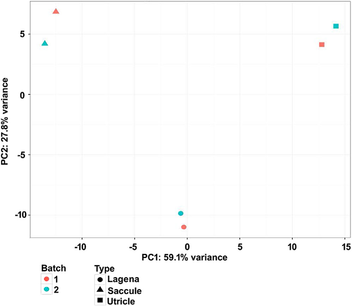
PCA of microarray expression levels across three different otolith organs in zebrafish. The x-axis represents the greatest source of variance across all samples and the y-axis represents the second greatest. Each symbol represents the results of one microarray hybridization, as indicated in the legend. Red symbols represent RNA samples from the first batch and blue symbols represent RNA samples from the second batch. Six samples are well grouped by different otolith organs, indicating that there is less variance between samples of the same otolith organs than between two different batches. The two greatest principal components account for 86.9% (59.1% + 27.8%) of the variance.
Analysis of the transcripts obtained from this experiment allowed us to determine which genes are expressed in each otolith organ. The expression levels of the top 200 genes in the saccule, utricle, and lagena, are shown in Figures 2–4, respectively. Expression levels and abundance rankings for the top 200 genes in all the three otolith organs are also illustrated. These figures demonstrate that gene expression is largely conserved among all otolith organs. Some of the genes were abundantly expressed in all the three otoliths organs including runt-related transcription factor 1 (runx1), mitochondrial cytochrome oxidases including mt-co1, and mt-co3 as well as mitochondrial encoded NADH dehydrogenases such as mt-nd1–4.
Fig. 2.
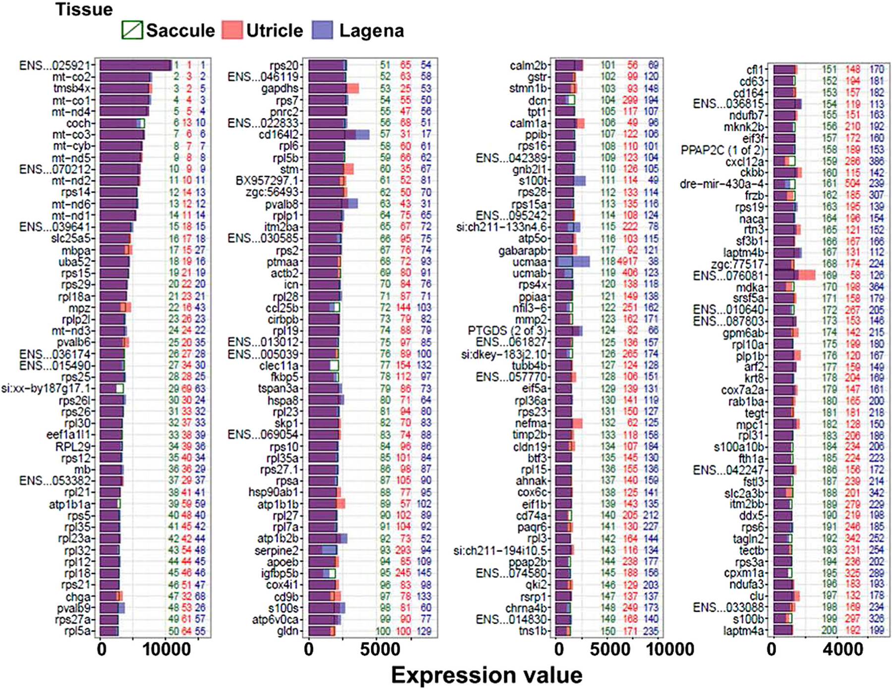
Expression levels of top 200 genes in the saccule. The bars show the fluorescent density of each gene after normalization. Numbers in green, red, and blue signify the abundance rank of the genes in the saccule, utricle, and lagena, respectively. In this and all subsequent figures, the last six digits of ENSMUST identification numbers are used.
Fig. 4.
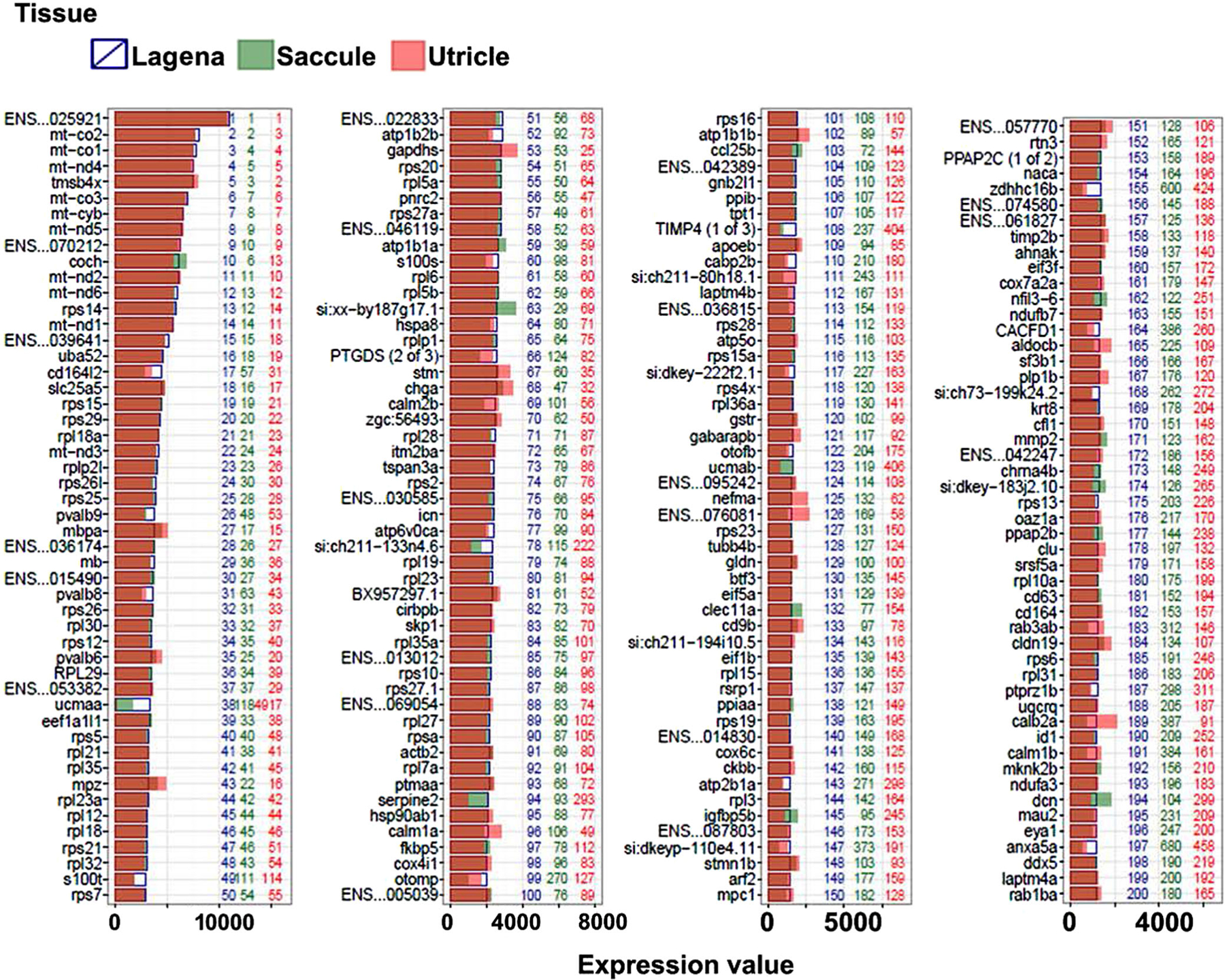
Expression levels of top 200 genes in the lagena. The bars show the fluorescent density of each gene after normalization. Numbers in green, red, and blue represent the abundance rank of the genes in the saccule, utricle, and lagena respectively.
Next, key differences in gene expression among the saccule, utricle and lagena were analyzed in order to understand the molecular basis underlying functional and developmental differences in these otolith organs. Transcript abundance in the saccule was compared with those of the utricle and lagena. Figure 5 illustrates the most differentially expressed genes in the saccule compared to the lagena (A) and utricle (B). We observed higher expression levels of wnt11r, cep41, prox1a, raraa, sema3e, gdf10a, otol1a, ctgfa, nr2f1a, and vwa2 in the saccule. These genes have also been demonstrated to be expressed in the otic vesicle in zebrafish in previous studies which serves to verify their relevance in hearing function of the saccule in zebrafish (Glasgow and Tomarev, 1998; Lee et al., 2012; Lu and DeSmidt, 2013; Pistocchi et al., 2008; Maier et al., 2014; http://zfin.org/). The functions of cep41, aldh1a2, otol1a, ctgfa, and nr2f1 (mouse orthologous genes) have been related to ear morphology and otic capsule development in mouse models (http://www.informatics.jax.org/). prox1a is widely expressed in the mouse inner ear, especially in the cochlea (Gray et al., 2004; Hartman et al., 2007; Hume et al., 2007; Dominguez-Frutos et al., 2009). Raraa has shown to be expressed in a wide variety of mouse ear compartments, such as in the vestibular component, the middle ear labyrinth, the organ of Corti, the limbus lamina spiralis, the spiral ligament, and the stria vascularis (Romand et al., 2002; Gray et al., 2004; Visel et al., 2004). Knockout mouse model for raraa orthologue showed hearing abnormalities (Lufkin et al., 1993). Sema3e, gdf10, and ctgf expression have been observed in the cochlea, otic capsule, pharyngotympanic tube, and tubotympanic recess of mice (Diez-Roux et al., 2011). Knockout mouse models for ctgf orthologue showed abnormalities in hearing and vestibular functions (Ivkovic et al., 2003; Doherty et al., 2010). Similar phenotypes were observed in a knockout mouse model (Qiu et al., 1997) for the nr2f1a orthologue that has been shown to be expressed in the cochlea (Jonk et al., 1994). In a patient with CHARGE syndrome and carrying a de novo balanced translocation involving Chromosomes 2 and 7, translocation breakpoints were mapped and Sema3e orthologue was identified within 200 kb of the breakpoint on 7q21.11 (Martin et al., 2001; Lalani et al., 2004). The chromosomal locations of human orthologues of cep41, cbln4, wnt11, masp1, aldh1a2, pla2g4c, sema3e, c1qtnf5, entpd5a, and apcdd1l have been associated with hereditary hearing loss in human patients (http://hereditaryhearingloss.org/). Figure 6 shows the differentially expressed genes in the utricle compared to the saccule (A) and lagena (B). Figure 7 shows the differentially expressed genes in the lagena compared to the saccule (A) and utricle (B). The comparisons show quite different gene expression profiles in three otolith organs, indicating different functional roles of these otolith organs in the zebrafish ear. The congruity between gene expression patterns in our study and others demonstrates the quality of the samples we collected from the three otolith organs in this study.
Fig. 5.
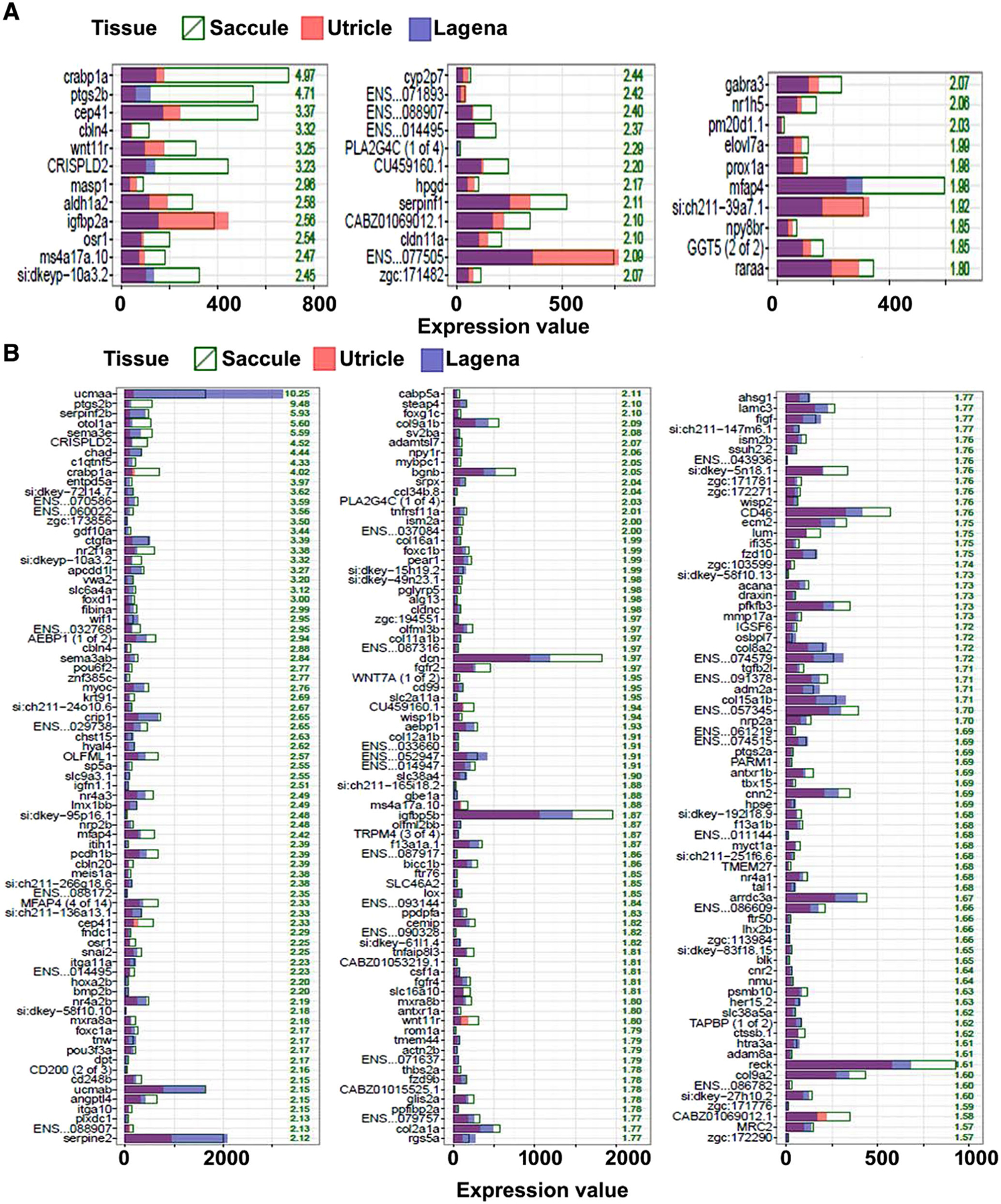
Differentially expressed genes in Saccule. The numerical values represent the fold difference in expression in the saccule versus the lagena (A) or the saccule versus the utricle (B).
Fig. 6.
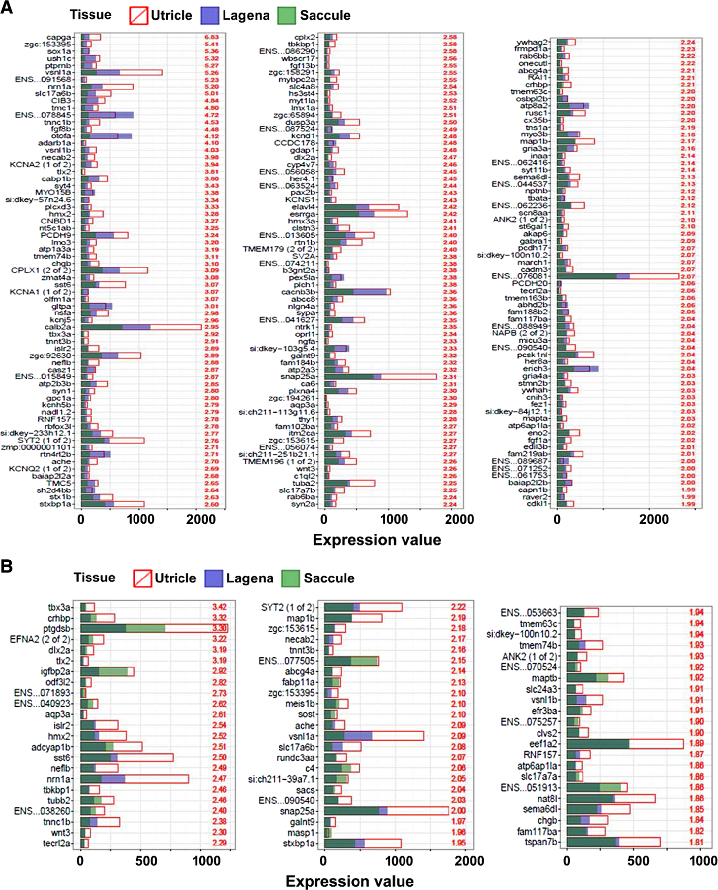
Differentially expressed genes in the utricle. The numerical values represent the fold difference in expression in the utricle versus the saccule (A) or the utricle versus the lagena (B).
Fig. 7.
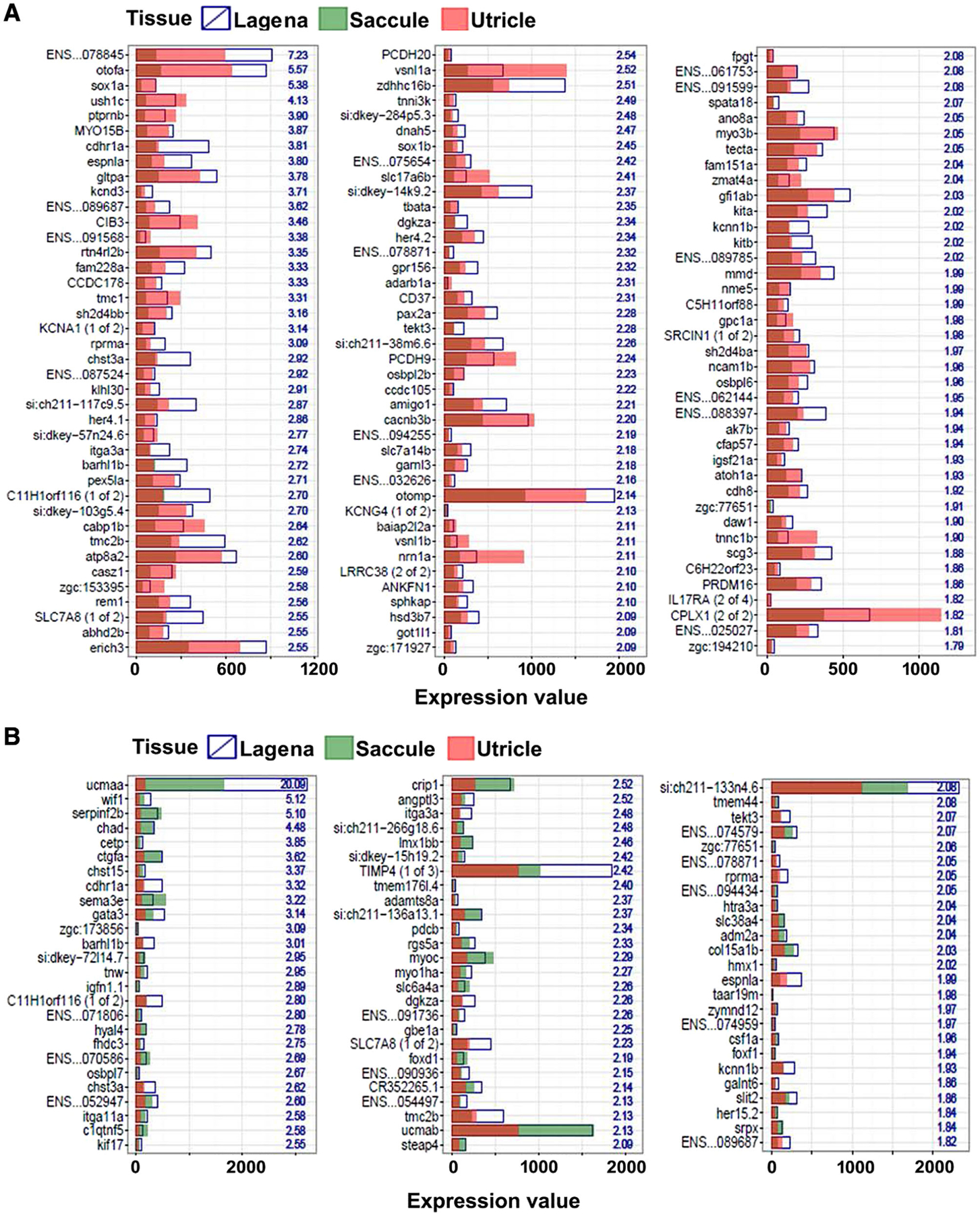
Differentially expressed genes in the lagena. The numerical values represent the fold difference in expression in the lagena versus the saccule (A) or the lagena versus the utricle (B).
Furthermore, we analyzed the differential expression of 96 transcripts to investigate their potential structural and functional contributions in the auditory system. We observed the expression of actn1, anxa6, arf1, argap17a, calm2a, calm2b, cdh23, dpysl2b, chd3, flnb, fscn2b, pls1, twf2b, ush1c, xirp2a, ywhaqa, and ywhaqb genes (Fig. 8). There are no studies available regarding the functional characterization and physiological relevance of these genes in the auditory system of zebrafish. However, some of these genes have been shown to be expressed in the mouse auditory system and implicated in maintaining the structure of stereocilia as well as in hearing. The expressions of calm2, cdh23, dpysl2b, flnb, pls1, twf2b, and ush1c orthologues have been observed in the mouse auditory system (Kamata et al., 1998; Di Palma et al., 2001; Ficker et al., 2004; Johnston et al., 2004; Visel et al., 2004; Lelli et al., 2009; Diez-Roux et al., 2011; Kamiya et al., 2014). Inner ear hair cells of Pls1-null mice have smaller and thinner stereocilia bundles and the mice develop progressive hearing loss at all frequencies (Taylor et al., 2015). Another gene USH1C was mapped to the PDZ73 gene to Chromosome 11p15.4-p15.1 by genomic sequence analysis and FISH (Scanlan et al., 1999). Mice with spontaneous hypomorphic mutations and knockout for USH1C showed behavioral, hearing, and vestibular abnormalities (Johnson et al., 2003; Lefevre et al., 2008; Grillet et al., 2009; Tian et al., 2010). Similarly, several phenotypes such as age-related hearing loss, degeneration of stereocilia, hair cell death, behavioral changes, and loss of harmonin in apex of hair cells have been observed in mouse models generated by chemical induction, knockouts, and spontaneous methods for cdh23 gene (Noben-Trauth et al., 1997; Di Palma et al., 2001; Zheng et al., 2005; Schwander et al., 2007; Bahloul et al., 2010; Manji et al., 2011; Han et al., 2014). In human studies, USH1C mutations were determined to be the cause of Usher syndrome type I in seven Acadians, one Pakistani, and one Canadian homozygous for the Acadian alleles (Ouyang et al., 2003), in two siblings from a Caucasian British family with hearing loss diagnosed at 4 years of age and retinitis pigmentosa (Saihan et al., 2011), and in 12 patients from 8 Israeli families of Yemenite Jewish origin with retinitis pigmentosa and late-onset hearing loss (Khateb et al., 2012). In a Cuban family, two different CDH23 mutations were identified (Bolz et al., 2001). Two nonsense and two frameshift mutations in the CDH23 gene in four families with USH1D (Bork et al., 2001), three novel mutations in 33 patients with type I Usher syndrome in whom USH1B and USH1C had been excluded (von Brederlow et al., 2002), a comprehensive catalog of 33 novel CDH23 mutations with recessive nonsyndromic deafness or Usher syndrome type I (Astuto et al., 2002), three families with Usher syndrome type I in which affected members carrying mutations in both CDH23 and PCDH15 (Zheng et al., 2005) have been observed.
Fig. 8.
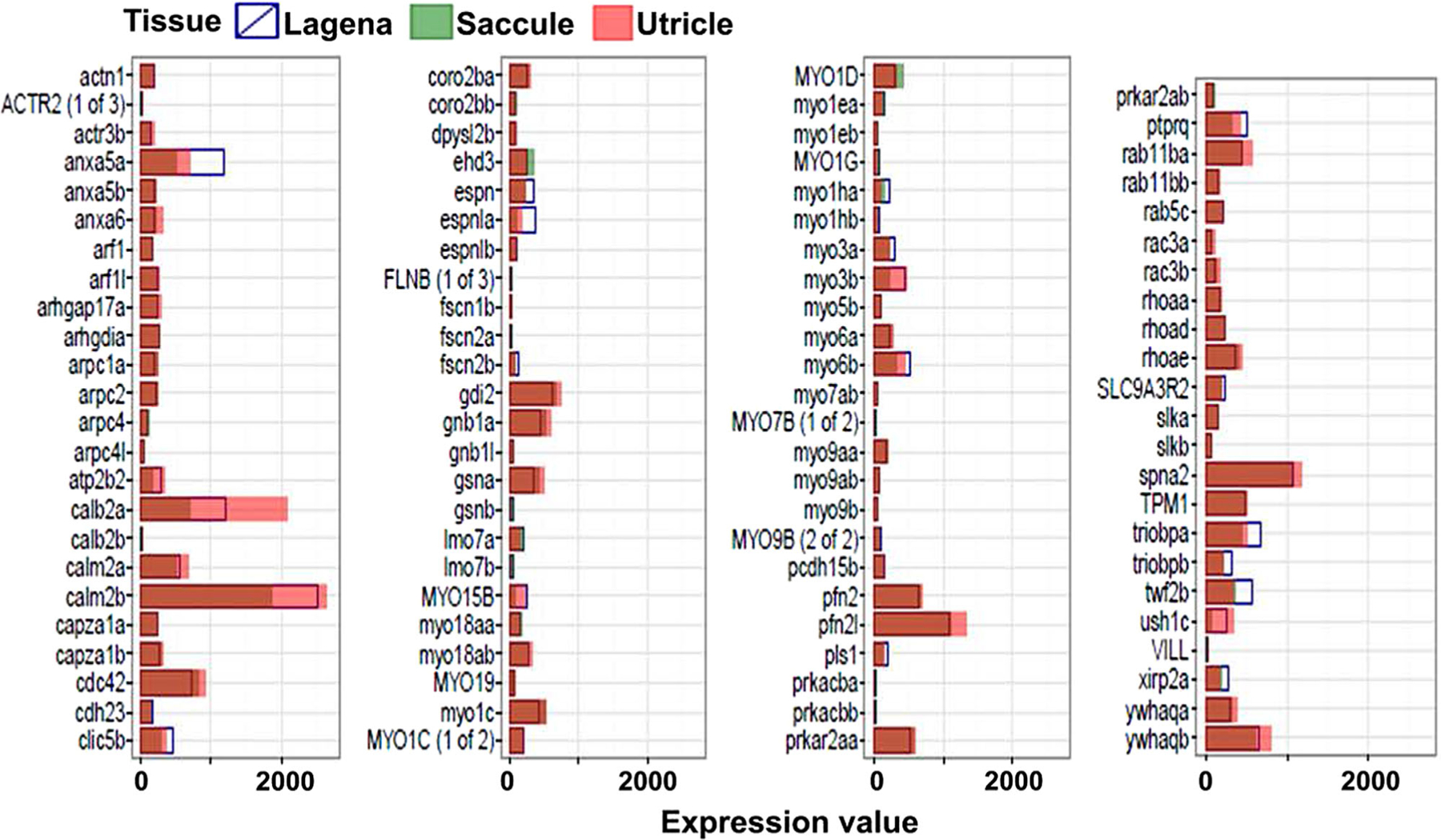
Expression level of genes associated with to stereocilia structure in the saccule, utricle and lagena.
Clic4, slc17a8, tmc1, tmc2a, tmc2b, and trpv4 have been demonstrated to be highly expressed in zebrafish in saccular, lagenar, utricular, crista, and neuromast hair cells, which is in agreement with the findings of the present study (Fig. 9) (Amato et al., 2012; Einhorn et al., 2012; Gabashvili et al., 2007; R. Maeda et al., 2014; Mangos et al., 2007; Obholzer et al., 2008). In case of mice, Clic5 has been shown to express in saccular and utricular maculae (Gagnon et al., 2006). Mice homozygous for a spontaneous mutation in Clic5 exhibit head bobbing and circling behavior, inability to swim, and complete deafness by 7–8 months of age caused by dysmorphic stereocilia and progressive hair cell degeneration. Slc17a8 is not expressed in the auditory system; however, studies on mouse models show changes in behavior, hearing, vestibular ear, and nervous system, (Gras et al., 2008; Seal et al., 2008). Tmc1 expression has been shown in otic capsule, macula of utricle, utricle, and cochlea of mice by in situ hybridization (ISH) (Diez-Roux et al., 2011; Kawashima et al., 2011). Spontaneous mutation and knockout of Tmc1 in mouse models have shown hearing defects. In the mouse, tmc2 is expressed in the utricular macula, utricle, and cochlea. Reverse genetic experiments reveal that deletion of Tmc1 in mice caused deafness, whereas Tmc2 deletion did not cause any distinct phenotype. Interestingly, targeted deletion of both genes yielded mice with auditory and vestibular malfunction due to loss of mechanotransduction activity of inner ear hair cells (Kawashima et al., 2011).
Fig. 9.
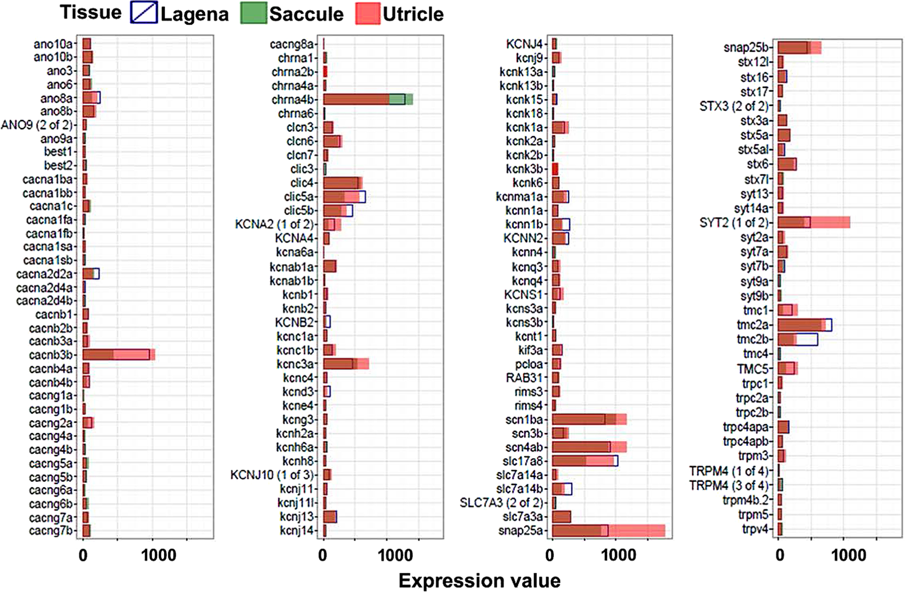
Expression level of genes associated with to Ion channels in the saccule, utricle and lagena.
In humans, a truncating mutation in the CLIC5 was found to be the cause of autosomal recessive deafness-103 in two siblings of Turkish decent (Seco et al., 2016). By genomic sequence analysis, tmc1 gene was mapped to the Chromosome 9q13-q21 (Kurima et al., 2002). A Caucasian family in North American was also found to have hearing loss associated with the 9q13-q21 chromosome location. Ten Pakistani families were found to have mutations in tmc1 and were diagnosed with DFNB7/11 (Kitajiri et al., 2007), as were seven Turkish families (Hilgert et al., 2008) and members of a Chinese family that were diagnosed with DFNA36 (Zhao et al., 2014). By genomic sequence analysis, the tmc2 gene was mapped to Chromosome 20p13 (Kurima et al., 2002) and TRPV4 gene to Chromosome 12q24.1 (Liedtke et al., 2000).
The information about the genes involved in regulating cell cycle can provide important clues about the development and maintenance of the zebrafish inner ear. The genes related to cell cycle in the saccule, utricle, and lagena are shown in Figure 10. We observed the expression of brca2, ccnb2, cdkn1ba, cdkn1bb, cdkn1ca, gadd45aa, mad2l1, mcm4, notch2, and pmp22b in all the three otolith organs. The expression of cdkn1b has been seen in the cochlea, cochlear duct epithelium, and organ of Corti of mice (Nagahama et al., 2001; Chen et al., 2002; Hartman et al., 2007), and Notch2 in the mouse otocyst (Hamada et al., 1999). Ccnb2 has been mapped to the Chromosome 15q22.2. Cdkn1c has been associated with Beckwith–Wiedemann Syndrome (BWS) in nine unrelated Japanese patients and detected mutations in two BWS patients (Hatada et al., 1996). A study of patients with BWS identified multiple mutations in the Cdkn1c gene which presumably caused this syndrome (Lam et al., 1999). By sequencing this gene, they were able to identify five probands of this gene. Three probands were found among 7 familial cases where 2 probands were found among 54 sporadic cases. Patients with BWS have a reduced expression of Cdkn1c which indicates that the disease is caused by haploinsufficiency. (Algar et al., 1999) Another study examined 7 families with hereditary BWS identified 32 individuals with BWS and observed mutations in Cdkn1c (Algar et al., 2000). Another study (Romanelli et al., 2010) identified 7 novel CDKN1C mutations in 8 out of the 50 BWS patients. The MAD2L1 gene has been mapped to 5q23-q31 by fluorescence ISH (Xu et al., 1997).
Fig. 10.
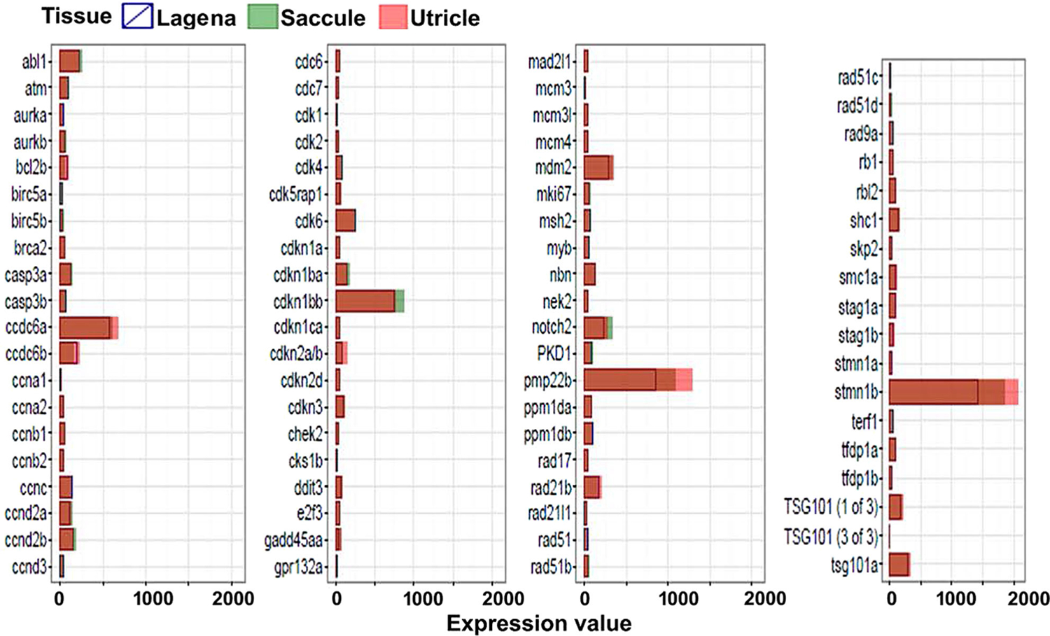
Expression level of genes associated with cell cycle in the saccule, utricle and lagena.
Figure 11 shows the expression of over 80 genes that have been implicated in human deafness or related to hearing in animal models. col11a1a, col11ab, col11a2, col4a5, dfna5b, eya1, eya2, grhl2a, grhl2b, kcnq4, otofa, otofb, and slc26a5 are highly expressed in the zebrafish otic vesicle, hair cells, and semicircular canal (Croushore et al., 2005; Blasiole et al., 2006; Albert et al., 2007; Xiao and Baier, 2007; Baas et al., 2009; Yokoi et al., 2009; Fang et al., 2010; Hoffman et al., 2010; Janicke et al., 2010; Landgraf et al., 2010; Wu et al., 2014; Chatterjee et al., 2015; Wang et al., 2015). col11a1 has been shown to express in the inner ear and otic capsule of mice (Yoshioka et al., 1995; Diez-Roux et al., 2011) and spontaneous mutation mouse models show both auditory and vestibular abnormalities. Although col11a2 expression has not been observed in the auditory system of mice, a knockout model still seems to show vestibular abnormalities (Li et al., 2001). Abundant Dfna5 expression has been observed in the cochlea and inner ear of mice (Maeda et al., 2001) and Dfna5 knockout mice develop hearing abnormalities (Van Laer et al., 2005). Eya1 is expressed in otocyst, otocyst epithelium, inner ear, otic capsule, and vestibule (Abdelhak et al., 1997; Lillevali et al., 2004; Ozaki et al., 2004). Spontaneous Eya1 knockout and targeted mouse models show hearing abnormalities (Johnson et al., 1999; Xu et al., 1999; Ahmed et al., 2012). Grhl2 expression in mice has been observed in the inner ear, cochlea, middle ear, pharyngotympanic tube (Visel et al., 2004; Diez-Roux et al., 2011). Similarly, Kcnq4 expression has been observed in the utricle (Holt et al., 2007), and knockout mouse models for this gene show hearing abnormalities (Kharkovets et al., 2006). Otof expression has shown to be expressed in the inner ear of mice (Wilson et al., 2005; Schwander et al., 2007). Otoferlin deficient mice (Otof −/−) are profoundly deaf demonstrating complete abrogation of exocytosis in inner hair cells (Roux et al., 2006). Slc26a5 expression has been observed in the cochlea of mice (Lelli et al., 2009) and radiation, targeted, and knockout models show hearing abnormalities (Liberman et al., 2002; Gao et al., 2007; Palos et al., 2008). The mutations in SLC26A5 cause deafness in 2 Japanese siblings (Mutai et al., 2013), col11a2 in American, Dutch, Iranian, Tunisian, and Turkish families (Chakchouk et al., 2015), DFNA5 in 2 Dutch families, 2 Chinese families, 1 Iranian family, and a 328-member cohort study (Van Laer et al., 1997; Van Camp et al., 2002; Yu et al., 2004; Van Laer et al., 2008). An autosomal dominant form of progressive, non-syndromic sensorineural hearing loss was identified in an American family in which a specific GRHL2 mutation cosegregated with affected patients (Peters et al., 2002). Similarly, in a large five-generation family, the GRHL2 mutation was shown to cause deafness (Vona et al., 2013). A heterozygous mutation in KCNQ4 has been identified in a large deaf family, and it was concluded that KCNQ4 related hearing loss is intrinsic to outer hair cells and KCNQ4 forms heteromeric channels with KCNQ3 (Kubisch et al., 1999). A study on three families from the Netherlands and Belgium (Van Camp et al., 2002) and two from Indonesia and the United States (Coucke et al., 1994) found missense mutations in KCNQ4 gene. KCNQ4 was also shown to be the cause of deafness in a five-generation American family (Talebizadeh et al., 1999), Dutch family (Van Hauwe et al., 2000), and four-generation Spanish family (Mencía et al., 2008). Three Indian siblings with OTOF mutation suffered profound hearing loss (Yasunaga et al., 2000), and one out of four Druze families had a novel OTOF mutation (Adato et al., 2000). In one Cuban family, two Spanish families, and eight sporadic Spanish patients, an OTOF mutation was associated with nonsyndromic sensorineural hearing loss (Migliosi et al., 2002). The screening for the same OTOF mutation was extended to 289 additional unrelated families, finding 15 new cases, 9 of which were homozygous and 6 of which were heterozygous (Rodríguez-Ballesteros et al., 2003). Thus, the deafness genes that we identified in zebrafish have implications in human hearing loss validating the use of zebrafish models to identify and characterize human deafness genes.
Fig. 11.
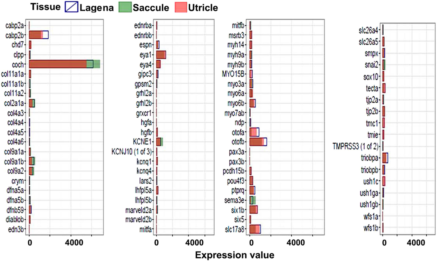
Expression level of genes associated with deafness in the saccule, utricle and lagena.
To further confirm our microarray data, we performed real-time quantitative PCR (qRT-PCR). We used the top differentially expressed genes in three otolith organs from our microarray data. We computed the log2-fold difference between the expression of genes in saccule and lagena using utricle as the normalizer. As shown in Figure 12, we observed that the differentially expressed genes identified by the two techniques, qRT-PCR and microarray, are highly consistent. These differentially expressed genes may provide valuable information to understand different biological properties (such as structural and functional differences) of three otolith organs.
Fig. 12.
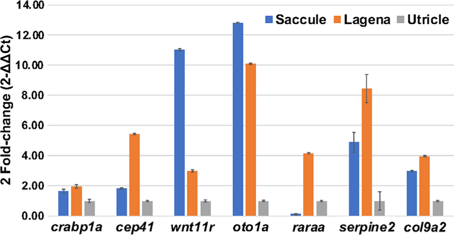
Expression levels of genes in the saccule and lagena using qRT-PCR; values from the utricle were used for normalization.
Remarkably, zebrafish and mammalian hair cells share many fundamental features at the cellular and molecular level (Coffin et al., 2004). Several genetic studies have demonstrated that molecules required by the auditory and vestibular system of zebrafish are also important in mice and humans (Nicolson, 2005; Yariz et al., 2012; Diaz-Horta et al., 2014; Grati et al., 2015; Bensaid et al., 2016; Varshney et al., 2016; DeSmidt et al., 2019; Zou et al., 2019). The inner ear of mammals is encased in the bony labyrinth deep inside the temporal bone, which prevents the sensory hair cells from being easily accessed. Contrarily, the hair cells of zebrafish larvae are found not only in the inner ear, but also within a lateral-line system that runs along exterior surface of the fish. Although the zebrafish uses hair cells in the lateral line system to detect water currents, the hair cells are remarkably similar in structure and function to the hair cells of the inner ear of mammals. Furthermore, zebrafish larvae are optically transparent. The lateral line contains a series of neuromasts, mechanoreceptors that form an interconnected network along the head and body of the fish. Because the hair cells of the lateral line are located on the external surface of the zebrafish, treatment and subsequent observation of live hair cells make the lateral line system particularly amenable to hair cell research. This access holds the promise of providing relatively straightforward pharmacodynamic assays and hair cell structure and in vivo functional studies. In addition, hair cells of zebrafish can mature within 24 hr (Kindt et al., 2012; Dow et al., 2015), whereas mammalian hair cells develop over several weeks (Kraus and Aulbach-Kraus, 1981; Romand and Varela-Nieto, 2003). The rapid development of zebrafish hair cells allows the entire maturation process of the hair cells to be recorded within one imaging session. Furthermore, several genetic techniques have been well established using the zebrafish model. For example, transgenic modifications of the zebrafish genome can allow certain gene products such as fluorescent markers to be expressed in a cell- or tissue-specific manner (Kwan et al., 2007). The fluorescent markers can easily be used in the transparent larvae, which enables visualization of hair cell structures in vivo and allows for cellular functions to be dynamically imaged in living cells.
In summary, the present investigation is the first transcriptomic study investigating the quantitative expression differences among the saccule, utricle and lagena of adult zebrafish at the genomic level. Although the expression of all the genes investigated is conserved among three otolith organs, there are few genes that are highly differentially expressed in the saccule, utricle and lagena. These differential expressed genes may impart unique structures and physiological functions to these otolith organs. This data set provides a unique tool to the auditory and neuroscience community to explore the functional roles of these genes in future studies employing CRISPR/Cas9 genome editing.
Fig. 3.
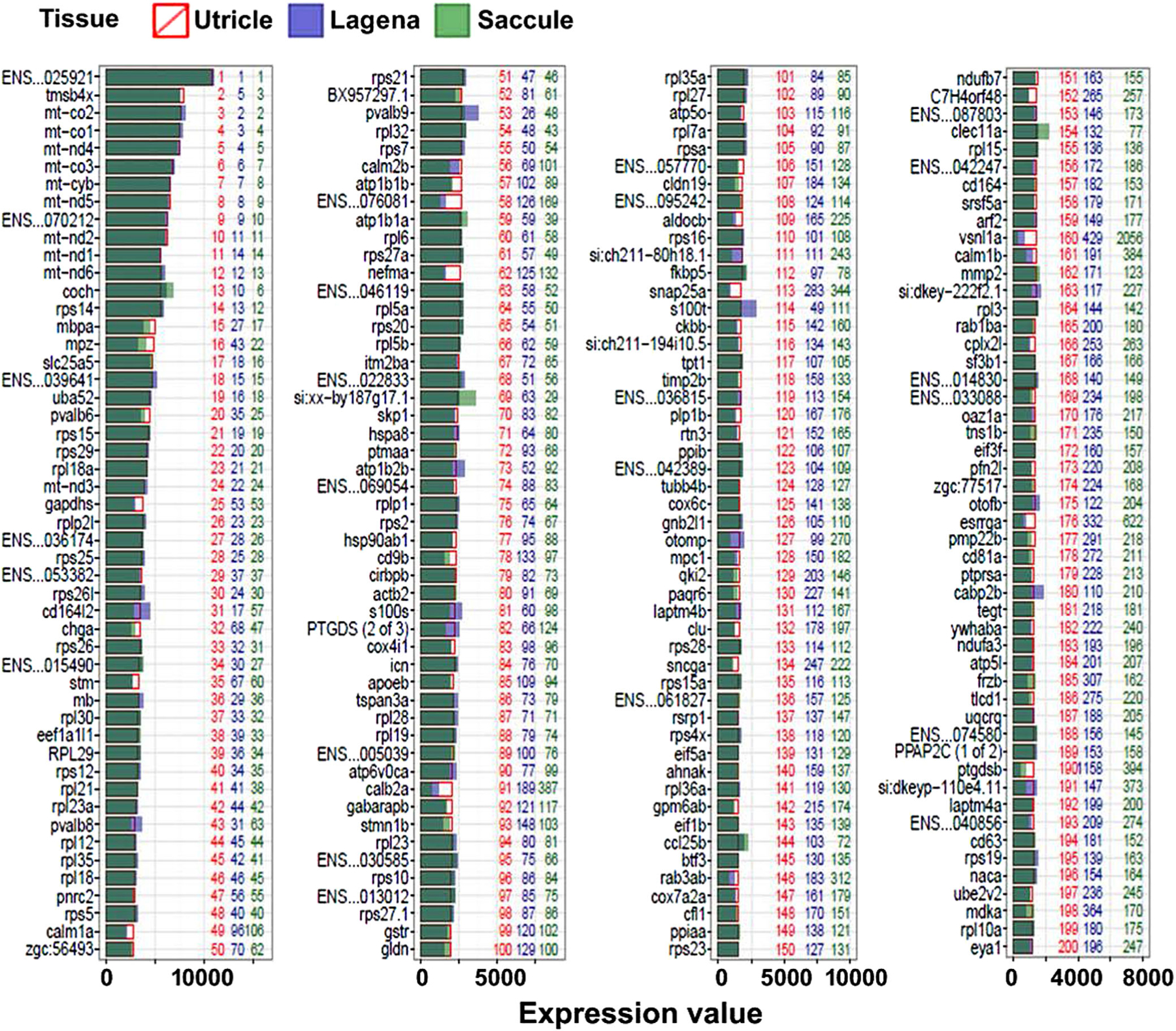
Expression levels of top 200 genes in the utricle. The bars show the fluorescent density of each gene after normalization. Numbers in green, red, and blue represent the abundance rank of the genes in the saccule, utricle, and lagena respectively.
ACKNOWLEDGMENTS
This work is supported by NIH/NIDCD grants R01 DC005575 and R01 DC012115 to Dr. Xuezhong Liu, University of Miami Provost’s Research Award and Gabelli Fellowship to Dr. Zhongmin Lu, and NIH/NIGMS grant P20GM103636 to Dr. Gaurav K. Varshney.
Grant sponsor: National Institute of General Medical Sciences; Grant number: P20GM103636; Grant sponsor: National Institute on Deafness and Other Communication Disorders; Grant number: R01 DC005575 R01 DC012115; Grant sponsor: University of Miami Provost’s Research Award and Gabelli Fellowship.
Literature Cited
- Abdelhak S, Kalatzis V, Heilig R, Compain S, Samson D, Vincent C, Levi-Acobas F, Cruaud C, Le Merrer M, Mathieu M, et al. 1997. Clustering of mutations responsible for branchio-oto-renal (BOR) syndrome in the eyes absent homologous region (eyaHR) of EYA1. Hum Mol Genet 6:2247–2255. [DOI] [PubMed] [Google Scholar]
- Adato A, Raskin L, Petit C, Bonne-Tamir B. 2000. Deafness heterogeneity in a Druze isolate from the Middle East: novel OTOF and PDS mutations, low prevalence of GJB2 35delG mutation and indication for a new DFNB locus. Eur J Hum Genet 8:437–442. [DOI] [PubMed] [Google Scholar]
- Ahmed M, Wong EY, Sun J, Xu J, Wang F, Xu PX. 2012. Eya1-Six1 interaction is sufficient to induce hair cell fate in the cochlea by activating Atoh1 expression in cooperation with Sox2. Dev Cell 22: 377–390. [DOI] [PMC free article] [PubMed] [Google Scholar]
- Albert JT, Winter H, Schaechinger TJ, Weber T, Wang X, He DZ, Hendrich O, Geisler HS, Zimmermann U, Oelmann K, et al. 2007. Voltage-sensitive prestin orthologue expressed in zebrafish hair cells. J Physiol 580:451–461. [DOI] [PMC free article] [PubMed] [Google Scholar]
- Algar E, Brickell S, Deeble G, Amor D, Smith P. 2000. Analysis of CDKN1C in Beckwith–Wiedemann syndrome. Hum Mutat 15: 497–508. [DOI] [PubMed] [Google Scholar]
- Algar EM, Deeble GJ, Smith PJ. 1999. CDKN1C expression in Beckwith–Wiedemann syndrome patients with allele imbalance. J Med Genet 36:524–531. [PMC free article] [PubMed] [Google Scholar]
- Amato V, Vina E, Calavia MG, Guerrera MC, Laura R, Navarro M, De Carlos F, Cobo J, Germanà A, Vega JA. 2012. TRPV4 in the sensory organs of adult zebrafish. Microsc Res Tech 75:89–96. [DOI] [PubMed] [Google Scholar]
- Astuto LM, Bork JM, Weston MD, Askew JW, Fields RR, Orten DJ, Ohliger SJ, Riazuddin S, Morell RJ, Khan S, et al. 2002. CDH23 mutation and phenotype heterogeneity: a profile of 107 diverse families with Usher syndrome and nonsyndromic deafness. Am J Hum Genet 71:262–275. [DOI] [PMC free article] [PubMed] [Google Scholar]
- Baas D, Malbouyres M, Haftek-Terreau Z, Le Guellec D, Ruggiero F. 2009. Craniofacial cartilage morphogenesis requires zebrafish col11a1 activity. Matrix Biol 28:490–502. [DOI] [PubMed] [Google Scholar]
- Bahloul A, Michel V, Hardelin JP, Nouaille S, Hoos S, Houdusse A, England P, Petit C. 2010. Cadherin-23, myosin VIIa and harmonin, encoded by Usher syndrome type I genes, form a ternary complex and interact with membrane phospholipids. Hum Mol Genet 19: 3557–3565. [DOI] [PMC free article] [PubMed] [Google Scholar]
- Bang PI, Sewell WF, Malicki JJ. 2001. Morphology and cell type heterogeneities of the inner ear epithelia in adult and juvenile zebrafish (Danio rerio). J Comp Neurol 438:173–190. [DOI] [PubMed] [Google Scholar]
- Barta CL, Liu H, Chen L, Giffen KP, Li Y, Kramer KL, Beisel KW, He DZ. 2018. RNA-seq transcriptomic analysis of adult zebrafish inner ear hair cells. Sci Data 5:180005. [DOI] [PMC free article] [PubMed] [Google Scholar]
- Bensaid M, Grati M, Zou B, Chakchouk I, Ma Q, Yao Q, Hammami B, Yan D, Mittal R, Ghorbel A, et al. 2016. A mutation in SLC22A4encoding organic cation and ergothioneine transporter of cochlea strial endothelium causes human recessive non-syndromic hearing loss DFNB60. Hum Genet 135:513–524. [DOI] [PMC free article] [PubMed] [Google Scholar]
- Berriz GF, Roth FP. 2008. The Synergizer service for translating gene, protein and other biological identifiers. Bioinformatics 24: 2272–2273. [DOI] [PMC free article] [PubMed] [Google Scholar]
- Bever MM, Fekete DM. 2002. Atlas of the developing inner ear in zebrafish. Dev Dyn 223:536–543. [DOI] [PubMed] [Google Scholar]
- Blasiole B, Canfield VA, Vollrath MA, Huss D, Mohideen MA, Dickman JD, Cheng KC, Fekete DM, Levenson R. 2006. Separate Na,K-ATPase genes are required for otolith formation and semicircular canal development in zebrafish. Dev Biol 294:148–160. [DOI] [PubMed] [Google Scholar]
- Bolz H, von Brederlow B, Ramírez A, Bryda EC, Kutsche K, Nothwang HG, Seeliger M, del C-Salcedó CM, Vila MC, Molina OP, et al. 2001. Mutation of CDH23, encoding a new member of the cadherin gene family, causes Usher syndrome type 1D. Nat Genet 27:108–112. [DOI] [PubMed] [Google Scholar]
- Bork JM, Peters LM, Riazuddin S, Bernstein SL, Ahmed ZM, Ness SL, Polomeno R, Ramesh A, Schloss M, Srisailpathy CR, et al. 2001. Usher syndrome 1D and nonsyndromic autosomal recessive deafness DFNB12 are caused by allelic mutations of the novel cadherin-like gene CDH23. Am J Hum Genet 68:26–37. [DOI] [PMC free article] [PubMed] [Google Scholar]
- Carvalho BS, Irizarry RA. 2010. A framework for oligonucleotide microarray preprocessing. Bioinformatics 26:2363–2367. [DOI] [PMC free article] [PubMed] [Google Scholar]
- Chakchouk I, Grati M, Bademci G, Bensaid M, Ma Q, Chakroun A, Foster J 2nd, Yan D, Duman D, Diaz-Horta O, et al. 2015. Novel mutations confirm that COL11A2 is responsible for autosomal recessive non-syndromic hearing loss DFNB53. Mol Genet Genomics 290:1327–1334. [DOI] [PMC free article] [PubMed] [Google Scholar]
- Chatterjee P, Padmanarayana M, Abdullah N, Holman CL, LaDu J, Tanguay RL, Johnson CP. 2015. Otoferlin deficiency in zebrafish results in defects in balance and hearing: rescue of the balance and hearing phenotype with full-length and truncated forms of mouse otoferlin. Mol Cell Biol 35:1043–1054. [DOI] [PMC free article] [PubMed] [Google Scholar]
- Chen P, Johnson JE, Zoghbi HY, Segil N. 2002. The role of Math1 in inner ear development: Uncoupling the establishment of the sensory primordium from hair cell fate determination. Development 129:2495–2505. [DOI] [PubMed] [Google Scholar]
- Coffin A, Kelley M, Manley GA, Popper AN. 2004. Evolution of sensory hair cells. In: Manley GA, Fay RR, Popper AN, editors. Evolution of the vertebrate auditory system. New York, NY: Springer. p 55–94. [Google Scholar]
- Coucke P, Van Camp G, Djoyodiharjo B, Smith SD, Frants RR, Padberg GW, Darby JK, Huizing EH, Cremers CW, Kimberling WJ, et al. 1994. Linkage of autosomal dominant hearing loss to the short arm of chromosome 1 in two families. N Engl J Med 331:425–431. [DOI] [PubMed] [Google Scholar]
- Croushore JA, Blasiole B, Riddle RC, Thisse C, Thisse B, Canfield VA, Robertson GP, Cheng KC, Levenson R. 2005. Ptena and ptenb genes play distinct roles in zebrafish embryogenesis. Dev Dyn 234:911–921. [DOI] [PMC free article] [PubMed] [Google Scholar]
- DeSmidt AA, Zou B, Yan D, Grati M, Yan D, Mittal R, Yao Q, Richmond MT, Denyer S, Liu XZ, et al. 2019. Zebrafish model for non-syndromic X-linked sensorineural deafness, DFNX1. Anat Rec 10.1002/ar.24115. [DOI] [PubMed]
- Di Palma F, Holme RH, Bryda EC, Belyantseva IA, Pellegrino R, Kachar B, Steel KP, Noben-Trauth K. 2001. Mutations in Cdh23, encoding a new type of cadherin, cause stereocilia disorganization in waltzer, the mouse model for Usher syndrome type 1D. Nat Genet 27:103–107. [DOI] [PubMed] [Google Scholar]
- Diaz-Horta O, Subasioglu-Uzak A, Grati M, DeSmidt DA, Foster J II, Cao L, Tokgoz-Yilmaz S, Duman D, Cengiz FB, Abad C, et al. 2014. FAM65B is a membrane-associated protein of hair cell stereocilia required for hearing. Proc Natl Acad Sci USA 111: 9864–9868. [DOI] [PMC free article] [PubMed] [Google Scholar]
- Diez-Roux G, Banfi S, Sultan M, Geffers L, Anand S, Rozado D, Magen A, Canidio E, Pagani M, Peluso I, et al. 2011. A high-resolution anatomical atlas of the transcriptome in the mouse embryo. PLoS Biol 9:e1000582. [DOI] [PMC free article] [PubMed] [Google Scholar]
- Doherty HE, Kim HS, Hiller S, Sulik KK, Maeda N. 2010. A mouse strain where basal connective tissue growth factor gene expression can be switched from low to high. PLoS One 5:e12909. [DOI] [PMC free article] [PubMed] [Google Scholar]
- Dominguez-Frutos E, Vendrell V, Alvarez Y, Zelarayan LC, Lopez-Hernandez I, Ros M, Schimmang T. 2009. Tissue-specific requirements for FGF8 during early inner ear development. Mech Dev 126:873–881. [DOI] [PubMed] [Google Scholar]
- Dow E, Siletti K, Hudspeth AJ. 2015. Cellular projections from sensory hair cells form polarity-specific scaffolds during synaptogenesis. Genes Dev 29:1087–1094. [DOI] [PMC free article] [PubMed] [Google Scholar]
- Einhorn Z, Trapani JG, Liu Q, Nicolson T. 2012. Rabconnectin3alpha promotes stable activity of the H+ pump on synaptic vesicles in hair cells. J Neurosci 32:11144–11156. [DOI] [PMC free article] [PubMed] [Google Scholar]
- Fang M, Adams JS, McMahan BL, Brown RJ, Oxford JT. 2010. The expression patterns of minor fibrillar collagens during development in zebrafish. Gene Expr Patterns 10:315–322. [DOI] [PMC free article] [PubMed] [Google Scholar]
- Ficker M, Powles N, Warr N, Pirvola U, Maconochie M. 2004. Analysis of genes from inner ear developmental-stage cDNA subtraction reveals molecular regionalization of the otic capsule. Dev Biol 268:7–23. [DOI] [PubMed] [Google Scholar]
- Gabashvili IS, Sokolowski BH, Morton CC, Giersch AB. 2007. Ion channel gene expression in the inner ear. J Assoc Res Otolaryngol 8:305–328. [DOI] [PMC free article] [PubMed] [Google Scholar]
- Gagnon LH, Longo-Guess CM, Berryman M, Shin JB, Saylor KW, Yu H, Gillespie PG, Johnson KR. 2006. The chloride intracellular channel protein CLIC5 is expressed at high levels in hair cell stereocilia and is essential for normal inner ear function. J Neurosci 26:10188–10198. [DOI] [PMC free article] [PubMed] [Google Scholar]
- Gao J, Wang X, Wu X, Aguinaga S, Huynh K, Jia S, Matsuda K, Patel M, Zheng J, Cheatham M, et al. 2007. Prestin-based outer hair cell electromotility in knockin mice does not appear to adjust the operating point of a cilia-based amplifier. Proc Natl Acad Sci USA 104:12542–12547. [DOI] [PMC free article] [PubMed] [Google Scholar]
- Glasgow E, Tomarev SI. 1998. Restricted expression of the homeobox gene prox 1 in developing zebrafish. Mech Dev 76:175–178. [DOI] [PubMed] [Google Scholar]
- Gleason MR, Nagiel A, Jamet S, Vologodskaia M, Lopez-Schier H, Hudspeth AJ. 2009. The transmembrane inner ear (Tmie) protein is essential for normal hearing and balance in the zebrafish. Proc Natl Acad Sci USA 106:21347–21352. [DOI] [PMC free article] [PubMed] [Google Scholar]
- Go W, Bessarab D, Korzh V. 2010. atp2b1a regulates Ca(2+) export during differentiation and regeneration of mechanosensory hair cells in zebrafish. Cell Calcium 48:302–313. [DOI] [PubMed] [Google Scholar]
- Gras C, Amilhon B, Lepicard EM, Poirel O, Vinatier J, Herbin M, Dumas S, Tzavara ET, Wade MR, Nomikos GG, et al. 2008. The vesicular glutamate transporter VGLUT3 synergizes striatal acetylcholine tone. Nat Neurosci 11:292–300. [DOI] [PubMed] [Google Scholar]
- Grati M, Chakchouk I, Ma Q, Bensaid M, Desmidt A, Turki N, Yan D, Baanannou A, Mittal R, Driss N, et al. 2015. A missense mutation in DCDC2 causes human recessive deafness DFNB66, likely by interfering with sensory hair cell and supporting cell cilia length regulation. Hum Mol Genet 24:2482–2491. [DOI] [PMC free article] [PubMed] [Google Scholar]
- Gray PA, Fu H, Luo P, Zhao Q, Yu J, Ferrari A, Tenzen T, Yuk DI, Tsung EF, Cai Z, et al. 2004. Mouse brain organization revealed through direct genome-scale TF expression analysis. Science 306: 2255–2257. [DOI] [PubMed] [Google Scholar]
- Grillet N, Xiong W, Reynolds A, Kazmierczak P, Sato T, Lillo C, Dumont RA, Hintermann E, Sczaniecka A, Schwander M, et al. 2009. Harmonin mutations cause mechanotransduction defects in cochlear hair cells. Neuron 62:375–387. [DOI] [PMC free article] [PubMed] [Google Scholar]
- Haddon C, Lewis J. 1996. Early ear development in the embryo of the zebrafish, Danio rerio. J Comp Neurol 365:113–128. [DOI] [PubMed] [Google Scholar]
- Hamada Y, Kadokawa Y, Okabe M, Ikawa M, Coleman JR, Tsujimoto Y. 1999. Mutation in ankyrin repeats of the mouse Notch2 gene induces early embryonic lethality. Development 126:3415–3424. [DOI] [PubMed] [Google Scholar]
- Han HW, Chou CM, Chu CY, Cheng CH, Yang CH, Hung CC, Hwang PP, Lee SJ, Liao YF, Huang CJ. 2014. The Nogo-C2/Nogo receptor complex regulates the morphogenesis of zebrafish lateral line primordium through modulating the expression of dkk1b, a Wnt signal inhibitor. PLoS One 9:e86345. [DOI] [PMC free article] [PubMed] [Google Scholar]
- Hartman BH, Hayashi T, Nelson BR, Bermingham-McDonogh O, Reh TA. 2007. Dll3 is expressed in developing hair cells in the mammalian cochlea. Dev Dyn 236:2875–2883. [DOI] [PubMed] [Google Scholar]
- Hatada I, Ohashi H, Fukushima Y, Kaneko Y, Inoue M, Komoto Y, Okada A, Ohishi S, Nabetani A, Morisaki H, et al. 1996. An imprinted gene p57KIP2 is mutated in Beckwith–Wiedemann syndrome. Nat Genet 14:171–173. [DOI] [PubMed] [Google Scholar]
- Hilgert N, Alasti F, Dieltjens N, Pawlik B, Wollnik B, Uyguner O, Delmaghani S, Weil D, Petit C, Danis E, et al. 2008. Mutation analysis of TMC1 identifies four new mutations and suggests an additional deafness gene at loci DFNA36 and DFNB7/11. Clin Genet 74:223–232. [DOI] [PMC free article] [PubMed] [Google Scholar]
- Hoffman GG, Branam AM, Huang G, Pelegri F, Cole WG, Wenstrup RM, Greenspan DS. 2010. Characterization of the six zebrafish clade B fibrillar procollagen genes, with evidence for evolutionarily conserved alternative splicing within the pro-alpha1 (V) C-propeptide. Matrix Biol 29:261–275. [DOI] [PMC free article] [PubMed] [Google Scholar]
- Holt JR, Stauffer EA, Abraham D, Geleoc GS. 2007. Dominant-negative inhibition of M-like potassium conductances in hair cells of the mouse inner ear. J Neurosci 27:8940–8951. [DOI] [PMC free article] [PubMed] [Google Scholar]
- Hume CR, Bratt DL, Oesterle EC. 2007. Expression of LHX3 and SOX2 during mouse inner ear development. Gene Expr Patterns 7: 798–807. [DOI] [PMC free article] [PubMed] [Google Scholar]
- Hwang WY, Fu Y, Reyon D, Maeder ML, Tsai SQ, Sander JD, Peterson RT, Yeh JR, Joung JK. 2013. Efficient genome editing in zebrafish using a CRISPR-Cas system. Nat Biotechnol 31:227–229. [DOI] [PMC free article] [PubMed] [Google Scholar]
- Ivkovic S, Yoon BS, Popoff SN, Safadi FF, Libuda DE, Stephenson RC, Daluiski A, Lyons KM. 2003. Connective tissue growth factor coordinates chondrogenesis and angiogenesis during skeletal development. Development 130:2779–2791. [DOI] [PMC free article] [PubMed] [Google Scholar]
- Janicke M, Renisch B, Hammerschmidt M. 2010. Zebrafish grainyhead-like1 is a common marker of different non-keratinocyte epidermal cell lineages, which segregate from each other in a Foxi3-dependent manner. Int J Dev Biol 54:837–850. [DOI] [PMC free article] [PubMed] [Google Scholar]
- Jao LE, Wente SR, Chen W. 2013. Efficient multiplex biallelic zebrafish genome editing using a CRISPR nuclease system. Proc Natl Acad Sci USA 110(34):13904–13909. [DOI] [PMC free article] [PubMed] [Google Scholar]
- Johnson KR, Cook SA, Erway LC, Matthews AN, Sanford LP, Paradies NE, Friedman RA. 1999. Inner ear and kidney anomalies caused by IAP insertion in an intron of the Eya1 gene in a mouse model of BOR syndrome. Hum Mol Genet 8:645–653. [DOI] [PubMed] [Google Scholar]
- Johnson KR, Gagnon LH, Webb LS, Peters LL, Hawes NL, Chang B, Zheng QY. 2003. Mouse models of USH1C and DFNB18: phenotypic and molecular analyses of two new spontaneous mutations of the Ush1c gene. Hum Mol Genet 12:3075–3086. [DOI] [PMC free article] [PubMed] [Google Scholar]
- Johnston AM, Naselli G, Niwa H, Brodnicki T, Harrison LC, Góñez LJ. 2004. Harp (harmonin-interacting, ankyrin repeat-containing protein), a novel protein that interacts with harmonin in epithelial tissues. Genes Cells 9:967–982. [DOI] [PubMed] [Google Scholar]
- Jonk LJ, de Jonge ME, Pals CE, Wissink S, Vervaart JM, Schoorlemmer J, Kruijer W. 1994. Cloning and expression during development of three murine members of the COUP family of nuclear orphan receptors. Mech Dev 47:81–97. [DOI] [PubMed] [Google Scholar]
- Kamata T, Subleski M, Hara Y, Yuhki N, Kung H, Copeland NG, Jenkins NA, Yoshimura T, Modi W, Copeland TD. 1998. Isolation and characterization of a bovine neural specific protein (CRMP-2) cDNA homologous to unc-33, a C. elegans gene implicated in axonal outgrowth and guidance. Brain Res Mol Brain Res 54:219–236. [DOI] [PubMed] [Google Scholar]
- Kamiya K, Michel V, Giraudet F, Riederer B, Foucher I, Papal S, Perfettini I, Le Gal S, Verpy E, Xia W, et al. 2014. An unusually powerful mode of low-frequency sound interference due to defective hair bundles of the auditory outer hair cells. Proc Natl Acad Sci USA 111:9307–9312. [DOI] [PMC free article] [PubMed] [Google Scholar]
- Kawashima Y, Géléoc GS, Kurima K, Labay V, Lelli A, Asai Y, Makishima T, Wu DK, Della Santina CC, Holt JR, et al. 2011. Mechanotransduction in mouse inner ear hair cells requires transmembrane channel-like genes. J Clin Invest 121:4796–4809. [DOI] [PMC free article] [PubMed] [Google Scholar]
- Kharkovets T, Dedek K, Maier H, Schweizer M, Khimich D, Nouvian R, Vardanyan V, Leuwer R, Moser T, Jentsch TJ. 2006. Mice with altered KCNQ4 K+ channels implicate sensory outer hair cells in human progressive deafness. EMBO J 25:642–652. [DOI] [PMC free article] [PubMed] [Google Scholar]
- Khateb S, Zelinger L, Ben-Yosef T, Merin S, Crystal-Shalit O, Gross M, Banin E, Sharon D. 2012. Exome sequencing identifies a founder frameshift mutation in an alternative exon of USH1C as the cause of autosomal recessive retinitis pigmentosa with late-onset hearing loss. PLoS One 7:e51566. [DOI] [PMC free article] [PubMed] [Google Scholar]
- Kierzek R, Turner DH, Kierzek E. 2015. Microarrays for identifying binding sites and probing structure of RNAs. Nucleic Acids Res. 43(1): 1–12. 10.1093/nar/gku1303. [DOI] [PMC free article] [PubMed] [Google Scholar]
- Kindt KS, Finch G, Nicolson T. 2012. Kinocilia mediate mechanosensitivity in developing zebrafish hair cells. Dev Cell 23: 329–341. [DOI] [PMC free article] [PubMed] [Google Scholar]
- Kitajiri S, Makishima T, Friedman TB, Griffith AJ. 2007. A novel mutation at the DFNA36 hearing loss locus reveals a critical function and potential genotype-phenotype correlation for amino acid-572 of TMC1. Clin Genet 71:148–152. [DOI] [PubMed] [Google Scholar]
- Kraus HJ, Aulbach-Kraus K. 1981. Morphological changes in the cochlea of the mouse after the onset of hearing. Hear Res 4:89–102. [DOI] [PubMed] [Google Scholar]
- Kubisch C, Schroeder BC, Friedrich T, Lütjohann B, El-Amraoui A, Marlin S, Petit C, Jentsch TJ. 1999. KCNQ4, a novel potassium channel expressed in sensory outer hair cells, is mutated in dominant deafness. Cell 96:437–446. [DOI] [PubMed] [Google Scholar]
- Kurima K, Peters LM, Yang Y, Riazuddin S, Ahmed ZM, Naz S, Arnaud D, Drury S, Mo J, Makishima T, et al. 2002. Dominant and recessive deafness caused by mutations of a novel gene, TMC1, required for cochlear hair-cell function. Nat Genet 30:277–284. [DOI] [PubMed] [Google Scholar]
- Kwan KM, Fujimoto E, Grabher C, Mangum BD, Hardy ME, Campbell DS, et al. 2007. The Tol2kit: a multisite gateway-based construction kit for Tol2 transposon transgenesis constructs. Dev Dyn 236:3088–3099. [DOI] [PubMed] [Google Scholar]
- Lalani SR, Safiullah AM, Molinari LM, Fernbach SD, Martin DM, Belmont JW. 2004. SEMA3E mutation in a patient with CHARGE syndrome. J Med Genet 41:e94. [DOI] [PMC free article] [PubMed] [Google Scholar]
- Lam WW, Hatada I, Ohishi S, Mukai T, Joyce JA, Cole TR, Donnai D, Reik W, Schofield PN, Maher ER. 1999. Analysis of germline CDKN1C (p57KIP2) mutations in familial and sporadic Beckwith–Wiedemann syndrome (BWS) provides a novel genotype-phenotype correlation. J Med Genet 36:518–523. [PMC free article] [PubMed] [Google Scholar]
- Landgraf K, Bollig F, Trowe MO, Besenbeck B, Ebert C, Kruspe D, Kispert A, Hänel F, Englert C. 2010. Sipl1 and Rbck1 are novel Eya1-binding proteins with a role in craniofacial development. Mol Cell Biol 30:5764–5775. [DOI] [PMC free article] [PubMed] [Google Scholar]
- Lee JE, Silhavy JL, Zaki MS, Schroth J, Bielas SL, Marsh SE, Olvera J, Brancati F, Iannicelli M, Ikegami K, et al. 2012. CEP41 is mutated in Joubert syndrome and is required for tubulin glutamylation at the cilium. Nat Genet 44:193–199. [DOI] [PMC free article] [PubMed] [Google Scholar]
- Lefevre G, Michel V, Weil D, Lepelletier L, Bizard E, Wolfrum U, Hardelin JP, Petit C. 2008. A core cochlear phenotype in USH1 mouse mutants implicates fibrous links of the hair bundle in its cohesion, orientation and differential growth. Development 135: 1427–1437. [DOI] [PubMed] [Google Scholar]
- Lelli A, Asai Y, Forge A, Holt JR, Geleoc GS. 2009. Tonotopic gradient in the developmental acquisition of sensory transduction in outer hair cells of the mouse cochlea. J Neurophysiol 101: 2961–2973. [DOI] [PMC free article] [PubMed] [Google Scholar]
- Li SW, Takanosu M, Arita M, Bao Y, Ren ZX, Maier A, Prockop DJ, Mayne R. 2001. Targeted disruption of Col11a2 produces a mild cartilage phenotype in transgenic mice: comparison with the human disorder otospondylomegaepiphyseal dysplasia (OSMED). Dev Dyn 222:141–152. [DOI] [PubMed] [Google Scholar]
- Liang J, Burgess SM. 2009. Gross and fine dissection of inner ear sensory epithelia in adult zebrafish (Danio rerio). J Vis Exp 27:pii:1211. [DOI] [PMC free article] [PubMed] [Google Scholar]
- Liberman MC, Gao J, He DZ, Wu X, Jia S, Zuo J. 2002. Prestin is required for electromotility of the outer hair cell and for the cochlear amplifier. Nature 419:300–304. [DOI] [PubMed] [Google Scholar]
- Liedtke W, Choe Y, Martí-Renom MA, Bell AM, Denis CS, Sali A, Hudspeth AJ, Friedman JM, Heller S. 2000. Vanilloid receptor-related osmotically activated channel (VR-OAC), a candidate vertebrate osmoreceptor. Cell 103:525–535. [DOI] [PMC free article] [PubMed] [Google Scholar]
- Lillevali K, Matilainen T, Karis A, Salminen M. 2004. Partially overlapping expression of Gata2 and Gata3 during inner ear development. Dev Dyn 231:775–781. [DOI] [PubMed] [Google Scholar]
- Lu Z, DeSmidt AA. 2013. Early development of hearing in zebrafish. J Assoc Res Otolaryngol 14:509–521. [DOI] [PMC free article] [PubMed] [Google Scholar]
- Lufkin T, Lohnes D, Mark M, Dierich A, Gorry P, Gaub MP, LeMeur M, Chambon P. 1993. High postnatal lethality and testis degeneration in retinoic acid receptor alpha mutant mice. Proc Natl Acad Sci USA 90:7225–7229. [DOI] [PMC free article] [PubMed] [Google Scholar]
- Maeda R, Kindt KS, Mo W, Morgan CP, Erickson T, Zhao H, Clemens-Grisham R, Barr-Gillespie PG, Nicolson T. 2014. Tip-link protein protocadherin 15 interacts with transmembrane channel-like proteins TMC1 and TMC2. Proc Natl Acad Sci USA 111: 12907–12912. [DOI] [PMC free article] [PubMed] [Google Scholar]
- Maeda Y, Fukushima K, Kasai N, Maeta M, Nishizaki K. 2001. Quantification of TECTA and DFNA5 expression in the developing mouse cochlea. Neuroreport 12:3223–3226. [DOI] [PubMed] [Google Scholar]
- Mangos S, Liu Y, Drummond IA. 2007. Dynamic expression of the osmosensory channel trpv4 in multiple developing organs in zebrafish. Gene Expr Patterns 7:480–484. [DOI] [PMC free article] [PubMed] [Google Scholar]
- Manji SS, Miller KA, Williams LH, Andreasen L, Siboe M, Rose E, Bahlo M, Kuiper M, Dahl HH. 2011. An ENU-induced mutation of Cdh23 causes congenital hearing loss, but no vestibular dysfunction, in mice. Am J Pathol 179:903–914. [DOI] [PMC free article] [PubMed] [Google Scholar]
- Martin DM, Sheldon S, Gorski JL. 2001. CHARGE association with choanal atresia and inner ear hypoplasia in a child with a de novo chromosome translocation t(2;7)(p14;q21.11). Am J Med Genet 99: 115–119. [DOI] [PubMed] [Google Scholar]
- McDermott BM Jr, Baucom JM, Hudspeth AJ. 2007. Analysis and functional evaluation of the hair-cell transcriptome. Proc Natl Acad Sci USA 104:11820–11825. [DOI] [PMC free article] [PubMed] [Google Scholar]
- Mencía A, González-Nieto D, Modamio-Høybjør S, Etxeberría A, Aránguez G, Salvador N, Del Castillo I, Villarroel A, Moreno F, Barrio L, et al. 2008. A novel KCNQ4 pore-region mutation (p. G296S) causes deafness by impairing cell-surface channel expression. Hum Genet 123: 41–53. [DOI] [PubMed] [Google Scholar]
- Migliosi V, Modamio-Høybjør S, Moreno-Pelayo MA, Rodríguez-Ballesteros M, Villamar M, Tellería D, Menéndez I, Moreno F, DelCastillo I 2002. Q829X, a novel mutation in the gene encoding otoferlin (OTOF), is frequently found in Spanish patients with prelingual non-syndromic hearing loss. J Med Genet 39:502–506. [DOI] [PMC free article] [PubMed] [Google Scholar]
- Musso G, Mosimann C, Panakova D, Burger A, Zhou Y, Zon LI, MacRae CA. 2015. Generating and evaluating a ranked candidate gene list for potential vertebrate heart field regulators. Genom Data 6:199–201. [DOI] [PMC free article] [PubMed] [Google Scholar]
- Mutai H, Suzuki N, Shimizu A, Torii C, Namba K, Morimoto N, Kudoh J, Kaga K, Kosaki K, Matsunaga T. 2013. Diverse spectrum of rare deafness genes underlies early-childhood hearing loss in Japanese patients: a cross-sectional, multi-center next-generation sequencing study. Orphanet J Rare Dis 8:172. [DOI] [PMC free article] [PubMed] [Google Scholar]
- Nagahama H, Hatakeyama S, Nakayama K, Nagata M, Tomita K, Nakayama K. 2001. Spatial and temporal expression patterns of the cyclin-dependent kinase (CDK) inhibitors p27Kip1 and p57Kip2 during mouse development. Anat Embryol (Berl) 203: 77–87. [DOI] [PubMed] [Google Scholar]
- Nicolson T 2005. The genetics of hearing and balance in zebrafish. Annu Rev Genet 39:9–22. [DOI] [PubMed] [Google Scholar]
- Noben-Trauth K, Zheng QY, Johnson KR, Nishina PM. 1997. mdfw: a deafness susceptibility locus that interacts with deaf waddler (dfw). Genomics 44:266–272. [DOI] [PubMed] [Google Scholar]
- Obholzer N, Wolfson S, Trapani JG, Mo W, Nechiporuk A, Busch-Nentwich E, Seiler C, Sidi S, Söllner C, Duncan RN, et al. 2008. Vesicular glutamate transporter 3 is required for synaptic transmission in zebrafish hair cells. J Neurosci 28:2110–2118. [DOI] [PMC free article] [PubMed] [Google Scholar]
- Ouyang XM, Hejtmancik JF, Jacobson SG, Xia XJ, Li A, Du LL, Newton V, Kaiser M, Balkany T, Nance WE, et al. 2003. USH1C: a rare cause of USH1 in a non-Acadian population and a founder effect of the Acadian allele. Clin Genet 63:150–153. [DOI] [PubMed] [Google Scholar]
- Ozaki H, Nakamura K, Funahashi J, Ikeda K, Yamada G, Tokano H, Okamura HO, Kitamura K, Muto S, Kotaki H, et al. 2004. Six1 controls patterning of the mouse otic vesicle. Development 131: 551–562. [DOI] [PubMed] [Google Scholar]
- Palos F, García-Rendueles ME, Araujo-Vilar D, Obregon MJ, Calvo RM, Cameselle-Teijeiro J, Bravo SB, Perez-Guerra O, Loidi L, Czarnocka B, et al. 2008. Pendred syndrome in two Galician families: insights into clinical phenotypes through cellular, genetic, and molecular studies. J Clin Endocrinol Metab 93:267–277. [DOI] [PMC free article] [PubMed] [Google Scholar]
- Parinov S, Kondrichin I, Korzh V, Emelyanov A. 2004. Tol2 transposon-mediated enhancer trap to identify developmentally regulated zebrafish genes in vivo. Dev Dyn 231:449–459. [DOI] [PubMed] [Google Scholar]
- Peters LM, Anderson DW, Griffith AJ, Grundfast KM, San Agustin TB, Madeo AC, Friedman TB, Morell RJ. 2002. Mutation of a transcription factor, TFCP2L3, causes progressive autosomal dominant hearing loss, DFNA28. Hum Mol Genet 11:2877–2885. [DOI] [PubMed] [Google Scholar]
- Pistocchi A, Gaudenzi G, Carra S, Bresciani E, Del Giacco L, Cotelli F. 2008. Crucial role of zebrafish prox1 in hypothalamic catecholaminergic neurons development. BMC Dev Biol 8:27. [DOI] [PMC free article] [PubMed] [Google Scholar]
- Platt C 1993. Zebrafish inner ear sensory surfaces are similar to those in goldfish. Hear Res 65:133–140. [DOI] [PubMed] [Google Scholar]
- Qiu Y, Pereira FA, DeMayo FJ, Lydon JP, Tsai SY, Tsai MJ. 1997. Null mutation of mCOUP-TFI results in defects in morphogenesis of the glossopharyngeal ganglion, axonal projection, and arborization. Genes Dev 11:1925–1937. [DOI] [PMC free article] [PubMed] [Google Scholar]
- Riley BB, Moorman SJ. 2000. Development of utricular otoliths, but not saccular otoliths, is necessary for vestibular function and survival in zebrafish. J Neurobiol 43:329–337. [DOI] [PubMed] [Google Scholar]
- Rodríguez-Ballesteros M, del Castillo FJ, Martín Y, Moreno-Pelayo MA, Morera C, Prieto F, Marco J, Morant A, Gallo-Terán J, Morales-Angulo C, et al. 2003. Auditory neuropathy in patients carrying mutations in the otoferlin gene (OTOF). Hum Mutat 22: 451–456. [DOI] [PubMed] [Google Scholar]
- Romand R, Hashino E, Dolle P, Vonesch JL, Chambon P, Ghyselinck NB. 2002. The retinoic acid receptors RARalpha and RARgamma are required for inner ear development. Mech Dev 119:213–223. [DOI] [PubMed] [Google Scholar]
- Romand R, Varela-Nieto I. 2003. Development of auditory and vestibular systems-3: molecular development of the inner ear, Vol. 57. Amsterdam: Elsevier. p 262–291. [Google Scholar]
- Romanelli V, Belinchón A, Benito-Sanz S, Martínez-Glez V, Gracia-Bouthelier R, Heath KE, Campos-Barros A, García-Miñaur S, Fernandez L, Meneses H, et al. 2010. CDKN1C (p57(Kip2)) analysis in Beckwith-Wiedemann syndrome (BWS) patients: Genotype-phenotype correlations, novel mutations, and polymorphisms. Am J Med Genet A 152A: 1390–1397. [DOI] [PubMed] [Google Scholar]
- Roux I, Safieddine S, Nouvian R, Grati M, Simmler MC, Bahloul A, Perfettini I, Le Gall M, Rostaing P, Hamard G, et al. 2006. Otoferlin, defective in a human deafness form, is essential for exocytosis at the auditory ribbon synapse. Cell 127:277–289. [DOI] [PubMed] [Google Scholar]
- Saihan Z, Stabej Ple Q, Robson AG, Rangesh N, Holder GE, Moore AT, Steel KP, Luxon LM, Bitner-Glindzicz M, Webster AR. 2011. Mutations in the USH1C gene associated with sector retinitis pigmentosa and hearing loss. Retina 31:1708–1716. [DOI] [PubMed] [Google Scholar]
- Scanlan MJ, Williamson B, Jungbluth A, Stockert E, Arden KC, Viars CS, Gure AO, Gordan JD, Chen YT, Old LJ. 1999. Isoforms of the human PDZ-73 protein exhibit differential tissue expression. Biochim Biophys Acta 1445:39–52. [DOI] [PubMed] [Google Scholar]
- Schwander M, Sczaniecka A, Grillet N, Bailey JS, Avenarius M, Najmabadi H, Steffy BM, Federe GC, Lagler EA, Banan R, et al. 2007. A forward genetics screen in mice identifies recessive deafness traits and reveals that pejvakin is essential for outer hair cell function. J Neurosci 27:2163–2175. [DOI] [PMC free article] [PubMed] [Google Scholar]
- Seal RP, Akil O, Yi E, Weber CM, Grant L, Yoo J, Clause A, Kandler K, Noebels JL, Glowatzki E, et al. 2008. Sensorineural deafness and seizures in mice lacking vesicular glutamate transporter 3. Neuron 57:263–275. [DOI] [PMC free article] [PubMed] [Google Scholar]
- Seco CZ, Giese AP, Shafique S, Schraders M, Oonk AM, Grossheim M, Oostrik J, Strom T, Hegde R, van Wijk E, et al. 2016. Novel and recurrent CIB2 variants, associated with non-syndromic deafness, do not affect calcium buffering and localization in hair cells. Eur J Hum Genet 24:542–549. [DOI] [PMC free article] [PubMed] [Google Scholar]
- Smedley D, Haider S, Durinck S, Pandini L, Provero P, Allen J, Arnaiz O, Awedh MH, Baldock R, Barbiera G, et al. 2015. The BioMart community portal: an innovative alternative to large, centralized data repositories. Nucleic Acids Res 43:W589–W598. [DOI] [PMC free article] [PubMed] [Google Scholar]
- Talebizadeh Z, Kelley PM, Askew JW, Beisel KW, Smith SD. 1999. Novel mutation in the KCNQ4 gene in a large kindred with dominant progressive hearing loss. Hum Mutat 14:493–501. [DOI] [PubMed] [Google Scholar]
- Taylor R, Bullen A, Johnson SL, Grimm-Günter EM, Rivero F, Marcotti W, Forge A, Daudet N. 2015. Absence of plastin 1 causes abnormal maintenance of hair cell stereocilia and a moderate form of hearing loss in mice. Hum Mol Genet 24:37–49. [DOI] [PMC free article] [PubMed] [Google Scholar]
- Tian C, Liu XZ, Han F, Yu H, Longo-Guess C, Yang B, Lu C, Yan D, Zheng QY. 2010. Ush1c gene expression levels in the ear and eye suggest different roles for Ush1c in neurosensory organs in a new Ush1c knockout mouse. Brain Res 1328:57–70. [DOI] [PMC free article] [PubMed] [Google Scholar]
- Van Camp G, Coucke PJ, Akita J, Fransen E, Abe S, De Leenheer EM, Huygen PL, Cremers CW, Usami S. 2002. A mutational hot spot in the KCNQ4 gene responsible for autosomal dominant hearing impairment. Hum Mutat 20:15–19. [DOI] [PubMed] [Google Scholar]
- Van Hauwe P, Coucke PJ, Ensink RJ, Huygen P, Cremers CW, Van Camp G. 2000. Mutations in the KCNQ4 K+ channel gene, responsible for autosomal dominant hearing loss, cluster in the channel pore region. Am J Med Genet 93:184–187. [DOI] [PubMed] [Google Scholar]
- Van Laer L, Pfister M, Thys S, Vrijens K, Mueller M, Umans L, Serneels L, Van Nassauw L, Kooy F, Smith RJ, et al. 2005. Mice lacking Dfna5 show a diverging number of cochlear fourth row outer hair cells. Neurobiol Dis 19:386–399. [DOI] [PubMed] [Google Scholar]
- Van Laer L, Van Camp G, van Zuijlen D, Green ED, Verstreken M, Schatteman I, Van de Heyning P, Balemans W, Coucke P, Greinwald JH, et al. 1997. Refined mapping of a gene for autosomal dominant progressive sensorineural hearing loss (DFNA5) to a 2-cM region, and exclusion of a candidate gene that is expressed in the cochlea. Eur J Hum Genet 5:397–405. [PubMed] [Google Scholar]
- Van Laer L, Van Eyken E, Fransen E, Huyghe JR, Topsakal V, Hendrickx JJ, Hannula S, Mäki-Torkko E, Jensen M, Demeester K, et al. 2008. The grainyhead like 2 gene (GRHL2), alias TFCP2L3, is associated with age-related hearing impairment. Hum Mol Genet 17:159–169. [DOI] [PubMed] [Google Scholar]
- Varshney GK, Pei W, Burgess SM. 2016. Using zebrafish to study human deafness and hearing regeneration. Genet Deaf 20: 110–131. [Google Scholar]
- Visel A, Thaller C, Eichele G. 2004. GenePaint.org: an atlas of gene expression patterns in the mouse embryo. Nucleic Acids Res 32: D552–D556. [DOI] [PMC free article] [PubMed] [Google Scholar]
- von Brederlow B, Bolz H, Janecke A, La O, Cabrera A, Rudolph G, Lorenz B, Schwinger E, Gal A. 2002. Identification and in vitro expression of novel CDH23 mutations of patients with Usher syndrome type 1D. Hum Mutat 19:268–273. [DOI] [PubMed] [Google Scholar]
- Vona B, Nanda I, Neuner C, Muller T, Haaf T. 2013. Confirmation of GRHL2 as the gene for the DFNA28 locus. Am J Med Genet A 161A:2060–2065. [DOI] [PMC free article] [PubMed] [Google Scholar]
- Wang J, Wu Y, Zhao F, Wu Y, Dong W, Zhao J, Zhu Z, Liu D. 2015. Fgf-signaling-dependent Sox9a and Atoh1a regulate otic neural development in zebrafish. J Neurosci 35:234–244. [DOI] [PMC free article] [PubMed] [Google Scholar]
- Whitfield TT. 2002. Zebrafish as a model for hearing and deafness. J Neurobiol 53:157–171. [DOI] [PubMed] [Google Scholar]
- Wilson L, Ching YH, Farias M, Hartford SA, Howell G, Shao H, Bucan M, Schimenti JC. 2005. Random mutagenesis of proximal mouse chromosome 5 uncovers predominantly embryonic lethal mutations. Genome Res 15:1095–1105. [DOI] [PMC free article] [PubMed] [Google Scholar]
- Wu C, Sharma K, Laster K, Hersi M, Torres C, Lukas TJ, Moore EJ. 2014. Kcnq1–5 (Kv7.1–5) potassium channel expression in the adult zebrafish. BMC Physiol 14:1. [DOI] [PMC free article] [PubMed] [Google Scholar]
- Xiao T, Baier H. 2007. Lamina-specific axonal projections in the zebrafish tectum require the type IV collagen Dragnet. Nat Neurosci 10:1529–1537. [DOI] [PubMed] [Google Scholar]
- Xu L, Deng HX, Yang Y, Xia JH, Hung WY, Siddque T. 1997. Assignment of mitotic arrest deficient protein 2 (MAD2L1) to human chromosome band 5q23.3 by in situ hybridization. Cytogenet Cell Genet 78:63–64. [DOI] [PubMed] [Google Scholar]
- Xu PX, Adams J, Peters H, Brown MC, Heaney S, Maas R. 1999. Eya1-deficient mice lack ears and kidneys and show abnormal apoptosis of organ primordia. Nat Genet 23:113–117. [DOI] [PubMed] [Google Scholar]
- Yao Q, DeSmidt AA, Tekin M, Liu X, Lu Z. 2016. Hearing assessment in zebrafish during the first week postfertilization. Zebrafish 13:79–86. [DOI] [PMC free article] [PubMed] [Google Scholar]
- Yariz KO, Duman D, Zazo Seco C, Dallman J, Huang M, Peters TA, Sirmaci A, Lu N, Schraders M, Skromne I, et al. 2012. Mutations in OTOGL, encoding the inner ear protein otogelin-like, cause moderate sensorineural hearing loss. Am J Hum Genet 91:872–882. [DOI] [PMC free article] [PubMed] [Google Scholar]
- Yasunaga S, Grati M, Chardenoux S, Smith TN, Friedman TB, Lalwani AK, Wilcox ER, Petit C. 2000. OTOF encodes multiple long and short isoforms: genetic evidence that the long ones underlie recessive deafness DFNB9. Am J Hum Genet 67:591–600. [DOI] [PMC free article] [PubMed] [Google Scholar]
- Yokoi H, Yan YL, Miller MR, BreMiller RA, Catchen JM, Johnson EA, Postlethwait JH. 2009. Expression profiling of zebrafish sox9 mutants reveals that Sox9 is required for retinal differentiation. Dev Biol 329:1–15. [DOI] [PMC free article] [PubMed] [Google Scholar]
- Yoshioka H, Iyama K, Inoguchi K, Khaleduzzaman M, Ninomiya Y, Ramirez F. 1995. Developmental pattern of expression of the mouse alpha 1 (XI) collagen gene (Col11a1). Dev Dyn 204:41–47. [DOI] [PubMed] [Google Scholar]
- Yu Y, Wylie-Sears J, Boscolo E, Mulliken JB, Bischoff J. 2004. Genomic imprinting of IGF2 is maintained in infantile hemangioma despite its high level of expression. Mol Med 10:117–123. [DOI] [PMC free article] [PubMed] [Google Scholar]
- Zamora LY, Lu Z. 2013. Alcohol-induced morphological deficits in the development of octavolateral organs of the zebrafish (Danio rerio). Zebrafish 10:52–61. [DOI] [PMC free article] [PubMed] [Google Scholar]
- Zhao Y, Wang D, Zong L, Zhao F, Guan L, Zhang P, Shi W, Lan L, Wang H, Li Q, et al. 2014. A novel DFNA36 mutation in TMC1 orthologous to the Beethoven (Bth) mouse associated with autosomal dominant hearing loss in a Chinese family. PLoS One 9: e97064. [DOI] [PMC free article] [PubMed] [Google Scholar]
- Zheng QY, Yan D, Ouyang XM, Du LL, Yu H, Chang B, Johnson KR, Liu XZ. 2005. Digenic inheritance of deafness caused by mutations in genes encoding cadherin 23 and protocadherin 15 in mice and humans. Hum Mol Genet 14:103–111. [DOI] [PMC free article] [PubMed] [Google Scholar]
- Zou B, Grati M, DeSmidt AA, Mittal R, Yan D, Richmond M, Tekin M, Liu XZ, Lu Z. 2019. The generation of zebrafish mariner model using the CRIPSPR/Cas9 system. Anat Rec. 10.1002/ar.24221. [DOI] [PubMed]
- Zou B, Mittal R, Grati M, Lu Z, Shu Y, Tao Y, Feng Y, Xie D, Kong W, Yang S, et al. 2015. The application of genome editing in studying hearing loss. Hear Res 327:102–108. [DOI] [PMC free article] [PubMed] [Google Scholar]