Abstract
We report a rare case of benign metastasizing leiomyoma in the heart of a 45-year-old woman 2 years after a uterine leiomyoma had been discovered during hysterectomy. Computed tomograms at presentation showed a large mixed cystic mass in the pelvis and bilateral lung nodules suggestive of metastatic disease. A large cardiac mass, attached to the chordae of the tricuspid valve and later shown to be histopathologically consistent with uterine leiomyoma, was successfully resected through a right atriotomy. This case suggests that benign metastasizing leiomyoma should be considered in the differential diagnosis of right-sided cardiac tumors.
Keywords: Echocardiography, heart neoplasms/diagnosis/pathology/secondary/surgery, heart ventricles/pathology, leiomyoma, neoplasm metastasis, uterine neoplasms/diagnosis
Although uterine leiomyomas (fibroids) affect one third of premenopausal women, benign metastasizing leiomyomas are rare.1 We report the highly unusual case of a woman with a uterine leiomyoma that metastasized to the heart and lungs.
Case Report
A 45-year-old woman presented for evaluation of a newly detected precordial murmur. Her medical history included supracervical hysterectomy 2 years previously, during which a uterine leiomyoma had been discovered.
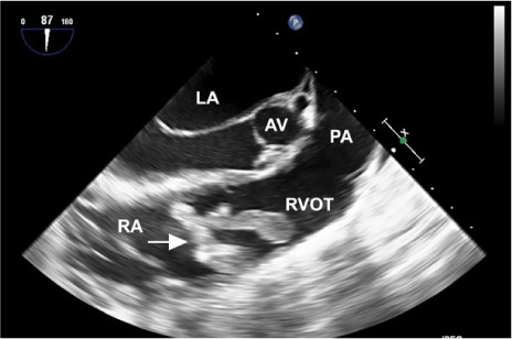
At the current presentation, a transthoracic echocardiogram showed a large right ventricular mass. Chest computed tomograms showed multiple, bilateral pulmonary nodules suggestive of metastatic disease. The largest nodule, located within the right lower lobe of the lung, was 2.1 cm long; 2 smaller nodules in the right lung base were 1.1 and 0.9 cm long; and a nodule in the left lung base was 1.2 cm long. Subsequent computed tomograms of the abdomen and pelvis showed a large (9.6 × 10 × 16 cm) heterogeneously enhancing, mixed cystic and solid mass, which raised concern for a cystic neoplasm. A transesophageal echocardiogram revealed a large, mobile, longitudinal mass attached to the tricuspid valve and protruding into the right ventricular outflow tract (Fig. 1). Cardiac magnetic resonance images showed a multilobulated lesion within the right ventricle. A biopsy sample of the pelvic mass showed characteristics consistent with benign leiomyoma.
Fig. 1.

Transesophageal echocardiogram shows a large, mobile, longitudinal mass (arrow) protruding into the right ventricular outflow tract (RVOT)
AV = aortic valve; LA = left atrium; PA = pulmonary artery; RA = right atrium
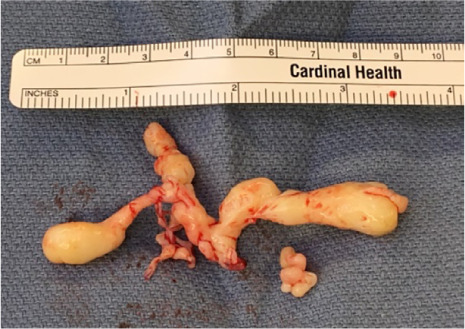
Given the size of the cardiac mass and the associated risk of embolization, a right atriotomy was performed. A 9-cm-long tumor was found to be attached to the chordae of the tricuspid valve and was resected (Fig. 2).
Fig. 2.

Photograph shows the 9-cm-long resected cardiac tumor.
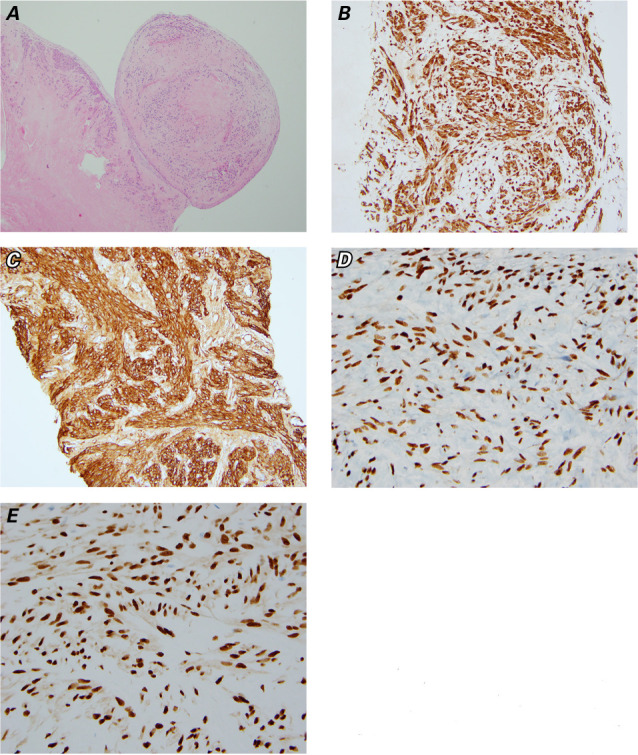
Histopathologic examination of the resected cardiac tumor revealed necrosis, but no atypia. Immunohistochemical staining was positive for smooth muscle actin, desmin, estrogen, and progesterone receptors—findings consistent with uterine leiomyoma (Fig. 3). Ki-67 immunostaining revealed a proliferation index of <2%. Although no lung biopsy samples were taken, the patient's lung nodules were assumed to be metastases. On postoperative day 10, the patient was prescribed progestin receptor modulator therapy and discharged from the hospital.
Fig. 3.

Photomicrographs of specimens from the resected cardiac tumor show A) endocardium undermined by a nodular mass (H&E, orig. ×40) and strong positive immunohistochemical reactivity for B) desmin (orig. ×200), C) smooth muscle actin (orig. ×200), D) estrogen receptors (orig. ×400), and E) progesterone receptors (orig. ×400).
At her 3-month follow-up visit, the size of the lung nodules had remained stable, but the pelvic mass had increased from 16 cm to 19 cm in the craniocaudal dimension. The patient also showed symptoms of right ventricular (RV) failure. At her last known follow-up visit, 3 years after presentation, the RV failure symptoms were well controlled by a daily oral regimen of loop diuretics. However, the pelvic mass had continued growing—to 21 cm in the anteroposterior dimension, 28 cm in the transverse, and 36 cm in the craniocaudal—resulting in a mass effect on the ureters that caused bilateral hydronephrosis and had necessitated bilateral nephrostomy tube placement.
Discussion
Benign metastasizing leiomyoma, a rare tumor characterized by extrauterine smooth muscle tumor growth, typically affects women of reproductive age.1 It usually occurs in the lungs and lymph nodes, but may also occur in skeletal muscle, liver, bones, brain, and heart.2 Three types of cardiac leiomyoma presentation have been reported: intravenous leiomyomatosis with intracardiac extension, primary intracardiac leiomyoma, and benign metastasizing leiomyoma.3 Intravenous leiomyomatosis, the most common of these presentations, is marked by the extension of smooth muscle tumor from the uterus, through the pelvic veins, and then into the inferior vena cava (IVC) and right side of the heart.3 In contrast, benign metastasizing leiomyomas do not extend from the IVC into the right heart, and they attach to the endothelial surface of the heart. In most reported cases, patients had undergone surgical resection of uterine leiomyomas before presenting with the benign metastasizing tumor.2 Benign metastasizing leiomyoma involving the heart was first reported by Takemura and colleagues in 1996,4 and at least 5 other cases have been reported since then (Table I).1,3,5–7
TABLE I.
Summary of Case Reports of Benign Metastasizing Leiomyoma in the Heart
| Reference | Patient Age (yr) | Noncardiac Metastasis Site(s) | Cardiac Tumor Size (cm)* | Location in Heart | Epicardial Involvement |
|---|---|---|---|---|---|
| Takemura G, et al.4 (1996) | 44 | Lungs | 4.7 | Right ventricle | No |
| Galvin SD, et al.6 (2010) | 41 | None | 3.5 | Right ventricle | No |
| Cai A, et al.1 (2014) | 37 | Lungs, liver, peritoneum, retroperitoneum, muscles | NR | Right ventricle | Yes |
| Consamus EN, et al.3 (2014) | 55 | Lungs | 4 | Right atrium | No |
| Williams M, et al.5 (2016) | 51 | Lungs | 4.5 | Right ventricle | Yes |
| Meddeb M, et al.7 (2018) | 36 | Lungs | 4.7 | Right ventricle | No |
| Current case | 45 | Lungs | 9 | Right ventricle | No |
NR = not reported
*Maximum length
The pathogenesis of benign metastasizing leiomyoma is poorly understood. It is thought to arise from tumor embolization through venous channels during hysterectomy.2 Of note, our patient had previously undergone a technically challenging hysterectomy complicated by morcellation of the uterus and by excessive bleeding that necessitated gonadal vein embolization.
When we treated the patient, the results of histopathologic examination and immunohistochemical staining of the tumor were consistent with uterine leiomyoma. Notably, the tumor did not involve the IVC and was attached to the chordae of the tricuspid valve. Consequently, we diagnosed benign metastasizing leiomyoma in the heart and lungs.
How best to treat benign metastasizing leiomyoma is not known, and it varies depending on the anatomic site involved and the associated symptoms.1 Tumor size may also be important. Our patient's heart tumor is by far the largest reported so far (9 cm, as compared with 3–5 cm in the previous reports). A benign metastasizing leiomyoma in the heart can pose life-threatening risks of embolization, arrhythmia, and obstruction and therefore is usually an indication for surgical resection. On the other hand, such tumors greatly depend on estrogen and progesterone and thus could theoretically be controlled by medical or surgical castration. However, the reported response of the tumors to hormonal suppression has been mixed.5 In our patient, the cardiac tumor responded suboptimally to hormonal therapy, as evidenced by the continued growth of her pelvic tumor.
Conclusion
Although rare, benign metastasizing leiomyoma in the heart should be considered in the differential diagnosis of right-sided cardiac tumors, especially in patients with a history of uterine fibroids. Future research is needed to better understand the pathogenesis and treatment of such tumors.
References
- 1.Cai A, Li L, Tan H, Mo Y, Zhou Y. Benign metastasizing leiomyoma: case report and review of the literature. Herz. 2014;39(7):867–70. doi: 10.1007/s00059-013-3904-1. [DOI] [PubMed] [Google Scholar]
- 2.Pacheco-Rodriguez G, Taveira-DaSilva AM, Moss J. Benign metastasizing leiomyoma. Clin Chest Med. 2016;37(3):589–95. doi: 10.1016/j.ccm.2016.04.019. [DOI] [PubMed] [Google Scholar]
- 3.Consamus EN, Reardon MJ, Ayala AG, Schwartz MR, Ro JY. Metastasizing leiomyoma to heart. Methodist DeBakey Cardiovasc J. 2014;10(4):251–4. doi: 10.14797/mdcj-10-4-251. [DOI] [PMC free article] [PubMed] [Google Scholar]
- 4.Takemura G, Takatsu Y, Kaitani K, Ono M, Ando F, Tanada S, et al. Metastasizing uterine leiomyoma: a case with cardiac and pulmonary metastasis. Pathol Res Pract. 1996;192(6):622–33. doi: 10.1016/S0344-0338(96)80116-6. [DOI] [PubMed] [Google Scholar]
- 5.Williams M, Salerno T, Panos AL. Right ventricular and epicardial tumors from benign metastasizing uterine leiomyoma. J Thorac Cardiovasc Surg. 2016;151(2):e21–4. doi: 10.1016/j.jtcvs.2015.09.059. [DOI] [PubMed] [Google Scholar]
- 6.Galvin SD, Wademan B, Chu J, Bunton RW. Benign metastasizing leiomyoma: a rare metastatic lesion in the right ventricle. Ann Thorac Surg. 2010;89(1):279–81. doi: 10.1016/j.athoracsur.2009.06.050. [DOI] [PubMed] [Google Scholar]
- 7.Meddeb M, Chow RD, Whipps R, Haque R. The heart as a site of metastasis of benign metastasizing leiomyoma: case report and review of the literature. Case Rep Cardiol. 2018;2018:7231326. doi: 10.1155/2018/7231326. [DOI] [PMC free article] [PubMed] [Google Scholar]


