

A 42‐year‐old black man was admitted to the hospital because of acute dyspnea of a few hours' duration. He had a 6‐year history of uncontrolled hypertension and admitted to salt avidity as well as noncompliance with his medication. His blood pressure was 200/139 mm Hg with a heart rate of 88 beats per minute and a respiration rate of 32 breaths per minute. He had bilateral diffuse rales and a laterally displaced point of maximal impulse; his neck veins were elevated to the jaw angle at 45°. There was no hepatomegaly or abdominal masses, and he had 1+ ankle edema. Chest x‐ray films showed pulmonary edema and the electrocardiogram showed left ventricular hypertrophy. The patient received 80 mg of intravenous furosemide, was placed on intravenous nitroglycerin, and was admitted to the intensive care unit.
Pulmonary edema cleared by the next morning and serial troponin I levels were normal. An echocardiogram revealed left ventricular hypertrophy with an estimated ejection fraction of 55%. Blood urea nitrogen (BUN) level was 24 mg/dL (normal, <19 mg/dL) with a creatinine level of 1.9 mg/dL (normal, 0.7–1.3 mg/dL) and a potassium level of 3.6 mmol/L (normal, 3.5–5.0 mEq/L). An adenosine dual isotope scan showed no obstructive coronary disease, and results of magnetic resonance angiography of the renal arteries were negative for renal artery stenosis. Unenhanced computed tomography (CT) of the adrenal glands revealed bilateral adrenal masses measuring 5 cm × 3.5 cm on the right side and 5.2 cm × 3.0 cm on the left side; 1–4 Hounsfield units (HU) on the right and −7 HU on the left (Figure 1). Magnetic resonance imaging (MRI) of the adrenal glands revealed bilateral lobulated and circumscribed masses measuring 5.0 × 3.5 cm on the right and 5.0 × 3.0 cm on the left. The T2‐weighted image was homogeneous and hyperintense related to the liver (Figure 2).
Figure 1.
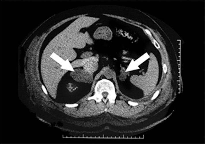
Computed tomography revealing large homogeneous bilateral adrenal masses with low attenuation: 5.0 × 3.5 cm and 1–4 Hounsfield units on the right; 5.2 × 3.0 cm and 7 Hounsfield units on the left.
Figure 2.
Appearance on magnetic resonance imaging of bilateral adrenal masses is hyperintense in relation to liver on T2‐weighted image, 5.0 × 3.5 cm on the right and 5.0 × 3.0 cm on the left.
Levels of fractional plasma metanephrines were 48 pg/mL (normal, ≤82 pg/mL); 24‐hour urine total metanephrines, 224 µg/24 h (normal, 90–690 µg/24 h); 24‐hour urine catecholamines, 84 µg/24 h (normal, 0–135 µg/24 h); and a 24‐hour urine free cortisol, 46 µg/24 h (normal, 4–50 µg/24 h). Morning aldosterone and renin levels were 6.0 ng/dL (normal, ≤28 ng/dL) and 1.4 ng/mL/h (normal, 0.6–5.0 ng/mL/h), respectively. Twenty‐four‐hour urine aldosterone level was 6.5 mg/24 h (normal, 2.3–21 mg/24 h). BUN and creatinine levels improved slightly to 22 mg/dL and 1.7 mg/dL, respectively, before discharge, and blood pressure had decreased to 124/68 mm Hg on a regimen of chlorthalidone/atenolol, 25/100 mg; lisinopril, 40 mg; and felodipine, 10 mg/d. A follow‐up MRI study of the adrenal glands 9 months later showed no change.
DISCUSSION
This patient with admitted medication and dietary noncompliance presented in a hypertensive emergency with acute pulmonary edema. While medication nonadherence is the most common cause of a hypertensive emergency, 1 such a presentation also warrants an expedited workup to rule out secondary etiologies. These are more common in this group of patients compared with those with uncomplicated hypertension. Acute pulmonary edema may be a curable manifestation of bilateral renal artery stenosis, 2 but in this patient it was attributed to cardiac diastolic dysfunction and stage 3 chronic kidney disease due to hypertensive nephropathy. Large bilateral adrenal masses were discovered.
Pheochromocytoma was a high‐priority diagnostic consideration because of the size of the masses and the acute presentation. Of patients with adrenal incidentalomas, 5.1% have been proven to have pheochromocytoma, 3 but the prevalence of pheochromocytoma would be expected to be higher in patients with a hypertensive crisis with adrenal masses. Surveys of adrenal incidentaloma generally exclude patients with severe hypertension. 4 However, both plasma metanephrines and 24‐hour urine collections for metanephrines and catecholamines were well within the normal range during a hypertensive crisis in the present patient.Table I illustrates the prevalence of diagnoses in patients with an incidentally discovered adrenal mass. 5 An adrenal mass accompanied by hypertension can be a manifestation of pheochromocytoma, primary aldosteronism, or Cushing's syndrome. Approximately 10% of adrenal incidentalomas are either hyperfunctional or significantly autonomous. 3 Hormonal evaluations to rule out each of these entities are described in Table II.
Table I.
Prevalence of Diagnoses in Patients With an Incidentally Discovered Adrenal Mass
| Condition | Prevalence Per 10,000 Patients |
|---|---|
| Pheochromocytoma | 650 |
| Aldosterone‐producing adenoma | 700 |
| Glucocorticoid‐producing adenoma | 3.5 |
| Adrenal carcinoma | 5.8 |
| Reprinted with permission from Ross and Aron. 5 | |
Table II.
Screening Tests to Assess Hormonal Function of an Adrenal Mass
| Pheochromocytoma | Primary Hyperaldosteronism | Cushing's Syndrome |
|---|---|---|
| (1) 24‐hour urine fractionated metanephrines + catecholamines or (2) plasma‐free metanephrines. | Morning aldosterone/plasma renin activity ratio ≥20 when absolute aldosterone ≥15 ng/dL with potassium ≥3.5 µg/L. Studies can be performed on all drugs except spironolactone, eplerenone, or amiloride. | (1) Dexamethasone suppression test: 1 mg dexamethasone between 11 pm and midnight; 8 am cortisol level should be <2.5 µg/dL or (2) 24‐hour urine‐free cortisol level, 4–50 µg/24 h. |
More extensive hormonal testing of incidentally discovered adrenal masses may reveal evidence of subclinical hypercortisolism. Low dehydroepiandrosterone sulfate levels occurred in 43% and defective dexamethasone suppression occurred in 15% of 45 patients in one study, indicative of mild inhibition of the hypothalamic‐pituitary‐adrenal axis. 6 However, false‐positive results and lack of evidence to support silent hypercortisolism as a predictor of future Cushing's syndrome reduce the clinical relevance of more extensive testing. In this case, silent hypercortisolism would not explain his hypertensive crisis.
Adrenocortical carcinoma is associated with hypertension, and extra‐adrenal carcinoma metastatic to the adrenal glands is also a consideration when an adrenal mass is discovered. Size and imaging phenotype are the important considerations when assessing the malignant potential of an incidentally noted adrenal mass. A diameter >4 cm was shown to have a 90% sensitivity and a 24% specificity for the detection of adrenocortical carcinoma in a report of 887 patients with adrenal incidentalomas. 4 Only 3, 6, and 12 operations would be necessary to remove an adrenocortical carcinoma with lesion threshold sizes of 6, 5, and 4 cm, respectively. 3 While fewer than 5% of adrenal incidentalomas are adrenocortical carcinoma, 95% of carcinomas are larger than 4 cm, and 95% of cortical adenomas are smaller than 5 cm. Most experts would recommend resecting lesions >6 cm, depending on the patient's functional status. The recommendation is to look carefully at the characteristics of the mass on computerized imaging before deciding a strategy for intermediate‐size lesions, however.
The characteristics of the mass on computerized imaging, or imaging phenotype, provides an important assessment of intracytoplasmic fat content, which is high in adrenal adenomas and low in malignancy. High adrenal intracytoplasmic fat content is associated with low attenuation on unenhanced CT. The Hounsfield scale is a semiquantitative assessment of x‐ray attenuation on the CT imaging spectrum which ranges from air (black) to bone (white). Generally, HU correspond to anatomic elements as follows: air, 0 HU; adipose tissue, −20 to −150 HU; and kidney, 20–50 HU. An adrenal mass with fewer than 0 HU attenuation on unenhanced CT has a likelihood of being a benign adenoma approximating 100%. 3 In a large national Italian study group survey, the ability of CT phenotype to discriminate carcinoma was, in addition to size, highly significant, r=0.39, P<.0001. According to the National Institutes of Health consensus conference on the management of adrenal incidentaloma, “a homogenous mass with a low attenuation value (<10 HU) on CT is probably a benign adenoma.” 7 Pheochromocytomas are usually >25 HU. 8
Another phenotypic imaging feature used to distinguish adenomas from nonadenomas including carcinomas is the rapidity of washout of contrast medium on delayed contrast‐enhanced CT. Rapid washout is more consistent with adrenal adenoma. An absolute washout of more than 50% of contrast medium after 10 minutes was reported to be 100% sensitive and specific for adenoma in studies comparing patients with adenomas with those with carcinomas, pheochromocytomas, or metastatic disease. 8 Functional assays still need to be performed in these patients.
MRI characteristics have likewise been employed to decide the malignant potential of adrenal lesions, but many adrenal masses are indeterminate with conventional spin‐echo MRI. As with CT, mild enhancement with rapid washout of gadolinium‐DPTA‐enhanced MRI favors adenoma, whereas greater enhancement and a slower washout of contrast favor malignancy. Imaging phenotype features used to distinguish adrenal incidentalomas are summarized in Table III. 8
Table III.
Characteristics of Adrenal Incidentalomas on Imaging (Imaging Phenotype)
| Variable | Adrenocortical Adenoma | Adrenocortical Carcinoma | Pheochromocytoma | Metastasis |
|---|---|---|---|---|
| Size | Small, usually ≤3 cm in diameter | Large, usually >4 cm in diameter | Large, usually >3 cm in diameter | Variable, frequently <3 cm |
| Shape | Round or oval, with smooth margins | Irregular, with unclear margins | Round or oval, with clear margins | Heterogeneous, with mixed densities |
| Texture | Homogeneous | Heterogeneous, with mixed densities | Heterogeneous, with cystic areas | Heterogeneous, with mixed densities |
| Laterality | Usually solitary, unilateral | Usually solitary, unilateral | Usually solitary, unilateral | Often bilateral |
| Attenuation (density) on unenhanced CT | ≤10 HU | >10 HU (usually >25) | >10 HU (usually >25) | >10 HU (usually >25) |
| Vascularity on contrast‐enhanced CT Rapidity of washout of contrast medium | Not highly vascular ≥50% at 10 minutes | Usually vascular <50% at 10 minutes | Usually vascular <50% at 10 minutes | Usually vascular <50% at 10 minutes |
| Appearance on MRI | Isointense in relation to liver on T2‐weighted image | Hyperintense in relation to liver on T2‐weighted image | Markedly hyperintense in relation to liver on T2‐weighted image | Hyperintense in relation to liver on T2‐weighted image |
| Necrosis, hemorrhage, or calcifications | Rare | Common | Hemorrhage and cystic areas common | Occasional hemorrhage and cystic areas |
| Growth rate | Usually stable over time or very slow (<1 cm/y) | Usually rapid (>2 cm/y) | Usually slow (0.5–1.0 cm/y) | Variable, slow to rapid |
| Abbreviations: CT, computed tomography; HU, Hounsfield units; MRI magnetic resonance imaging. Reprinted with permission from Gross et al. 9 | ||||
Adrenal masses occur bilaterally in 15% of adrenal incidentalomas and the differential diagnosis includes metastatic disease, congenital adrenal hyperplasia, lymphoma, infection (tuberculosis, fungal), hemorrhage, adrenocorticotropic hormone‐dependent Cushing's syndrome, pheochromocytoma, amyloidosis, and infiltrative disease. 3 This patient did not have clinical, imaging, or biochemical evidence of any of these entities.
Iodocholesterol scintigraphy has been used to distinguish benign from malignant adrenal lesions, but it is not sensitive or specific for either lesion and it is not a simple procedure. Normally functioning or hyperfunctioning adrenal tissue should take up and process the radio‐labeled cholesterol. To perform this study, oral iodine needs to be administered for 2 days before and 14 days after the scan, and the scintigraphic images may take 5 to 7 days. Moreover, most of the successful diagnostic studies have been performed in patients with unilateral adrenal masses in which a normal adrenal gland is available for gamma camera comparison. Iodocholesterol uptake may simply reflect an enlarged adrenal gland. 6 Experience with iodocholesterol scintigraphy imaging of bilateral adrenal masses is very limited. 9
Negative hormonal assays at the time of a hypertensive emergency made pheochromocytoma, primary aldosteronism, and Cushing's syndrome unlikely diagnoses in this case. A sex hormone‐secreting adrenocortical tumor was felt to be clinically unlikely. Given the large size of these masses, their malignant potential was of concern. A washout study with CT enhancement was not performed, because of stage 3 kidney disease, and iodocholesterol scintigraphy was not performed, because its utility was felt to be minimal in evaluating bilateral adrenal masses in light of the benign imaging phenotype. The Hounsfield description suggested prominent intracytoplasmic fat content and a differential diagnosis of myelolipoma, lipidrich adenoma, or benign hyperplasia. Benign imaging phenotype in this case was very helpful because it permitted noninvasive follow‐up of these large adrenal masses. Follow‐up imaging was recommended instead of a fine needle aspiration biopsy, and 9 months later there was no MRI change.
According to the National Institutes of Health consensus conference on adrenal incidentaloma, lesions between 4 and 6 cm with a benign imaging phenotype can be managed by follow‐up rather than with adrenalectomy. 7 Fewer than 30% of incidentalomas increase in size and fewer than 20% become active biochemically after 10 years of follow‐up; 3% to 4% decrease in size. According to the conference summary, “in patients with tumors that remain stable on 2 imaging studies done at least 6 months apart and do not exhibit hormonal hypersecretion over 4 years, further follow‐up may not be warranted.” 7
References
- 1. Zampaglione B, Pascale C, Marchisio M, et al. Hypertensive urgencies and emergencies: prevalence and clinical presentation. Hypertension. 1996;27(1):144–147. [DOI] [PubMed] [Google Scholar]
- 2. Pickering TG, Devereux RB, James GD, et al. Recurrent pulmonary edema due to bilateral renal artery stenosis; treatment by angioplasty or surgical revascularization. Lancet. 1988;2(8610):551–552. [DOI] [PubMed] [Google Scholar]
- 3. Young WF. Endocrine incidentalomas, management approaches to adrenal incidentalomas, a view from Rochester, Minnesota. Endocrinol Metab Clin North Am. 2000;29:159–185. [DOI] [PubMed] [Google Scholar]
- 4. Angeli A, Osella G, Ali A, et al. Adrenal incidentaloma: an overview of clinical and epidemiological data from the National Italian Study Group. Horm Res. 1997;47:279–283. [DOI] [PubMed] [Google Scholar]
- 5. Ross NS, Aron DC. Hormonal evaluation of the patient with an incidentally discovered adrenal mass. N Engl J Med. 1990;323:1401–1405. [DOI] [PubMed] [Google Scholar]
- 6. Osella G, Terzolo M, Borretta G, et al. Endocrine evaluation of incidentally discovered adrenal masses (incidentalomas). J Clin Endocrinol Metab. 1994;79:1532–1539. [DOI] [PubMed] [Google Scholar]
- 7. Grumbach MM, Biller BMK, Braunstein GD, et al. Management of the clinically inapparent adrenal mass (“incidentaloma”). Ann Intern Med. 2003;138:424–429. [DOI] [PubMed] [Google Scholar]
- 8. Young WF. The incidentally discovered adrenal mass. N Engl J Med. 2007;356:601–610. [DOI] [PubMed] [Google Scholar]
- 9. Gross MD, Shapiro B, Bouffard A, et al. Distinguishing benign from malignant euadrenal masses. Ann Intern Med. 1988;109:613–618. [DOI] [PubMed] [Google Scholar]