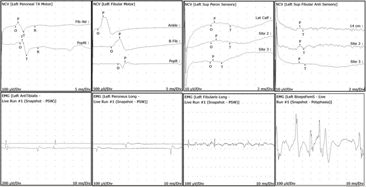
Figure 1. EMG/NCS left lower extremity representative waveforms.
EMG: electromyography; NCS: nerve conduction study
Top left two figures (NCS): Left peroneal/fibular motor nerve conduction block noted in popliteal regions indicating compressive neuropathy
Top right two figures (NCS): Left superficial peroneal/fibular sensory nerve conduction is poor and seen with inconsistent responses
Bottom left two figures (EMG): Left anterior tibialis muscle with positive sharp waves (PSWs) and fibrillations (Fibs). Left peroneus longus muscle with PSWs. These indicate acute denervation of the muscles
Bottom right two figures (EMG): Left peroneus/fibularis longus muscle with PSWs which indicates acute muscle denervation. Biceps femoris muscle with polyphasic potentials which indicate subacute denervation of muscle

