

Abstract
Introduction
Horseshoe kidneys are the most common fusion defect of the kidneys, which amounts to about 0.25% of the population. They are usually asymptomatic and are often identified incidentally. The horseshoe kidney can push the second and third part of the duodenum anteriorly, leading to an altered CBD course. Choledocholithiasis is seen in approximately 10‐15% of patients with cholelithiasis. Presently, the most preferred approach for managing CBD stones is ERCP. However, in ERCP failure cases, Laparoscopic CBD exploration is the primary treatment modality, with or without T-tube use, with all the advantages of minimally invasive surgery.
Case presentation and discussion
A 65-year-old female presented with complaints of pain in the right hypochondriac region for three months associated with nausea, jaundice, and loss of appetite and weight. Her USG abdomen showed cholelithiasis with dilated CBD with horseshoe kidney with severe hydronephrosis of the left kidney. They are usually asymptomatic and are often identified incidentally. In this patient, it was believed that the horseshoe kidney had pushed the second and third part of the duodenum anteriorly, leading to an altered CBD course leading to ERCP failure. MRCP confirmed cholelithiasis with choledocholithiasis with dilated CBD of 11.3 mm with horseshoe kidney. ERCP was attempted but was unsuccessful due to non-visualization of the papilla due to overcrowding of duodenal folds.
For patients with ERCP failure, laparoscopic CBD exploration is mandatory. For this patient, the CBD was cannulated with a guidewire, if needed, for repeat ERCP and was closed with T-tube in situ.
Conclusion
There are no particular preoperative indicators that can predict the failure of ERCP. However, in ERCP failure cases, laparoscopic CBD exploration (with or without T-tube use) is the primary treatment modality.
Abbreviations: LC, laparoscopic cholecystectomy; CBD, common bile duct; LCBDE, laparoscopic CBD exploration; USG, ultrasonography; MRCP, magnetic resonance cholangiopancreatography; ERCP, endoscopic retrograde cholangiopancreatography
Keywords: Choledocholithiasis, Choledochotomy, Guidewire, Horseshoe kidney, ERCP failure, Laparoscopic CBD exploration
Highlights
-
•
Horseshoe kidney causing altered CBD course leading to ERCP failure
-
•
Laparoscopic CBD exploration in patients with ERCP failure
-
•
Cannulation of CBD with guidewire for easier identification of papilla in case of repeat ERCP
-
•
One step laparoscopic cholecystectomy with laparoscopic CBD exploration vs. ERCP
-
•
Laparoscopic CBD exploration with T-tube insertion
1. Introduction
Choledocholithiasis or CBD stones are present in as many as 10‐15% of patients with symptomatic cholelithiasis [1]. CBD stones require extraction to relieve symptoms and prevent complications such as acute suppurative cholangitis, obstructive jaundice, hepatic abscess, and acute pancreatitis. CBD stones can be managed either surgically, by open or laparoscopic exploration, or by endoscopy (ERCP). Presently, the two most preferred approaches include single-step Laparoscopic Cholecystectomy (LC) with Laparoscopic CBD exploration (LCBDE) or LC with pre/post-operative ERCP with or without endoscopic sphincterotomy (two-stage).
Endoscopic retrograde cholangiography and endoscopic sphincterotomy have drastically changed the management of choledocholithiasis. ERCP has the advantage of clearing the CBD stones without surgery. The main disadvantage is that it is usually performed as a second procedure before or after LC. Patients may develop pancreatitis, duodenal perforation, and bleeding secondary to sphincterotomy [[2], [3], [4]]. LC with a concomitant LCBDE has the advantage of being a single-stage procedure that can treat both cholelithiasis and choledocholithiasis. Various trials have compared the outcomes of laparoscopic vs. endoscopic procedures [[5], [6], [7], [8]]. A 2013 Cochrane study reported no significant difference in the mortality and morbidity in patients with LCDBE and ERCP [8]. Both procedures have been proven effective, but LC + LCBDE has shown advantages in a lower technical failure rate, fewer procedures, shorter hospital stay duration, and lower hospital charges [9,10]. It has been seen that despite all the innovations, training programs, and improved imaging sources, failed biliary cannulation in ERCP occurs in 5‐20% of all cases [4].
This case was reported following the SCARE criteria [11].
2. Case report
A 65-year-old female presented to the outpatient clinic in August 2020 with complaints of pain in the right upper quadrant and epigastric region for three months. The pain was constant throughout the day but increased in intensity post meals, non-radiating, and relieved with medications. The pain was associated with nausea, loss of appetite, and weight. She had no fever, yellowish discoloration of the skin, chest pain, and changes in bowel or bladder habits. However, the patient reported yellowish discoloration of skin and eyes two months before presentation, which had improved spontaneously. Her history was significant for diabetes mellitus, for which she was on regular treatment for 18 years. The patient had no pertinent family or social history. Her vitals were stable on presentation, and she was admitted for further work-up.
On investigations, she had leukocytosis (11.6 K/mm3). All other blood investigations were within normal limits. Abdomen USG showed multiple GB calculi, largest measuring 8.5 mm, a dilated CBD in its entire extent measuring 9 mm in maximal luminal diameter at the porta.
USG also revealed a horseshoe kidney with severe hydronephrosis and parenchymal thinning in the left kidney. MRCP was done, which demonstrated a horseshoe kidney, and a distended GB with multiple intra-luminal filling defects, largest measuring 8.5 mm. CBD was found to be dilated with a luminal diameter of 11.3 mm at the porta. There was an abrupt distal cut-off with subtle hypo-isodensities seen in the dependent part of mid and distal CBD. (Fig. 1, Fig. 2, Fig. 3, Fig. 4).
Fig. 1.

MRCP showing multiple gall stones.
Fig. 2.

MRCP showing horseshoe shaped kidney (black arrow).
Fig. 3.

MRCP showing duodenum (black arrow) and CBD (red arrow) being pushed anteriorly by the horseshoe kidney.
Fig. 4.

MRCP showing CBD with abrupt distal cut-off.
The patient underwent ERCP on the third admission day, which was unsuccessful due to overcrowding of mucosal folds in the duodenum, leading to difficulty accessing D2. Papilla could not be located despite multiple attempts; hence, the procedure was aborted.
She was then planned for lap cholecystectomy with CBD exploration on the fourth day of admission.
3. Operative procedure
The surgery was performed under general anesthesia using aseptic precautions. Four ports were made; two 10 mm ports in the epigastrium & infra-umbilical region and two right lateral 5 mm ports. Proximal 1/3rd of the gall bladder was cleared off the liver bed and the fatty and fibrous tissues to achieve the critical view of safety (Fig. 5).
Fig. 5.

Dissected Calot's triangle.
The CBD was dissected up to 2-3 cm from the upper border of the duodenum. CBD was confirmed by aspiration of bile. A small incision of approx. 1 cm was given on the CBD (Fig. 6). CBD was visualized for its whole extent using a nephroscope (choledochoscope is not available at our institution). Sludge was visible in the distal end which was washed out with saline irrigation. A guidewire was passed through the nephroscope into the entire length of the CBD for easier visualization of the papilla, if needed, for repeat ERCP later on (Fig. 7).
Fig. 6.

Choledochotomy incision with T-tube (left) and guide wire (right) in-situ.
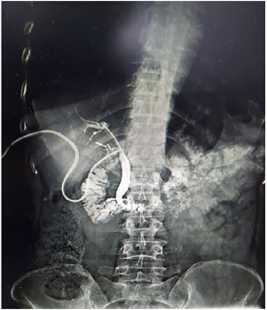
Fig. 7.

Post-operative x-ray showing T-tube (red arrow), drain (white arrow) and guide wire (black arrow) in-situ.
The T-tube was taken, and both the short horizontal limbs were cut to a length of 2 and 2.5 cm, respectively. The T-tube was introduced in the abdominal cavity by the epigastric port. The shorter horizontal limb was introduced towards the proximal part of the CBD, and the longer horizontal limb was introduced towards the distal part of the CBD at a safe distance from the ampulla of vater. The choledochotomy incision was sutured snugly around the t-tube with vicryl 3-0. The operation was concluded by performing cholecystectomy.
After completing the cholecystectomy, the t-tube catheter was brought out through the right subcostal port and fixed to the skin. The sub-hepatic drain placed was brought out of the right flank port, and the guidewire was brought out through the epigastric port. Care was taken not to dislodge the T-tube. The operation was concluded by deflating the pneumoperitoneum and closure of all the ports.
The patient was discharged on the fifth postoperative day, after drain removal, and was regularly followed up in the outpatient department. The t-tube was kept for 14 days for a full recovery. The guidewire was removed inadvertently by the patient. On the 7th post-operative day, the collection in the t-tube decreased to <50 ml/day, following which, the t-tube was clamped for 6 h alternating with periods of unclamping. The patient reported no complaints of abdominal discomfort, nausea, or vomiting. The t-tube was kept clamped for 24 h the next day.
T-tube cholangiogram was performed on the 10th post-operative day, which was normal (Fig. 8). The t-tube was kept clamped and was removed by gentle traction on the 14th post-op day. She had no complaints of abdominal pain, nausea or vomiting.
Fig. 8.
T-tube cholangiogram showing good opacification of common bile duct with no filling defect and normal drainage into the duodenum.
The patient was kept under regular follow-up with no fresh complaints. Her repeat LFT was within normal limits.
4. Discussion
In our case, the patient had to undergo LCBDE due to the failure of ERCP. The ultrasound and MRCP both revealed the incidental finding of horseshoe kidney. Horseshoe kidneys are the most common fusion defect of the kidneys, which amounts to about 0.25% of the population. It is found in approximately 1 in 500 in the average population, with a male preponderance of 2:1 [12,13]. They are usually asymptomatic and are often identified incidentally. In this patient, it was believed that the horseshoe kidney had pushed the second and third part of the duodenum anteriorly, leading to an altered CBD course with overcrowding of the duodenal folds, leading to failure of ERCP. In such cases, surgical CBD exploration becomes mandatory. Hence, a guidewire was left in the CBD for easy identification of papilla in case of future need of ERCP.
Surgical CBD exploration can be done either via lap or open technique. Open CBD exploration is associated with higher morbidity and mortality. LCBDE, in many studies, is equally, if not more, efficacious as compared to ERCP for choledocholithiasis management [8,9].
Laparoscopic CBD exploration can be performed either with a trans-cystic or Choledochotomy approach. The trans-cystic approach is ideal in patients with [14] a CBD diameter of <7 mm, stone location distal to the cystic duct/CBD junction, a cystic duct diameter > 4 mm, fewer than 6–8 stones in the CBD, stones <10 mm, and in case of lateral entrance of cystic duct to CBD.
The choledochotomy approach is more technically demanding and is done in the following conditions at present [15]:
-
•
Failed laparoscopic trans-cystic or preoperative endoscopic extraction
-
•
Narrow/tortuous cystic duct
-
•
Dilated CBD >7 mm
-
•
Stones>10 mm
-
•
Multiple stones
-
•
Stone location proximal to cystic duct/CBD junction
-
•
Distal or posterior cystic duct entrance to CBD
5. T-tube management
Traditionally, post LCBDE, especially in patients who underwent choledochotomy, T-tube is placed. It is thought to help decompress the biliary system, prevent biliary stricture, and treat any residual stones or sludge conveniently. It also helps in radiological visualization of the CBD and biliary system, post-operatively [16].
However, it is associated with a few complications and is also a source of discomfort in terms of longer period of management post-discharge. Complications include cholangitis, bile leak and bile peritonitis, biliary fistula, fluid and electrolyte imbalance, bile duct trauma during removal, dislocation of the tube, retention of a fragment of t-tube, and inclusion of t-tube in the suture [17,18].
6. Conclusion
Ideal management of cholelithiasis with choledocholithiasis varies from surgeon to surgeon and even has an institutional preference. The choice of procedure, be it pre or post-operative ERCP and LC or concomitant LC + LCBDE (with or without t-tube use), is highly variable and depends on the surgeon's expertise. However, there is no doubt that LCBDE becomes the procedure of choice in cases with failed ERCP. Despite a steep learning curve, in the hands of an experienced surgeon, LC and LCBDE in one seating can be a safe and feasible option with the advantage of minimally invasive surgery.
Sources of funding
This research did not receive any specific grant from funding agencies in the public, commercial, or not-for-profit sectors.
Ethical approval
This is a case report study and ethical approval is not required.
Consent
Written informed consent was obtained from the patient for publication of this case report and accompanying images. A copy of the written consent is available for review by the Editor of this journal.
Guarantor
Vimal Jain.
Declaration of competing interest
The authors of this work have nothing to disclose.
References
- 1.Verbesey J.E., Birkett D.H. Common bile duct exploration for choledocholithiasis. Surg. Clin. North Am. 2008;88:1315. doi: 10.1016/j.suc.2008.08.002. [DOI] [PubMed] [Google Scholar]
- 2.Freeman M.L., Nelson D.B., Sherman S., Haber G.B., Herman M.E., Dorsher P.J. Complications of endoscopic biliary sphincterotomy. N. Engl. J. Med. 1996;335:909e918. doi: 10.1056/NEJM199609263351301. [DOI] [PubMed] [Google Scholar]
- 3.Masci E., Toti G., Mariani A., Curioni S., Lomazzi A., Dinelli M. Complications of diagnostic and therapeutic ERCP: a prospective multicentre study. Am. J. Gastroenterol. 2001;96:417e423. doi: 10.1111/j.1572-0241.2001.03594.x. [DOI] [PubMed] [Google Scholar]
- 4.Williams E.J., Taylor S., Fairclough P., Hamlyn A., Logan R.F., Martin D., Riley S.A., Veitch P., Wilkinson M., Williamson P.R., Lombard M., BSG Audit of ERCP Are we meeting the standards set for endoscopy? Gut. 2007;56:821–829. doi: 10.1136/gut.2006.097543. [DOI] [PMC free article] [PubMed] [Google Scholar]
- 5.Bansal V.K., Misra M.C., Rajan K., Kilambi R., Kumar S., Krishna A. Single stage laparoscopic common bile duct exploration and cholecystectomy versus two-stage endoscopic stone extraction followed by laparoscopic cholecystectomy for patients with concomitant gallbladder stones and common bile duct stones: a randomized controlled trial. Surg. Endosc. 2014;28:875–885. doi: 10.1007/s00464-013-3237-4. [DOI] [PubMed] [Google Scholar]
- 6.Kharbutli Velanovich V. Management of preoperatively suspected choledocholithiasis: a decision analysis. J. Gastrointest. Surg. 2008;12(11):1973–1980. doi: 10.1007/s11605-008-0624-6. [DOI] [PubMed] [Google Scholar]
- 7.Zhu H.Y., Xu M., Shen H.J., Yang C., Li F., Li K.W., Shi W.J., Ji F. A meta-analysis of single-stage versus two-stage management for concomitant gallstones and common bile duct stones. Clin. Res. Hepatol. Gastroenterol. 2015;39(5):584–593. doi: 10.1016/j.clinre.2015.02.002. [DOI] [PubMed] [Google Scholar]
- 8.Dasari B.V.M., Tan C.J., Gurusamy K.S. Surgical versus endoscopic treatment of bile duct stones. Cochrane Database Syst. Rev. 2013;(12) doi: 10.1002/14651858.CD003327.pub4. [DOI] [PMC free article] [PubMed] [Google Scholar]
- 9.Rocha F.G. Surgical Common Bile Duct Exploration. https://www.uptodate.com/contents/common-bile-duct-exploration
- 10.Vindal A., Chander J., Lal P., Mahendra B. Comparison between intraoperative cholangiography and choledochoscopy for ductal clearance in laparoscopic CBD exploration: a prospective randomized study. Surg. Endosc. 2015;29:1030–1038. doi: 10.1007/s00464-014-3766-5. [DOI] [PubMed] [Google Scholar]
- 11.Agha R.A., Borrelli M.R., Farwana R., Koshy K., Fowler A., Orgill D.P., For the SCARE Group The SCARE 2018 statement: updating consensus Surgical CAse REport (SCARE) guidelines. Int. J. Surg. 2018;60:132–136. doi: 10.1016/j.ijsu.2018.10.028. [DOI] [PubMed] [Google Scholar]
- 12.Schiappacasse G., Aguirre J., Soffia P., Silva C.S., Zilleruelo N. CT findings of the main pathological conditions associated with horseshoe kidneys. Br. J. Radiol. 2015 Jan;88(1045):20140456. doi: 10.1259/bjr.20140456. [DOI] [PMC free article] [PubMed] [Google Scholar]
- 13.Glodny B., Petersen J., Hofmann K.J., Schenk C., Herwig R., Trieb T., Koppelstaetter C., Steingruber I., Rehder P. Kidney fusion anomalies revisited: clinical and radiological analysis of 209 cases of crossed fused ectopia and horseshoe kidney. BJU Int. 2009 Jan;103(2):224–235. doi: 10.1111/j.1464-410X.2008.07912.x. [DOI] [PubMed] [Google Scholar]
- 14.Zerey M., Haggerty S., Richardson W. Laparoscopic common bile duct exploration. Surg. Endosc. 2018;32:2603. doi: 10.1007/s00464-017-5991-1. [DOI] [PubMed] [Google Scholar]
- 15.Memon M.A., Hassaballa H., Memon M.I. Laparoscopic common bile duct exploration: the past, the present, and the future. Am. J. Surg. 2000;179:309. doi: 10.1016/s0002-9610(00)00346-9. [DOI] [PubMed] [Google Scholar]
- 16.Halstead W.S. Contributions to surgery of the bile passages especially of the common bile duct. Bull. Johns Hopkins Hosp. 1900;106:1–11. [Google Scholar]
- 17.Haq A., Morris J., Goddard C., Mahmud S., Nassar A.H. Delayed cholangitis resulting from a retained T-tube fragment encased within a stone. Surg. Endosc. 2002 Apr 1;16(4):714. doi: 10.1007/s00464-001-4235-5. [DOI] [PubMed] [Google Scholar]
- 18.Will V.L., Gibson K., Karihaloot C., Jorgensen J.O. Complication of biliary T-tube after choledochotomy. ANZ J. Surg. 2002;72(3):177–180. doi: 10.1046/j.1445-2197.2002.02308.x. [DOI] [PubMed] [Google Scholar]