

Abstract
Background:
Total hip (THA) and total knee arthroplasty (TKA) are becoming an increasingly standard procedure in the whole world. In conjunction with an aging population and increased prevalence of osteoporosis, proper management of periprosthetic, and interprosthetic fractures is of great interest to orthopedic surgeons. This study aims to report the clinical and radiographic outcomes, complications and reoperations of IFFs in geriatric patients.
Methods:
A retrospective single-institution case series study was conducted. Between 2011 and 2019, 83 patients underwent surgical treatment for periprosthetic femoral fractures. Thirteen fractures were identified as IFFs. Patient demographics and comorbidities were collected preoperatively, and fractures were classified with the Vancouver and AO unified classification system (AO-UCS).
Results:
We included 12 patients (13 hips) with IFFs (AO-UCS type IV.3 B (2/13) type IV.3 C (3/13), type IV.3 D (8/13)). The average patient age was 86.54 (range, 79-89) years. There were 10 females and 2 males. Perioperative morbidity has been identified in 10 of the 12 patients, and the 3-month and 1-year mortality were reported in 2 and 3 patients, respectively. Cerclage cables were used in 9 of 12 patients. One of 12 patients showed a local complication, with no documented implant failure or revision. Patients achieved complete union and returned to their preoperative ambulatory status, and full weight-bearing at an average of 5 (range, 2 to 7) months later.
Conclusion:
Management of IFF can be challenging because these fractures require extensive surgical expertise. Locking plate seems to be a valuable treatment option for geriatric patients with IFFs. Despite the complexity of this type of fracture, the overall complication and revision rate, as well as the radiographic outcome are good to excellent.
Level of Evidence:
Level III, Therapeutic study.
Keywords: interprosthetic fracture, femoral fractures, locking plate, geriatric trauma, total knee arthroplasty, total hip arthroplasty
Introduction
The use of total hip (THA) and total knee arthroplasty (TKA)) have spread worldwide as a result of an expansion of indications and an aging society.1,2 As the number of total hip arthroplasties (THAs) is increasing, the expected population’s percentage having more than 1 prosthetic implant, especially ipsilateral TKA and THA implants1,2,3,4 is also expected to increase. Although total hip (THA) and total knee arthroplasty (TKA) are safe and highly effective procedures, both are major surgical procedures with veritable risks of adverse outcomes including mortality, morbidity, and complications.5,6,7 However, the number of studies focusing on the association between age and complication rates, specifically in the primary TKA and THA populations, is quite limited.
In 1995 described Dave et al.8 the first case of an IFF. Furthermore, Kenny et al.9 stated that IFFs occur in 1.25% of patients who undergo hip and knee replacements. Although the IFF risk still low, with 240 IFFs annually in the US, these fractures are destined to rise.10 Among the most common risk factors, aging of the population, revision arthroplasty,11 osteoporosis12 or osteopenia as well as female sex13,14 and others represented the main clinical risk factors associated with augmented risk of IFF.
Their treatment is a difficult and demanding procedure and can have serious complications. According to the type of fracture, the prosthesis stability, bone quality, patient age, and clinical comorbidities, the treatment must be carefully determined and assessed.15-18 The treatment aims to restore length, axis, and rotation of the fractured femur, heal the fracture while preserving the adjacent prostheses’ function, and ensure sufficiently stable fixation to enable early mobilization and avoid complications.12,13,19
Referring to the complexity and challenges in many of these cases, experience in managing these injuries appears to be necessary and useful in clinical practice.
Therefore, locking plates has come into favor in recent years, but its outcome also seems to be affected by such challenging fractures.2 However, the outcomes of locking plates in patients with IFF have not yet been broadly reported.20-22
This study aimed to present our experience in managing of IFFs following ipsilateral hip and knee joint replacement, with particular emphasis on the individual treatment and outcome of the patients.
Materials and Methods
Institutional review board approval was obtained by the ethics committee of the University of Zurich (ID 2020-01906).
The authors reviewed the clinical records and trauma database of Zurich General Hospital, Stadtspital Waid, and identified all patients with peri- and interprosthetic fractures of the femur (n = 83) who were admitted to this level-I trauma center between 2011 and 2019.
Patients Selection
Patients with IFFs following ipsilateral hip and knee arthroplasty were sorted, and their dataset was examined for completeness and accuracy. There were 13 cases (n = 14 hips) identified who presented at this level 1 trauma center with IFFs. The involved patients were collected from the clinic database based on a computer query for peri- and interprosthetic fractures. Inclusion criteria were: interprosthetic femur fracture with well-fixed total hip and knee components. All operative reports were reviewed to extract the relevant surgical and implant-related data. Of the 14 IFFs, 1 patient was excluded because of loss to follow-up.
Clinical and Radiographic Assessment
Patient demographics of age, sex, comorbidities, injury mechanism, fracture side, and type were reviewed. Additionally, Barthel Index,23 ASA Score (American Society of Anesthesiologists Physical Status Classification System),24 mortality, and definitive weight-bearing status were assessed for all patients. Radiographically, IFFs around the THA and TKA were classified according to the OTA/AO (Orthopaedic Trauma Association/Arbeitsgemeinschaft Osteosynthese) unified classification system25,26 and Vancouver classification.27 All fractures were classified by an experienced arthroplasty surgeon and faculty member of the OTA/AO (MD) according the location and severity. The main difference between AO/OTA UCS C and D is that D type describes mainly interprosthetic fracture of the femur between THA and TKA, which is close to THA. Type C describes fracture of the femur distal to the implant and cement mantle. In a radiologic examination, 2 views (anteroposterior and lateral view) of the initially injured femur were taken. Additional oblique views, or computed tomography scans (CT) with coronal and sagittal reconstructions were also performed for assessing fracture pattern, displacement, and stability of the prosthetic components. The bone healing radiographic criteria for our patient collective based on plain radiographs were bridging of the fracture site by callus, trabeculae, or bone; bridging of the fracture site at 3 cortices; and obliteration of the fracture line or cortical continuity. IFF’s open or closed reduction and internal fixation was performed with the patient in the supine position with the fractured leg draped freely. The femur’s operative approaches were tailored to each patient based on the injury’s particular pattern, associated injuries, and soft tissue involvement.
For patients who had died before the point of analysis, the last documented residential status was used; the level of pain or mobilization was not evaluated. Written consent was obtained when feasible following the local ethics committee.
Clinical examination was performed at regular and consistent intervals for clinical and radiographic signs of a union at 2, 6, 12, and 24 weeks postoperatively and then yearly after that or until fracture union and full-weight-bearing status were met. Complications regarding infection, union, hardware or fixation failure, and revision surgery were reported.
Surgical Technique
All patients were treated with open reduction and internal fixation (ORIF) using a locked-plated construct and a lateral approach, and 2 experienced arthroplasty surgeons (MD, PF) performed all procedures. The plate was chosen according to the radiographic classification. For proximal femoral fractures, distal femoral locking plates were used from the contralateral side turned upside down. The plate’s distal aspect was placed over the greater trochanter, and locking screws were used to fix the plate in a proximal to distal fashion. IV.3 B fractures were secured with a plate that extended just beyond the tip of the hip stem. IV.3 C and IV.3 D fractures were treated using a locking plate that spanned the entire interprosthetic zone, effectively acting as a bridge plate. For fractures around the knee implant, a distal femoral locking plate was used. The number of plate holes ranged from 9 to 16. Fractures around long-stemmed implants were treated with bridge-plate implants. In insufficient bone stock or limited bone availability, cerclage was used as an adjunct to maintaining adequate reduction and fixation.
Statistical Analysis
Data was analyzed using the SPSS software v24.0 (IBM, New York, USA). Descriptive statistics were completed. For identifying interacting factors and correlations, multivariate analyzes (Spearman and Pearson / Point biserial analysis) were used. Chi-square and t-tests were also used for statistical analysis and comparison of those that developed complications versus those that did not, such as demographic data, contributing factors, and plate length. The alpha level was set at 0.05.
Results
Patients and Demographics
A total of 12 patients with an average age of 86.5 years (range 79-89years) at the time of injury were finally included in this study. Of the 12 patients, 10 were females and 2 were males with an average body mass index (BMI) of 27.1 kg/m2 (range 25.8-27.9 kg/m2). The mean length of hospital stay was 10 days (range 6-18 days).
A majority of patients had significant comorbidities (Table 1).
Table 1.
Perioperative Data and Outcomes.
| N cases | P value | |
|---|---|---|
| ASA scores | 0.361 | |
| I | 1 | – |
| II | 0 | – |
| III | 9 | – |
| IV | 2 | – |
| Barthel index | 77 (25-100) | 0.331 |
| Comorbidities and risk factors | 0.413 | |
| Cardiovascular or peripheral vascular disease | 9 | – |
| Respiratory disease | 2 | – |
| Diabetes | 6 | – |
| Osteoporosis | 5 | – |
| Plate design | LISS-plate = 10 | 0.387 |
| VA-LCP = 3 | ||
| Additional cerclages | 9 | 0.623 |
| Bone augmentation | 3 | 0.513 |
| Complications and revisions | – | |
| Hardware complication | 1 | – |
| Reoperation | 0 | – |
| Mortality | – | |
| 3 months | 2 | – |
| 1 year | 3 | – |
The majority of the patients (9/12) had a history of cardiovascular or peripheral vascular disease. Furthermore, 2 out of 12 patients were diagnosed with respiratory disease, and 6 patients were diagnosed with diabetes. Almost half of our patient population (5/12) was already on medication for osteoporosis treatment. Comorbidities did not influence the outcomes (p = 0.413).
The mechanism of injury for the majority of the patients (11/12) was low-energy fall, from a standing height. Each of the remaining 2 patients had a different fracture cause, with 1 falling 6 stairs, 1 with a fracture falling from a bicycle. Fractures were histologically not related to malignoma. No open fractures occurred.
Radiographic Outcomes
Fractures were classified as type IV.3 B, C, and D fractures, according to the AO/OTA classification using the unified classification system.25 All fractures were also classified as Vancouver type B1, B2, or C.27 The Vancouver classification and the AO/OTA classification did not show any differences in their groups (Table 2). There was 1 fracture around the knee implant. Additionally, there was 1 UCS IV.3-B3 fracture, which was not classified as a Vancouver B3, because of the fracture’s morphology. The specific case was a fracture of the proximal femur, with a loose prosthesis and without substantial bone loss or poor bone quality.
Table 2.
| Classification | Vancouver | AO/OTA-UCS | |||||
|---|---|---|---|---|---|---|---|
| Subtype | B1 | B2 | C | IV.3-B1 | IV.3-B3 | IV.3-C | IV.3-D |
| Number | 3 | 2 | 8 | 1 | 1 | 3 | 8 |
Two different plate designs were used: LISS-plate (Less Invasive Stabilization System, DePuy Synthes, Zuchwil, Switzerland; 10/13) and VA-LCP (Variable Angle—Locking Compression Plate, DePuy Synthes; 3/13). Plate length had a medium of 14 holes (range 9 to 16) and averaged 290 mm (range 197 to 360 mm). Additional cerclages wiring as an adjunct were utilized in 9 patients to optimize the reduction (Figures 1 and 2). In 3 of 13 IFFs, bone augmentation with cement was performed due to osteopenia. Plate design, additional cerclages, or bone augmentation did not influence nonunion formation (p = 0.387, p = 0.623, and p = 0.513, respectively).
Figure 1.
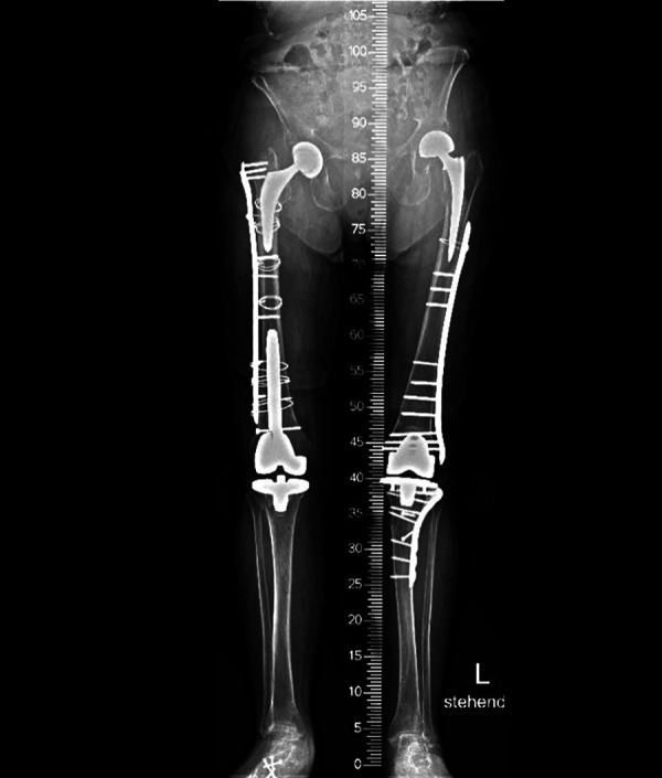
Locking plate fixation of bilateral interprosthetic femoral fractures in 88-year-old woman.
Figure 2.

Postoperative anteroposterior and lateral radiographs of interprosthetic femur fractures in an 86-year-old woman. Images show fracture healing after 6 months of fixation by applying a less invasive stabilization system (LISS) spanning the hip prosthesis.
Postoperatively, weight-bearing restrictions were defined individually depending on bone quality, expected stability of the osteosynthesis as well as the patients’ cognitive and physical abilities and their comorbidities: 4 of the 12 patients were restricted to no weight-bearing (wheelchair) for 6 to 8 weeks, while 7 were allowed partial and only 1 immediate full weight-bearing. Two patients were physically unable to perform partial weight-bearing and were therefore restricted to no weight-bearing.
The ASA score was documented for all patients: the most common ASA classification was grade III in 9 cases and grade IV in 2 cases, followed by 1 case grade I. Although there was an increasing trend to develop complications while the ASA scores worsened, there was no statistically significant difference in complications and outcomes (p = 0.163, p = 0.361).
The preoperative Barthel Index was available in 7 cases with a mean score of 77 (25-100). No statistically significant relationship was found between Barthel Index and complications or outcomes (p = 0.244, p = 0.331).
Complications and Revisions
12 of 13 fractures healed after the index procedure. No patient required additional surgery. One of 13 patients developed symptomatic hardware complications due to iliotibial band contact with the plate, which was painful, particularly during knee flexion/extension.
Two patients developed severe heterotopic ossification (HO) (Brooker 3/ grade C). HO was not correlated to complications.28
Five patients had previews revision surgeries (3 cases had revision TKA due to periprosthetic infection (PJI) and implant loosening and 2 revision THA because of PJI). Previous revision TJA did not influence the clinical and radiological outcomes (p = 0.876).
Mortality
Of 12 patients with IFF receiving surgical treatment, only 5 patients still live. The 3-month and 1-year mortality were reported in 2 and 3 patients, respectively, all unrelated to the implant surgery.
We were able to assess the residential status of 11 patients. All patients received either short-term rehabilitation or immediate placements in nursing homes after the hospitalization. Before the injury, 5 of the 12 patients lived independently in their own homes. Only 2 of these patients returned to their domestic environment, while 3 required long-term care in nursing and residential homes after discharge from the hospital or short-term rehabilitation. Data regarding postoperative mobility at the time of analysis were gathered from the patients directly or via the respective nursing facilities through telephone interviews and were available for 8 patients. Of these patients, 1 had persistent pain in the affected leg, 2 required a wheelchair for mobilization, and 3 of them used a wheeled walker. Only 2 of these patients were ambulating without any walking aid.
Discussion
As the population ages and the number of total hip and knee arthroplasty procedures increase, the incidence of IFFs are likely to increase.29 Their treatment in patients with ipsilateral stemmed THA and TKA can be challenging to treat surgically.30 IFFs present a rare occasion in clinical practice, and therefore their treatment is still associated with potential complications. The literature provides little recommendations and few data to guide treatment decisions and find appropriate concepts for adequate management of those injuries.9,20,30,31 IFF treatment options include fixation using locking plates, cerclage wires, autologous bone grafts and revision with a stemmed prosthesis.32 However, these treatment options are limited, mainly when there is severe bone loss in elderly patients.2,9
This study presents an analysis of the results in 13 IFFs of 12 patients with a follow-up of up to 9 years following a locking plate’s implantation. The overall outcome was good to excellent in 11 of the 12 patients with a low complication rat (only 1 patient). Morbidity was moderate to high as expected in this patient cohort.
In our study, low complication rates could be achieved by following the recommendations of Sah et al. and Mamczak et al.1,21 Critical aspects of the procedure include using long plates that span the entire interprosthetic zone, obtaining adequate fixation proximally and distally, avoiding excessive stripping of the fracture site, and obtaining anatomical or near anatomical reduction.2,30 Despite the high level of comorbidities in this population and the relatively poor bone stock, we observed unexpectedly no revisions in our cohort. Five patients had undergone previous revision procedures.
Operatively, achieving successful reduction and fixation is challenging due to limited bone availability (due to both implants) and patients commonly having poor bone quality.32 Using locking plates to treat IFFs is not new. Mamczak et al.21 retrospectively studied 20 patients with IFFs treated with plate fixation that spanned the interprosthetic zone and applied soft tissue preserving techniques without adjuvant bone grafts. Three defects of consolidation, loosening of a prosthetic stem, and a superficial infection were noted, while all the remaining patients were healed clinically and radiographically. All reported complications occurred in supracondylar fracture patterns. The authors concluded that IFFs tended to occur more frequently in the supracondylar region. The study we present had similar fracture union results, with full weight-bearing occurring an average of 4.02 months postoperatively. Separate analysis of implant stability process required to plan a type D UCS femur fracture was based on the plain radiographs as well as the bone quality. Intramedullary nailing was in our cases not possible due to the presence of both hip and knee arthroplasties. Because of the 2 prosthetic components, these diaphyseal fractures were best stabilized with a long locking compression plate bridging the fracture and extending as far proximally and distally as possible.
Our study showed similar epidemiological data and healing times to previous studies. Ehlinger et al.33 had 8 patients treated with locked plates spanning the femur, with healing in an average of 14 weeks. In contrast, Hou et al.20 assessed 13 IFFs classified separately according to the Vancouver classification. They reported fracture union and full weight bearing at an average of 4.7 months.
In our opinion, the current classification systems with the best compromise of effectiveness, simplicity, and high adoption rates, are the Vancouver and AO unified classification systems. We recognize that the unified classification system is perfect for IFFs, as it comprehensively addresses all fracture possibilities or guides all treatment options.
This study showed a satisfactory outcome following the individualized treatment of IFFs following ipsilateral hip and knee joint replacement. Compared to the current literature’s rare data, we had promising functional results and a high bony fusion rate. However, considering the complexity and challenges in many of these cases, IFFs require an adequate analysis of the fracture etiology and a suitable transfer into the best possible treatment concept.
Another significant issue is postoperative mortality. The overall rate of 1-year mortality in our patient cohort was 25% (3 of the 12 patients). These findings are corroborated by reports of appreciable mortality rates with IFFs. Soenen et al. reported 1 of 14 patients dying within 6 months of the fracture event 15. Platzer et al. documented 1 of 23 patients dying from cardiac arrest 15 days postoperatively,34 and Ehlinger et al. showed 1 death out of 8 patients 4 months after surgery.33 Several risk factors have been identified in our study, including advanced age, multiple medical comorbidities, and immobilization.
There are several limitations to our study that should be acknowledged. Firstly, it was a retrospective study with a small sample size (12 patients). Secondly, fracture geometry varied greatly, and no control was used, which compromises the analysis of confounding factors. However, there was no selection bias due to the retrospective study design, as the only factor deciding the inclusion of the patients was driven by the type and position of implant-related fractures. Finally, because the study spanned 9 years, treatment options have increased since the start of the study.
Conclusion
In conclusion, the reconstruction of IFFs with locking plates appears to be associated with a low complication rate and satisfactory results in various IFFs and compromised bone quality, as seen in this series of patients. We emphasize the extreme importance of considering an individual’s fracture type, bone quality, and prostheses to determine appropriate plate length to prevent possible mechanical failure and revision surgery. More extensive studies with strict inclusion criteria and patient-reported outcome measures are required to determine the real contribution to improved outcome performance.
Footnotes
Authors’ Note: The institutional review board and the local ethical committee approved the study protocol, BASEC-Nr. Req-2020-01906. Informed consent, which included surgical treatment and follow-up examinations was obtained from each patient. Level III, Therapeutic study. All authors have made substantial contributions to all of the following: (1) the conception and design of the study, or acquisition of data, or analysis and interpretation of data, (2) drafting the article or revising it critically for important intellectual content, (3) final approval of the version to be submitted, (4) being accountable for all aspects of the work in ensuring that questions related to the accuracy or integrity of any part of the work are appropriately investigated and resolved. Institutional review board approval was obtained by the ethics committee of the University of Zurich (ID 2020-01906).
Declaration of Conflicting Interests: The author(s) declared no potential conflicts of interest with respect to the research, authorship, and/or publication of this article.
Funding: The author(s) received no financial support for the research, authorship, and/or publication of this article.
ORCID iDs: Marios Loucas, MD  https://orcid.org/0000-0002-1576-8368
Rafael Loucas, MD
https://orcid.org/0000-0001-5729-2508
Michael Dietrich, MD
https://orcid.org/0000-0003-2512-3368
References
- 1. Sah AP, Marshall A, Virkus WV, Estok DM II, Della Valle CJ. Interprosthetic fractures of the femur: treatment with a single-locked plate. J Arthroplasty. 2010;25(2):280–286. doi:10.1016/j.arth.2008.10.008 [DOI] [PubMed] [Google Scholar]
- 2. Hoffmann MF, Lotzien S, Schildhauer TA. Clinical outcome of interprosthetic femoral fractures treated with polyaxial locking plates. Injury. 2016;47(4):934–938. doi:10.1016/j.injury.2015.12.026 [DOI] [PubMed] [Google Scholar]
- 3. Lehmann W, Rupprecht M, Hellmers N, et al. Biomechanical evaluation of peri- and interprosthetic fractures of the femur. J Trauma. 2010;68(6):1459–1463. doi:10.1097/TA.0b013e3181bb8d89. [DOI] [PubMed] [Google Scholar]
- 4. Liu J, Wilson L, Poeran J, et al. Trends in total knee and hip arthroplasty recipients: a retrospective cohort study. Reg Anesth Pain Med. 2019;44(9):854–859. doi:10.1136/rapm-2019-100678 [DOI] [PubMed] [Google Scholar]
- 5. Rupprecht M, Sellenschloh K, Grossterlinden L, et al. Biomechanical evaluation for mechanisms of periprosthetic femoral fractures. J Trauma. 2011;70(4):E62–66. doi:10.1097/TA.0b013e3181e99ff1 [DOI] [PubMed] [Google Scholar]
- 6. Springer BD, Berry DJ, Lewallen DG. Treatment of periprosthetic femoral fractures following total hip arthroplasty with femoral component revision. J Bone Joint Surg Am. 2003;85(11):2156–2162. doi:10.2106/00004623-200311000-00015 [DOI] [PubMed] [Google Scholar]
- 7. Abdel MP, Houdek MT, Watts CD, Lewallen DG, Berry DJ. Epidemiology of periprosthetic femoral fractures in 5417 revision total hip arthroplasties: a 40-year experience. Bone Joint J. 2016;98-b(4):468–474. doi:10.1302/0301-620x.98b4.37203 [DOI] [PubMed] [Google Scholar]
- 8. Dave DJ, Koka SR, James SE. Mennen plate fixation for fracture of the femoral shaft with ipsilateral total hip and knee arthroplasties. J Arthroplasty. 1995;10(1):113–115. doi:10.1016/S0883-5403(05)80111-1 [DOI] [PubMed] [Google Scholar]
- 9. Kenny P, Rice J, Quinlan W. Interprosthetic fracture of the femoral shaft. J Arthroplasty. 1998;13(3):361–364. doi:10.1016/s0883-5403(98)90187-5 [DOI] [PubMed] [Google Scholar]
- 10. Solarino G, Vicenti G, Moretti L, Abate A, Spinarelli A, Moretti B. Interprosthetic femoral fractures-a challenge of treatment. A systematic review of the literature. Injury. 2014;45(2):362–368. doi:10.1016/j.injury.2013.09.028 [DOI] [PubMed] [Google Scholar]
- 11. Abendschein W. Periprosthetic femur fractures—a growing epidemic. Am J Orthop (Belle Mead NJ). 2003;32(9 Suppl):34–36. [PubMed] [Google Scholar]
- 12. Franklin J, Malchau H. Risk factors for periprosthetic femoral fracture. Injury. 2007;38(6):655–660. doi:10.1016/j.injury.2007.02.049 [DOI] [PubMed] [Google Scholar]
- 13. Rorabeck CH, Taylor JW. Classification of periprosthetic fractures complicating total knee arthroplasty. Orthop Clin North Am. 1999;30(2):209–214. doi:10.1016/s0030-5898(05)70075-4 [DOI] [PubMed] [Google Scholar]
- 14. Culp RW, Schmidt RG, Hanks G, Mak A, Esterhai JL, Jr, Heppenstall RB. Supracondylar fracture of the femur following prosthetic knee arthroplasty. Clin Orthop Relat Res. 1987;(222):212–222. [PubMed] [Google Scholar]
- 15. Soenen M, Migaud H, Bonnomet F, Girard J, Mathevon H, Ehlinger M. Interprosthetic femoral fractures: analysis of 14 cases. Proposal for an additional grade in the Vancouver and SoFCOT classifications. Orthop Traumatol Surg Res. 2011;97(7):693–698. doi:10.1016/j.otsr.2011.07.009 [DOI] [PubMed] [Google Scholar]
- 16. Leonidou A, Moazen M, Skrzypiec DM, Graham SM, Pagkalos J, Tsiridis E. Evaluation of fracture topography and bone quality in periprosthetic femoral fractures: a preliminary radiographic study of consecutive clinical data. Injury. 2013;44(12):1799–1804. doi:10.1016/j.injury.2013.08.010 [DOI] [PubMed] [Google Scholar]
- 17. McGraw IW, Spence SC, Baird EJ, Eckhardt SM, Ayana GE. Incidence of periprosthetic fractures after hip hemiarthroplasty: are uncemented prostheses unsafe? Injury. 2013;44(12):1945–1948. doi:10.1016/j.injury.2013.07.023 [DOI] [PubMed] [Google Scholar]
- 18. Hoffmann MF, Jones CB, Sietsema DL, Koenig SJ, Tornetta P, III. Outcome of periprosthetic distal femoral fractures following knee arthroplasty. Injury. 2012;43(7):1084–1089. doi:10.1016/j.injury.2012.01.025 [DOI] [PubMed] [Google Scholar]
- 19. Zuurmond RG, Pilot P, Verburg AD. Retrograde bridging nailing of periprosthetic femoral fractures. Injury. 2007;38(8):958–964. doi:10.1016/j.injury.2006.12.011 [DOI] [PubMed] [Google Scholar]
- 20. Hou Z, Moore B, Bowen TR, et al. Treatment of interprosthetic fractures of the femur. J Trauma. 2011;71(6):1715–1719. doi:10.1097/TA.0b013e31821dd9f1 [DOI] [PubMed] [Google Scholar]
- 21. Mamczak CN, Gardner MJ, Bolhofner B, Borrelli J, Jr, Streubel PN, Ricci WM. Interprosthetic femoral fractures. J Orthop Trauma. 2010;24(12):740–744. doi:10.1097/BOT.0b013e3181d73508 [DOI] [PubMed] [Google Scholar]
- 22. Su ET, Kubiak EN, Dewal H, Hiebert R, Di Cesare PE. A proposed classification of supracondylar femur fractures above total knee arthroplasties. J Arthroplasty. 2006;21(3):405–408. doi:10.1016/j.arth.2005.05.022 [DOI] [PubMed] [Google Scholar]
- 23. Mahoney FI, Barthel DW. Functional evaluation: the Barthel index. Md State Med J. 1965;14:61–65. [PubMed] [Google Scholar]
- 24. Saklad M. Grading of patients for surgical procedures. Anesthesiology. 1941;2(3):281–284. [Google Scholar]
- 25. Muller M, Allgower M, Schneider R, Willeneger H. Manual of Internal fixation: The Comprehensive Classification of fractures of Long Bones. Springer Verlag; 1991. [Google Scholar]
- 26. Duncan CP, Haddad FS. The Unified Classification System (UCS): improving our understanding of periprosthetic fractures. Bone Joint J. 2014;96-b(6):713–716. doi:10.1302/0301-620x.96b6.34040 [DOI] [PubMed] [Google Scholar]
- 27. Duncan CP, Masri BA. Fractures of the femur after hip replacement. Instr Course Lect. 1995;44:293–304. [PubMed] [Google Scholar]
- 28. Brooker AF, Bowerman JW, Robinson RA, Riley LH, Jr. Ectopic ossification following total hip replacement. Incidence and a method of classification. J Bone Joint Surg Am. 1973;55(8):1629–1632. [PubMed] [Google Scholar]
- 29. Abdelaziz H, Saleri S, Mau H, et al. Interprosthetic femoral sleeves in revision arthroplasty: a 20-year experience. J Arthroplasty. 2019;34(7):1423–1429. doi:10.1016/j.arth.2019.02.055 [DOI] [PubMed] [Google Scholar]
- 30. Rozell JC, Delagrammaticas DE, Schwarzkopf R. Interprosthetic femoral fractures: management challenges. Orthop Res Rev. 2019;11:119–128. doi:10.2147/ORR.S209647 [DOI] [PMC free article] [PubMed] [Google Scholar]
- 31. Della Valle CJ, Tejwani N, Koval KJ. Interprosthetic fracture of the femoral shaft treated with a percutaneously inserted dynamic condylar screw: case report. J Trauma. 2003;54(3):602–605. doi:10.1097/01.Ta.0000046444.21003.06 [DOI] [PubMed] [Google Scholar]
- 32. Velickovic M, Periprosthetic HT. Interprosthetic, and refractures after interprosthetic femoral fractures—extension of the currently used Vancouver, Rorabeck and Platzer classification. J ortho Rehab Surg. 2018;2(1):1–8. [Google Scholar]
- 33. Ehlinger M, Czekaj J, Adam P, Brinkert D, Ducrot G, Bonnomet F. Minimally invasive fixation of type B and C interprosthetic femoral fractures. Orthop Traumatol Surg Res. 2013;99(5):563–569. doi: 10.1016/j.otsr.2013.01.011 [DOI] [PubMed] [Google Scholar]
- 34. Platzer P, Schuster R, Luxl M, et al. Management and outcome of interprosthetic femoral fractures. Injury. 2011;42(11):1219–1225. doi:10.1016/j.injury.2010.08.020 [DOI] [PubMed] [Google Scholar]