Abstract
Background:
Irregularities in the number of teeth can also occur in deciduous and in permanent dentition.
Objective:
The aim of this article is to report the case of a seven years old child and a 27 years old male patient affected by a numeric dental anomaly.
Methods:
This paper has shown the pathologic condition characterized by the presence of supernumerary tooth (mesiodens) and supernumerary canine as well as supplementary premolars in a non-syndromic patients. Clinical and instrumental examinations were made to perform a correct orthodontic examination and diagnosis. A young patient was affected by numeric dental anomaly in the upper jaw. An adult patient was affected by numeric anomaly in both jaws, supplementary premolars in lower jaw and a supernumerary canine in lower and upper jaw.
Discussion:
The aim of surgical-orthodontic treatment was extraction of the erupted supernumerary teeth to obtain the physiologic eruption and placement of the permanent ones.
Conclusion:
Therapy of supernumerary/ supplementary teeth is the extraction. But also, an excess tooth in the dentition can be left as a replacement tooth, due to a previously lost permanent tooth from the dentition, if its biological value and potential is sufficient to complete the dentition both functionally and aesthetically.
Keywords: Supplemental and supernumerary teeth, dental anomaly, mesiodens
1. BACKGROUND
The process of tooth growth and development, both in deciduous and permanent dentition, is susceptible the action of a whole range of genetic, systemic and local factors, which can endanger it and lead to the appearance of certain irregularities. Irregularities in the number of teeth can also occur in deciduous and in permanent dentition (1, 2).
Genetic control of dental growth and development represents a whole series of processes, which, for easier understanding and monitoring, can be divided into two directions: one that follows the shape, size and position of each tooth, and the other, which refers to specific enamel and dentin formation processes. Genes, associated with early tooth positioning and development, participate in the function of regulating morphogenesis. Their mutations result in various developmental disorders (1).
The long-term, perennial process of tooth development is, for descriptive reasons, divided into several phases: the phase of initiation, proliferation, histomorphologic differentiation, apposition (mineralization), eruption and resorption of new teeth. Developmental irregularities can occur under the influence of the environment (pathological general and local factors) or various hereditary factors, at any stage of development. The divisions of irregularities in tooth development are numerous. According to the clinical picture, there are irregularities in: eruption, number, size, shape, position, color and structure of teeth (3). Hyperdontia is an anomaly of an increased number of teeth (1). Excess teeth can occur in both dentitions but are five times more common in permanent dentition (1% to 3.5%). In 98% of cases, they occur in the frontal part of the maxilla, of which 75% are mesiodens. They occur almost twice as often in boys (1: 0.4) (3).
Prevalence according to type and location varies: upper lateral incisors represent 50%, mesiodens 36%, central upper incisor 11% and premolars 3%.
Single supernumerary teeth represent a 76-86% percentage, double supernumerary teeth correspond to 12-23% of cases, and four supernumerary molars or distomolars account for 18% of cases. Multiple supernumerary teeth represent less than 1% of all cases (4).
The terminology used to denote supernumerary teeth is based on their localization. Thus, if the excess tooth is located in the frontal part of the maxilla, then it is called mesiodens, distally from the wisdom tooth is distomolar, and orally or vestibularly from the dentition peridens (peripremolar, perimolar). Excess teeth may be normally shaped (dentes supernumeraria) or may differ in appearance (dentes accessoria). They are often hypoplastic and can be placed inside or outside the dentition. The appearance of excessive teeth can interfere with the eruption and proper establishment of other teeth, create new retention sites for food and make it difficult to maintain adequate oral hygiene. Cystic formations can often develop around unexplained excess teeth (3).
A classification of numeric dental anomalies was published by Tomes (1873), who defined the following.
Supplemental: tooth characterized by the same form and function of adjacent teeth with no anatomical differences.
Supernumerary: tooth characterized by an atypical anatomic form; often these teeth are smaller than normal.
Bush classification (1897) analyzed the different morphology of supernumerary teeth:
Conic: tooth of a small volume and conic form, its root is short and palatine.
Tubercolate: tooth with several cusps. Its root is short and hooked (5).
2. OBJECTIVE
The aim of this article is to report the case of a seven years old child and a 27 years old male patient affected by a numeric dental anomaly.
3. CASE REPORTS
3.1. Mesiodens - Case 1
A seven years old girl was brought by worried mother in dental office. A thorough general examination was carried out to rule out the presence of any syndrome. Medical and family histories were non-contributory. Intraoral examination revealed permanent dentition with a supernumerary tooth, similar shaped as an permanent incisor only smaller in size an rotated, present medial to 11 and displacing 21 palataly. The case was discussed with orthodontist who suggested an immediate extraction and will be followed up after eruption, further orthodontic treatment will be then planned and conducted.
After radiographic examination no other tooth anomaly was present so it was proceeded to extraction.
On X-ray it is visible that tooth 21 is rotated and unable to erupt due to mesiodens blockage.
3.2. Supernumerary and supplementary teeth - Case 2
The patient reports to the dental office with the problem, as he states, narrowness between the teeth, food decay and the inability to adequately maintain oral hygiene. Due to the same problem in area of the right lower premolars, he lost tooth 45, which was previously extracted as the patient states.
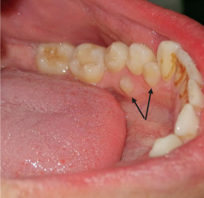
Currently in the mouth on the lower right side the clinical finding is neat, the dentition is also neat, because the excess tooth has taken the place of tooth 45 and thus formed a full dentition. On the lower left side in the area of the premolars, there are two supernumerary teeth, smaller compared to the permanent premolars.
Clinical examination revealed the presence of one redundant tooth between teeth 33 and 34, and one redundant tooth between teeth 34 and 35, which did not erupt completely. On tooth 35, probably due to the impossibility of cleaning, a filling is present on the occlusal-distal surface of the tooth. In the therapy, the extraction of both redundant teeth was performed without major difficulties and the need to send them to an oral surgeon. On the radiological image, another redundant tooth can be seen, which is not visible in the oral cavity on clinical examination. Its position is high in the maxillary bone, and it is superimposed with the tip of the root of tooth 23. For this redundant tooth, the patient was referred to an orthodontist for further treatment.
4. DISCUSSION
Presence of supernumerary tooth in normal dentition is not unusual phenomenon but the presence of multiple supernumerary teeth in individuals without any syndromic disorder is not common. Literature shows that single supernumerary tooth is the most common (6).
It is essential to enumerate and identify the teeth present clinically and radiographically before a definitive diagnosis and treatment plan regarding supernumerary teeth can be formulated. Radiographs played an important role to rule out the presence of impacted supernumerary teeth or other associated anomalies (7).
Salcido Garcia et al in their study reported supernumerary premolars as the second most common supernumerary teeth after mesiodens with a reported incidence of 0.09–0.64% (8).
In our case, report supernumerary premolars had a higher incidence then mesiodens.
Yusof et al in his study observed, the premolar region of the mandible is the preferred site of multiple supernumerary teeth occurring in the absence of any syndrome, and that was as well presented in our case 2. (9)
Most of the time, supernumerary teeth are asymptomatic but as always problems may appear that include periodontitis, dilacerations, dentigerous cyst formation, root resorption of adjacent teeth, occlusive disturbance and no aesthetic appearance (10).
Cortés-Bretón Brinkmann et al in their study revealed that premolars were the most frequently seen type of supernumerary tooth and constituted 45.5 percent of the sample (11). In our case 2 we had 3 supernumarary premolars in lower jaw and the other patient had only one supernumerary tooth which was mesiodens.
In their review of literature Solares and Romero stated that supernumerary premolars appear to be more common than previously estimated. They are also the most common supernumerary teeth in the mandibular arch (7%), and their incidence is 1% (1 in 157)-much higher than previously reported. Maxillary supernumerary premolars were found to occur at a lower rate (26%). The possible mechanisms of development are described, with a localized hyperactivity of the dental lamina being the most widely accepted theory (12).
Cortés-Bretón Brinkmann et al stated that the early diagnosis and follow-up of patients with multiple supernumerary teeth should help clinicians prevent the diseases associated with this kind of hyperodontia (11).
5. CONCLUSION
Whenever supernumerary teeth are diagnosed, single or multiple, treatment options should be reviewed carefully. Therapy of supernumerary/supplementary teeth is usually the extraction as both seen in case 1 and 2. But also, an excess tooth in the dentition can be left as a replacement tooth as we saw it the case 2, due to a previously lost permanent tooth from the dentition, if its biological value and potential is sufficient to complete the dentition both functionally and aesthetically.
Figure 1. An image of mesiodens in a seven year old patient.
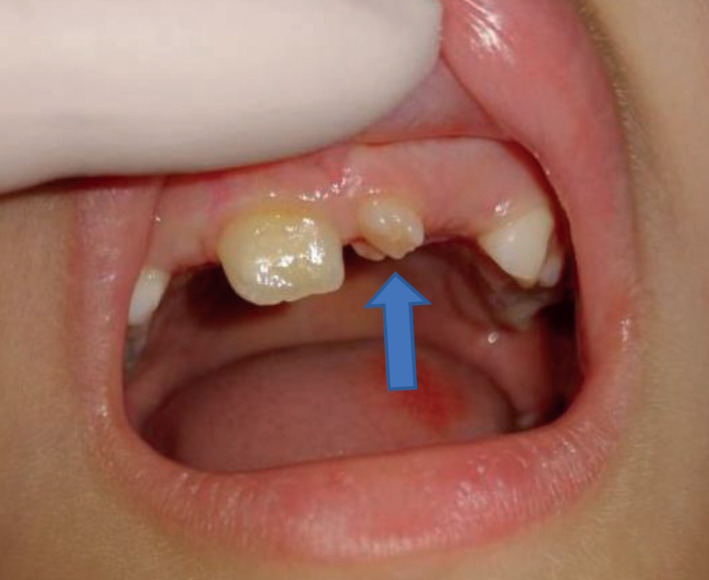
Figure 2. Radiographic image of a mesiodens, no other supernumerary teeth are visible.
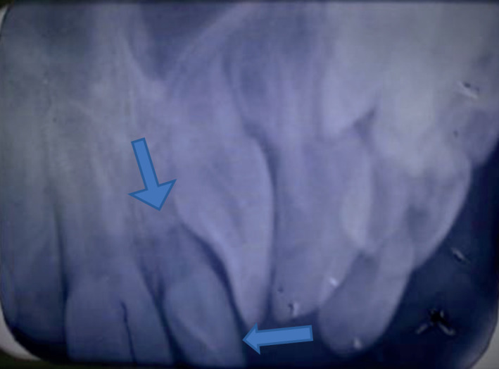
Figure 3. Mesiodens examined after extraction.
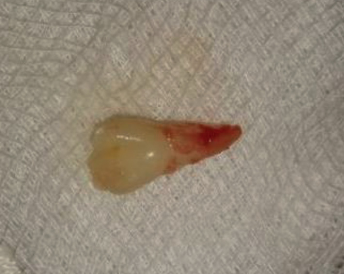
Figure 4. An image of orthopantomogram, showing supernumerary and suplementary teeth.
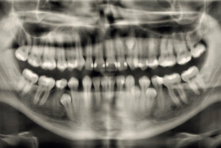
Figure 5. The supplementary tooth took place and function of a normal permanent premolar.
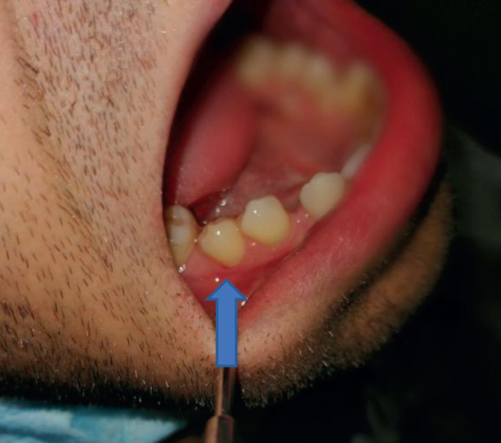
Figure 6. Supernumarary lower left premolar and a supernumerary canine.
Author’s contribution:
All authors were involved in all steps of preparation this article. Final proof reading was made by the first author.
Conflict of interest:
None declared.
Financial support and sponsorship:
Nil.
REFERENCES
- 1.Jurić H. Jastrebarsko: Naklada Slap; 2015. Dječja dentalna medicina. [Google Scholar]
- 2.Rakhshan V. Congenitally missing teeth (hypodontia): A review of the literature concerning the etiology, prevalence, risk factors, patterns and treatment. Dent Res J (Isfahan) 2015;12:1–13. doi: 10.4103/1735-3327.150286. [DOI] [PMC free article] [PubMed] [Google Scholar]
- 3.Gajić M, Lalić M. izdanje – Pančevo: Stomatološki fakultet; 2011. Dječija stomatologija.1. [Google Scholar]
- 4.Blanco Ballesteros G. Dientes múltiples supernumerarios. Reporte de un caso. Revista Estomatológica. 2005;13(1):13–18. [Google Scholar]
- 5.Lo Giudice G, Nigrone V, Longo A, Cicciù M. Supernumerary and supplemental teeth: case report. European Journal of Paediatric Dentistry. 2008;9:2. [PubMed] [Google Scholar]
- 6.Rajab LD, Hamdan MA. Supernumerary teeth: review of the literature and a survey of 152 cases. Int J Paediatr Dent. 2002 Jul;12(4):244–254. doi: 10.1046/j.1365-263x.2002.00366.x. [DOI] [PubMed] [Google Scholar]
- 7.Kumar A, Namdev R, Bakshi L, Dutta S. Supernumerary teeth: Report of four unusual cases. Contemp Clin Dent. 2012 Apr;3(Suppl 1):S71–77. doi: 10.4103/0976-237X.95110. [DOI] [PMC free article] [PubMed] [Google Scholar]
- 8.Salcido-García JF, Ledesma-Montes C, Hernández-Flores F, Pérez D, Garcés-Ortíz M. Frequency of supernumerary teeth in Mexican population. Med Oral Patol Oral Cir Bucal. 2004 Nov-Dec;9(5):407–409. [PubMed] [Google Scholar]
- 9.Yusof WZ. Non-syndrome multiple supernumerary teeth: literature review. J Can Dent Assoc. 1990 Feb;56(2):147–149. [PubMed] [Google Scholar]
- 10.Ansari AA, Malhotra S, Pandey RK, Bharti K. Non-syndromic multiple supernumerary teeth: report of a case with 13 supplemental teeth. BMJ Case Rep. 2013 Mar 6;2013:bcr2012008316. doi: 10.1136/bcr-2012-008316. [DOI] [PMC free article] [PubMed] [Google Scholar]
- 11.Cortés-Bretón Brinkmann J, Barona-Dorado C, Martínez-Rodriguez N, Martín-Ares M, Martínez-González JM. Nonsyndromic multiple hyperdontia in a series of 13 patients: epidemiologic and clinical considerations. J Am Dent Assoc. 2012 Jun;143(6):e16–24. doi: 10.14219/jada.archive.2012.0243. [DOI] [PubMed] [Google Scholar]
- 12.Solares R, Romero MI. Supernumerary premolars: a literature review. Pediatr Dent. 2004 Sep-Oct;26(5):450–458. [PubMed] [Google Scholar]


