Abstract
SUMMARY: Electrical injuries are becoming more common and are increasingly imaged with advanced technologies, such as MR imaging. However, the MR imaging findings of such injuries remain largely unstudied. We report a high-voltage electrical injury to the cerebral corticospinal tracts and document evolution on serial MR images.
As electrification increases worldwide, electrical injuries are also increasing. They occur almost exclusively in children and young men.1 Childhood injuries commonly occur in the household and involve low-voltage exposures.1 In young men, most incidents involve utility or construction workers.2,3 A smaller subset of this group experience electrical injury due to lightning strikes.4 Because increasing numbers of these injuries are being evaluated with MR imaging, understanding the imaging appearance of electrical injury is becoming more important both for its use in investigation of specific electrical injuries and for differentiation of electrically induced abnormalities from more ominous pathology discovered on routine examinations. We report a case of a high-voltage cerebral electrical injury with serial MR imaging examinations demonstrating partial resolution of the imaging abnormality over a 22-month period.
Case Report
A 44-year-old man experienced an electrical shock while working with a high-voltage arc welder. There was witnessed onset of aphasia and right hand weakness, which subsided within 20 minutes. The patient did not lose consciousness or experience seizures. He was evaluated at an outside emergency department and found to be hypertensive with physical examination normal. CT of the brain was normal. The emergency department staff diagnosed a transient ischemic attack. Five months after the accident, the patient presented to our tertiary care center and underwent his first brain MR imaging.
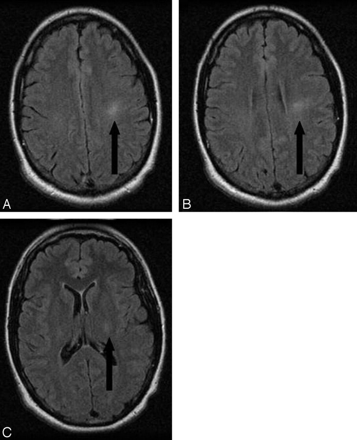
Contrast-enhanced brain MR imaging demonstrated atypical increased T2 signal intensity within the left corticospinal tract best seen on the fluid-attenuated inversion recovery (FLAIR) sequence (Fig 1). This nonenhancing signal intensity abnormality extended from the subcortical white matter of the precentral gyrus to the level of the posterior limb of the left internal capsule. In addition, a few punctuate foci of nonspecific T2 signal hyperintensity were seen within the left centrum semiovale anterior to the corticospinal tract signal intensity abnormality. There was no evidence of encephalomalacia or other specific findings of cerebral ischemia. The right corticospinal tract showed no abnormalities. At this point, diagnostic considerations included atypical Wallerian degeneration, low-grade glioma, and electrical injury.
Fig 1.
Axial FLAIR images (TE = 140 ms; TR = 11,002; inversion time = 140 ms) obtained at levels of the centrum semiovale (A), corona radiata (B), and posterior limb of the internal capsule (C) demonstrate increased T2 signal intensity within the left corticospinal tract 5 months after the electrocution injury.
MR imaging obtained 7, 13, and 22 months after the injury demonstrated significant partial resolution of the left corticospinal tract T2 signal intensity abnormality without evidence of subsequent volume loss within the left corticospinal tract or brain stem to support Wallerian degeneration, leading us to conclude that the patient's imaging findings represented a transient cerebral electrocution injury.
Discussion
The sporadic nature of electrical injury has made studying and evaluating treatments difficult. Most available literature consists of case studies and animal experimentation. Still, valuable principles concerning these injuries, and the important role that radiology plays in their investigation, can be derived.
The mechanisms by which electricity causes injury are not completely defined but include several pathways. The most obvious is thermal, with large quantities of heat produced by the current passing through the victim resulting in external and internal burns.5,6 No less important is electroporation, in which membrane proteins permanently change conformation and can no longer maintain transmembrane ion gradients, resulting in cell death.5–7 In addition, the actual physical forces involved in the injury, both direct (ie, a person thrown from the site of a lightning strike by the force of the strike itself) and indirect (ie, a person loses consciousness and falls from the ladder where they were working), can injure the victim.4 In 1995, Cherington4 categorized electrical injuries into 4 groups, which are useful in understanding electrical injuries, as well as in guiding appropriate imaging.
Immediate and Transient
These injuries are apparent immediately after the event. Examples include: loss of consciousness, amnesia, confusion, paresthesias, and weakness/paralysis. The limb clumsiness and aphasia affecting our patient fall into this category. These injuries are the most common but often resolve within minutes to hours. Imaging has traditionally included plain films and CT examinations and is almost universally negative. Case reports with acute neurologic MR imaging examinations have demonstrated T2 abnormalities consistent with neurologic symptoms.8,9 Follow-up imaging is rare, but when obtained, the T2 abnormalities seem to partially resolve.
Immediate and Prolonged or Permanent
These injuries are less common but are debilitating and receive a disproportionate amount of neurologic imaging.4 Examples include: hemorrhage, chromolysis of pyramidal and other neurons, glial proliferation, and infarction.4 Factors postulated to contribute to this category include the length and severity of the insult, as well as the path of the electricity.9 MR imaging offers superior soft tissue characterization and can document stability or progression of these lesions.
As in our patient, remote electrical insults without residual clinical symptoms may result in persistent T2 abnormalities that could potentially be confused with other pathologies, such as a low-grade neoplasm. A careful history and sequential imaging are helpful in arriving at the diagnosis of electrical injury.
Delayed and Progressive
This category may be difficult to define clinically, because differentiating between it and the immediate and prolonged or permanent category may not be possible. These injuries are also debilitating and receive a disproportionate amount of imaging, usually MR imaging.4 Numerous conditions have been reported, including basal ganglia disorders, myelopathy, and motor system diseases.4 Other coincidental, and perhaps treatable, conditions must be ruled out by imaging, because these injuries carry a poor prognosis.
Event-Associated Injuries
This category is the most obvious and uses the full gamut of radiologic services. Examples include trauma or hypoxic injures due to ventricular fibrillation.4 Because victims of electrical injury are often otherwise healthy individuals, recognition and treatment of secondary conditions play important roles in determining their outcome.
Conclusion
Electrical injuries are increasingly common and occur primarily in young men as a result of occupational hazards. MR imaging is important for initial evaluation of electrical injuries involving the central nervous system and ongoing follow-up. Radiologists should consider the diagnosis of electrical injury when encountering confluent white matter signal intensity abnormality in patients with appropriate clinical history.
Footnotes
Paper previously presented at: Annual Meeting of the American Society of Neuroradiology, June 13, 2007; Chicago, Ill.
References
- 1.Baker MD, Chiaviello C. Household electrical injuries in children. Epidemiology and identification of avoidable hazards. Am J Dis Child 1989;143:59–62 [DOI] [PubMed] [Google Scholar]
- 2.Taylor AJ, McGwin G Jr, Davis GC, et al. Occupational electrocutions in Jefferson County, Alabama. Occup Med 2002;52:102–06 [DOI] [PubMed] [Google Scholar]
- 3.Bailer AJ, Bena JF, Stayner LR, et al. External cause-specific summaries of occupational fatal injuries. Part I: an analysis of rates. Am J Ind Med 2003;43:237–50 [DOI] [PubMed] [Google Scholar]
- 4.Cherington M. Central nervous system complications of lightning and electrical injuries. Semin Neurol 1995;15:233–40 [DOI] [PubMed] [Google Scholar]
- 5.Arevalo JM, Lorente JA, Balseiro-Gomez J. Spinal cord injury after electrical trauma treated in a burn unit. Burns 1999;25:449–52 [DOI] [PubMed] [Google Scholar]
- 6.Hunt JL, Mason AD, Masterson TS, et al. The pathophysiology of acute electric injuries. J Trauma 1976;16:335–40 [DOI] [PubMed] [Google Scholar]
- 7.Freeman CB, Goyal M, Bourque PR. MR imaging findings in delayed reversible myelopathy from lightning strike. AJNR Am J Neuroradiol 2004;25:851–53 [PMC free article] [PubMed] [Google Scholar]
- 8.Isao T, Masaki F, Nakayama R, et al. Delayed brain atrophy after electrical injury. J Burn Care 2005;26:456–58 [DOI] [PubMed] [Google Scholar]
- 9.Kalita J, Jose M, Misra UK. Myelopathy and amnesia following accidental electrical injury. Spinal Cord 2002;40:253–55 [DOI] [PubMed] [Google Scholar]


