Abstract
SUMMARY: The spontaneous occurrence of acute Brown-Séquard syndrome is an extremely rare event, with most reported cases being secondary to spontaneous epidural hematomas and spinal cord ischemia. We report a rare case of Brown-Séquard syndrome from spontaneous intraspinal hemorrhage in a patient with multiple cavernous angiomas in the spinal cord secondary to craniospinal radiation in childhood. Postulated mechanisms leading to the condition include postradiation molecular changes and venous occlusion.
The spontaneous occurrence of acute hemispinal cord dysfunction or acute Brown-Séquard syndrome is an extremely rare event. Most reported cases have been secondary to spontaneous epidural hematomas and spinal cord ischemia.1,2 In rare instances, it has been caused by a spontaneous subarachnoid hematoma involving the cervical spine3 or as a complication of a ruptured spinal hemangioma.4 We report a rare case of Brown-Séquard syndrome from spontaneous intraspinal hemorrhage in a patient with multiple cavernous angiomas in the spinal cord secondary to craniospinal radiation in childhood.
Case Report
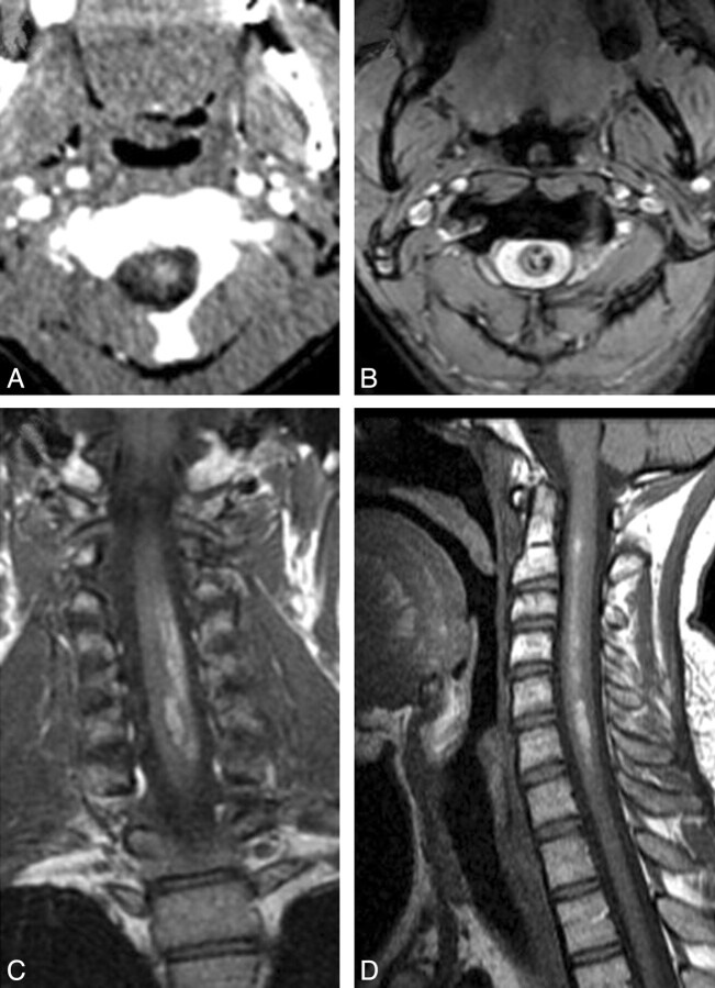
A 27-year-old man presented to the emergency department with 2 days of mild weakness in his left arm and leg and numbness and tingling in his right leg that rapidly worsened in the preceding few hours. His medical history included childhood acute lymphocytic leukemia, for which he was treated with chemotherapy and craniospinal radiation therapy between the ages of 3 and 8 years. This was complicated by panhypopituitarism requiring hormone replacement. At presentation, he was alert and oriented with intact cranial nerve function. He had diffuse severe weakness of his left upper and lower extremities (motor grades: deltoid, III; biceps, II; brachioradialis, II; interossei, I; iliopsoas, IV; gastrocnemius, IV; tibialis anterior, IV). The right upper and lower extremities had grade V power across all muscle groups. Sensory examination showed decreased pain and temperature sensation on the right arm and leg. He showed decreased reflexes (grade I) over the left triceps, brachioradialis, and triceps, and increased reflexes (grade III) at the left knee and ankle. His right extremities showed grade II reflexes across all tendons. His plantar reflexes were upgoing bilaterally. Initial CT and MR imaging of the cervical spine showed intramedullary blood components involving the cervical cord without significant cord swelling or edema (Fig 1A–D). Subsequent MR imaging of the brain revealed multiple indolent cavernous angiomas (images not shown). The patient was treated acutely with high-dose intravenous corticosteroids, managed as an inpatient, and later discharged to a rehabilitation unit.
Fig 1.
Axial CT scan at the level of the C2 vertebral body after intravenous contrast administration shows a small focus of hyperattenuation in the left portion of the spinal cord (A). This did not change from noncontrast CT, suggesting a nidus of calcification or blood. Gradient echo axial MR imaging at the C2 level shows abnormal foci of high and low signal intensity in the left side of the cord, the low signal intensity consistent with magnetic susceptibility effects of hemosiderin (B). T1-weighted coronal and sagittal MR images of the cervical cord depict irregular vertically distributed zones of high signal intensity consistent with the paramagnetic effects of methemoglobin (C and D). Note the long extent of the abnormality with no significant swelling of the cord.
Discussion
The description of clinical findings associated with lateral hemispinal cord dysfunction or Brown-Séquard syndrome is ascribed to Charles Édouard Brown-Séquard for his study of spinal pathways.5 In most cases, acute Brown-Séquard syndrome occurs secondary to penetrating trauma involving a lateral half of the spine. On occasion, it arises spontaneously as a complication of spontaneous spinal epidural hematomas and ischemia.1,2 In our patient, the intraspinal hemorrhage arose from postradiation cavernous angiomas. There was no personal or family history of a bleeding disorder and no evidence of coagulopathy on laboratory testing. His clinical manifestation followed a biphasic pattern with mild symptoms for 2 days followed by rapid progression in the preceding hours before presentation. This presentation is likely because of a second hemorrhage.
Although the occurrence of postradiation cavernomas is well described in the brain, especially after radiation therapy in childhood,6 its occurrence in the spine is rare.7 Although its exact pathogenesis remains obscure, several mechanisms have been postulated including children with acute lymphocytic leukemia (a frequent reason for craniospinal irradiation) being inherently prone to the development of cavernomas, radiation-induced growth of pre-existing tiny cavernomas, and radiation-induced fibrosis and venous outflow occlusion as well as release of hypoxia inducible factors and vasculo-endothelial growth factor.6 Bleeds from cavernous angiomas are often mild. The lack of significant cord swelling or edema in our patient ruled out a major bleed. Hemorrhage from a postradiation cavernous angioma resulting in spontaneous acute Brown-Séquard syndrome is an extremely rare event. In addition to ours, 1 previous report has documented this occurrence.7
Conclusion
Brown-Séquard syndrome can rarely occur as a complication of spontaneous intraspinal hemorrhages in patients with cavernous angiomas. This cause should be considered in patients with a remote history of radiation therapy. Accurate diagnosis and management of such patients depends on additional imaging studies as well as a careful history and physical examination.
References
- 1.Egido Herrero JA, Saldana C, Jimenez A, et al. Spontaneous cervical epidural hematoma with Brown-Séquard syndrome and spontaneous resolution. Case report. J Neurosurg Sci. 1992;36:117–19 [PubMed] [Google Scholar]
- 2.Decroix JP, Ciaudo-Lacroix C, Lapresle J. Brown-Séquard syndrome caused by a spinal cord infarction. Rev Neurol (Paris) 1984;140:585–86 [PubMed] [Google Scholar]
- 3.Okuno S, Morimoto T, Sakaki T. A case of spontaneous subarachnoid hematoma of the high cervical spine presenting as Brown-Séquard's syndrome. No Shinkei Geka. 2001;29:851–55 [PubMed] [Google Scholar]
- 4.Ehsan T, Henderson JM, Manepalli AN. Epidural hematoma producing Brown-Séquard syndrome: a case due to ruptured hemangioma with magnetic resonance imaging findings. J Neuroimaging. 1996;6:62–63 [DOI] [PubMed] [Google Scholar]
- 5.Goody W. Some aspects of the life of Dr. C.E. Brown-Séquard. Proc R Soc Med 1964;57:189–92 [PMC free article] [PubMed] [Google Scholar]
- 6.Heckl S, Aschoff A, Kunze S. Radiation-induced cavernous hemangiomas of the brain: a late effect predominantly in children. Cancer 2002;94:3285–91 [DOI] [PubMed] [Google Scholar]
- 7.Maraire JN, Abdulrauf SI, Berger S, et al. De novo development of a cavernous malformation of the spinal cord following spinal axis radiation. Case report. J Neurosurg 1999;90(2 Suppl):234–38 [DOI] [PubMed] [Google Scholar]


