Abstract
BACKGROUND AND PURPOSE: High-attenuation areas (HDAs) called pseudo-subarachnoid hemorrhages (SAHs) may develop in some patients resuscitated from cardiopulmonary arrest (CPA), though no hemorrhage has occurred. We investigated the imaging characteristics and clinical significance of this phenomenon.
MATERIALS AND METHODS: CT images of consecutive patients resuscitated from nontraumatic CPA were reviewed and classified into cases with pseudo-SAH (pseudo-SAH[+] group, n = 9), those without pseudo-SAH (pseudo-SAH[−] group, n = 28), and those with true SAH (SAH-CPA group, n = 8). Typical patients with SAH (SAH group, n = 13) and 20 healthy individuals were also extracted as control groups. The degree of brain edema was scored visually as none, mild, or severe, and the CT values of the HDAs and brain parenchyma were measured. These parameters were compared among the groups. We also compared the prognosis between the pseudo-SAH(+) and pseudo-SAH(−) groups.
RESULTS: On CT, pseudo-SAH was associated with severe brain edema, whereas there was mild or no edema without pseudo-SAH. The CT values of the HDAs in the pseudo-SAH(+) group were significantly lower than those of the CPA-SAH and SAH groups (P < .0001). The brain parenchyma of the pseudo-SAH(+) group had the lowest CT values among all of the groups (P < .0001). The prognosis of the pseudo-SAH(+) group was significantly poorer than that of the pseudo-SAH(−) group in terms of both clinical outcome (P = .02) and survival (P = .046).
CONCLUSION: The findings of pseudo-SAH have several imaging characteristics differing from SAH and predict a poor prognosis. This provides important information that can be used for deciding treatment strategies.
In patients with hypoxic encephalopathy after resuscitation from cardiopulmonary arrest (CPA) or patients with severe head trauma, marked brain edema may develop in the acute or subacute phase. Several investigators have reported that head CT images of such patients sometimes show high-attenuation areas (HDAs) along the cisterns or cortical sulci, mimicking subarachnoid hemorrhage (SAH), which was ruled out at autopsy or after a lumbar puncture and CSF examination.1–12 These SAH-like findings were first described by Spiegel et al in 1986.1 They reported that 10 patients with marked brain edema associated with a brain tumor or cerebral infarction showed SAH-like HDAs along the interhemispheric fissure and tentorium cerebelli on CT; in all 10 patients, intracranial hemorrhage was ruled out at autopsy. In 1998, on reviewing head CT examinations of 100 comatose patients with brain edema, Avrahami et al2 found SAH-like findings along the cisterns and sulci in all of them and concluded that a CT diagnosis of SAH was unlikely. They proposed the term “pseudo-subarachnoid hemorrhage” (pseudo-SAH) for this phenomenon.
It has been speculated that the causal mechanism of such spurious findings is a combined effect of decreased CT attenuation of the brain parenchyma and distention of the superficial vessels as a consequence of elevated intracranial pressure associated with severe brain edema. However, no systematic investigation has examined pseudo-SAH in terms of its incidence, the difference from true SAH on CT, and its clinical significance.
This study investigated the mechanism leading to the development of the pseudo-SAH phenomenon and the points that distinguish it from true SAH and sought to clarify its clinical significance, including its ability to predict the prognosis, by comparing CT features among patients after resuscitation for CPA, of typical SAH, and in healthy individuals.
Materials and Methods
Patients
By searching the radiology report data base of our hospital from April 2003 to March 2007, we collected consecutive patients who were resuscitated from CPA and underwent head CT. Previous reports on pseudo-SAH excluded patients with head trauma, probably because CT differentiation between pseudo-SAH and true SAH was considered too difficult in trauma patients.2,3 Therefore, we also excluded patients with head trauma and enrolled 45 nontraumatic consecutive patients with CPA from 2 to 90 years of age (average, 54.3 years).
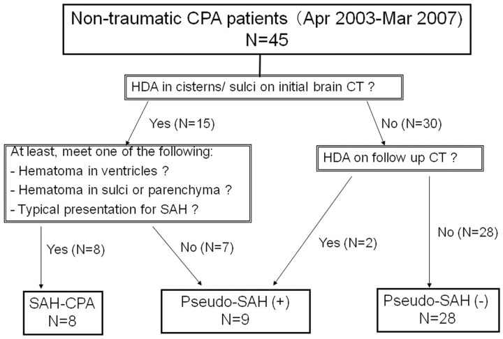
These 45 patients were classified into 3 groups: SAH-CPA, pseudo-SAH(+), and pseudo-SAH(−) groups (Fig 1). We reviewed CT images of all patients to check for HDAs along the cortical sulci or cisterns. The SAH-CPA group, in whom a true SAH was deemed to have developed primarily followed by CPA secondarily, consisted of the patients with such HDAs on the initial CT that met at least 1 of the following 3 criteria: 1) a hematoma in the ventricle system; 2) clumped HDAs in the subarachnoid space or brain parenchyma obviously suggesting a hematoma; or 3) a clinical presentation with the typical findings of SAH, including sudden-onset headache and impaired consciousness. The pseudo-SAH(+) group included patients in whom no intracranial HDAs were noted on the first CT but in whom HDAs appeared in the cisterns or sulci on the follow-up CT, and patients with such HDAs on the initial CT who failed to meet any of the criteria for the SAH-CPA group. The pseudo-SAH(−) group was defined as patients who showed no such HDAs in any CT images obtained during the clinical course. The number of patients in each group is shown in Fig 1. Eight patients met the criteria of the SAH-CPA group; all of them had intraventricular hematoma, 3 with subarachnoid clumped high-attenuation lesions and 1 with the typical clinical presentation. Ultimately, our subjects included 9 patients in the pseudo-SAH(+) group (17–73 years of age; average, 39.8 years), 8 patients in the SAH-CPA group (52–72 years of age; average, 65.4 years), and 28 patients in the pseudo-SAH(−) group (2–90 years of age; average, 56.1 years).
Fig 1.
Classification of nontraumatic CPA groups.
SAH and healthy control groups were established for comparison. Searching the radiology report data base for the same period as for the CPA group, we collected 13 patients for the SAH group that were diagnosed on CT as SAH without CPA, for whom a definitive diagnosis of SAH was confirmed at surgery (29–82 years of age; average, 57.2 years). The healthy control group consisted of 20 randomly selected patients diagnosed with normal brain CT findings (16–74 years of age; average, 50.5 years).
CT Examination
Brain CT images were obtained in the transverse plane parallel to the supraorbitomeatal or orbitomeatal line in 5-mm thicknesses for the infratentorial sections and in 7- to 8-mm thicknesses for the supratentorial sections with gapless scanning. The nonhelical conventional method was used with a tube voltage of 120 kV and a tube current of 200–500 mA. Excluding the SAH-CPA group, 37 patients with CPA in the pseudo-SAH(+) and pseudo-SAH(−) groups underwent a total of 80 CT examinations of the head, all of which were available for this retrospective analysis.
Measurement of CT Values
For all the cases, DICOM data were available for measuring CT values by using the DICOM image viewer (POP NET Server; ImageONE, Tokyo, Japan; http://www.imageone.co.jp/english/index.html).
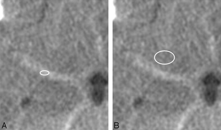
For patients in the pseudo-SAH(+), SAH-CPA, and SAH groups, the CT values of HDAs in the Sylvian vallecula were measured by defining regions of interest (the average area = 10 mm2) on the CT images where HDAs first appeared (Fig 2A). In 2 patients in the pseudo-SAH(+) group who underwent follow-up CT examinations after the appearance of the HDAs, the CT values were measured sequentially. One patient was studied 5 times from 5 to 25 days; another was studied 6 times from 8 to 268 days after the onset.
Fig 2.
Definition of regions of interest. A, An oval region of interest is defined on the HDA of the Sylvian vallecula, providing the CT number. B, A round or oval region of interest is defined in the brain parenchyma just ventral to the Sylvian vallecula.
In all patients, the CT values of the brain parenchyma (white matter) just ventral to the Sylvian vallecula were measured by using elliptic regions of interest (average area = 60 mm2, Fig 2B). All patients except 1 in the pseudo-SAH(+) group were not given contrast media within 24 hours before these CT images used for the region of interest analysis were obtained. The 1 pseudo-SAH(−) patient, in whom CT was obtained with contrast media, was excluded from this analysis.
We defined these regions of interest just by seeing a target section without knowledge of the group and diagnosis of the patients. The CT values of HDAs in the Sylvian vallecula were measured on every side that had HDAs. All patients in the pseudo-SAH(+) and the SAH-CPA groups and 10 of the SAH group had bilateral lesions, and the remaining 3 in the SAH group had unilateral lesions. The CT values of the brain parenchyma were measured bilaterally in all patients. Finally, the CT values of HDAs were obtained from 57 sides of 30 patients, and those of the brain parenchyma, from 156 sides of 78 patients.
The CT value was measured on the CT scan obtained on the day of symptom onset in all except 3 patients in the SAH-CPA and SAH groups, and in those 3 patients, it was measured on the second-day images, which were the initial examinations.
Evaluation Items and Statistical Analysis
For the pseudo-SAH(+) and pseudo-SAH(−) groups, the relationship between the presence or absence of the pseudo-SAH findings and the severity of brain edema was evaluated by reviewing all available CT studies. Brain edema was graded visually as none, mild, or severe, with “mild edema” showing obscuration of the gray-white matter junction or mild narrowing of the cortical sulci, “severe edema” being almost complete obliteration of the cortical sulci, and “none” being none of these findings. The grading was independently done by 2 radiologists at first, and then the inter-rater disagreements were corrected in consensus fashion. The inter-rater agreement was evaluated with κ statistics. The difference among the number of patients in each category was evaluated by using the χ2 test.
The mean CT values of the HDAs compared among the pseudo-SAH(+), SAH-CPA, and SAH groups and those of the brain parenchyma were compared among all 5 groups by using 1-way factorial analysis of variance (ANOVA) with the Bonferroni post hoc test.
The clinical outcomes of the pseudo-SAH(+) and pseudo-SAH(−) groups were evaluated according to the modified Rankin Scale. The scales were compared between the pseudo-SAH(+) and pseudo-SAH(−) groups by using the Mann-Whitney U test. The survival rate was evaluated by using the Kaplan-Meier method and the logrank test. The statistical analysis was conducted on a personal computer by using Stat View 5.0 (SAS Institute, Cary, NC). Results with P < .05 were considered statistically significant.
Results
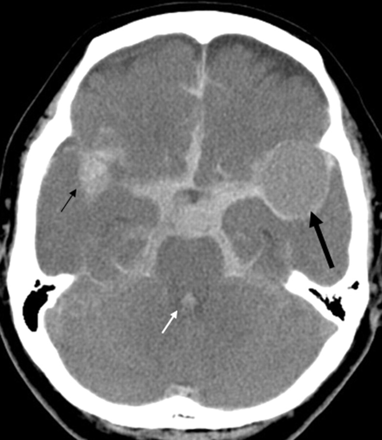
Typical examples from the pseudo-SAH(+), pseudo-SAH(−), and SAH-CPA groups are shown in Figs 3–5, respectively.
Fig 5.
A 72-year-old woman of the SAH-CPA group. She suddenly fell unconscious and experienced CPA. The first-day CT shows diffuse HDAs in the basal cisterns, Sylvian valleculae/fissures, and cerebral sulci. Note hematoma in the right Sylvian vallecula (small arrow) and in the fourth ventricle (white arrow). The brain shows diffuse subtle low attenuation with obliteration of the cerebral sulci. The CT values of the HDA and the brain are 49 and 29 HU, respectively. In the left Sylvian vallecula, there is a round filling defect in the hematoma that is thought to represent an aneurysm (large arrow).
Findings of pseudo-SAH were recognized in 9 (20%) of the 45 consecutive nontraumatic patients with CPA. At an autopsy that was done in only 1 patient in the pseudo-SAH(+) group, no SAH was detected.
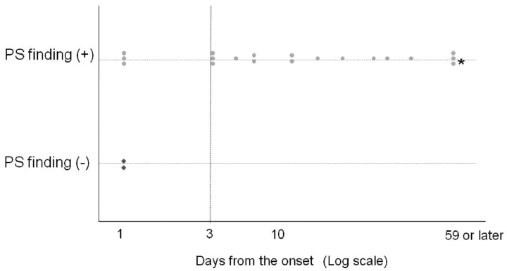
On the basis of whether the findings of pseudo-SAH were present, all 20 CT examinations conducted in the 9 patients in the pseudo-SAH(+) group were plotted against when the respective CT studies were performed (Fig 6). Findings of pseudo-SAH were not seen in 2 (40%) of 5 examinations conducted before the third day. By contrast, in all CT images obtained on the third day or later, the findings were observed, even until the 59th day or later. In the 2 patients in whom the sequential changes in CT values of the pseudo-SAH findings were evaluated, no specific tendency was recognized, though the values fluctuated slightly (between 31 and 45 HU) with time.
Fig 6.
The relationship between the days from the onset and the pseudo-SAH finding in patients eventually falling into the pseudo-SAH (+) group. Every dot indicates 1 occasion of CT examination. The asterisk indicates that these 3 examinations were performed on the 59th, 129th, and 268th days. PS indicates pseudo-SAH.
Substantial inter-rater agreements were found in the grades of brain edema scored by the 2 radiologists (κ = 0.71). With the final results obtained by consensus of the 2 raters, a statistically significant relationship was seen between the presence or absence of pseudo-SAH findings and the degree of brain edema (P < .0001, χ2 test, Table 1).
Table 1:
| Pseudo-SAH Finding | Degree of Cerebral Edema |
|||
|---|---|---|---|---|
| None | Mild | Severe | Total | |
| Positive | 0 | 0 | 19 | 19 |
| Negative | 35 | 22 | 4 | 61 |
| Total | 35 | 22 | 23 | 80 |
P < .0001 (χ2 test).
Numeric values in the cells represent the number of CT examinations.
There was a significant difference in the mean CT values of the HDAs among the pseudo-SAH(+), SAH-CPA, and SAH groups (Table 2; ANOVA, P < .0001). The mean CT value of the pseudo-SAH(+) group was significantly lower than those of the other 2 groups (Bonferroni test, P < .0001). The range of the CT values of the pseudo-SAH(+) group did not overlap those of the other groups, except a single patient in the SAH-CPA group, in whom the initial CT was performed on the second day after CPA. The average and distribution of the CT values were similar between the SAH-CPA and SAH groups.
Table 2:
CT values of hyperdense areas in the Sylvian vallecula*
| Groups | CT Values (HU) |
|||
|---|---|---|---|---|
| No. | Mean | SD | Range (Min-Max) | |
| Pseudo-SAH(+) | 18 | 37.6† | 3.3 | 30.0–42.0 |
| CPA-SAH | 14 | 56.6 | 7.8 | 41.0–67.0 |
| SAH | 23 | 53.7 | 5.1 | 42.0–60.0 |
Note:—No. indicates number of hemispheres; Min, minimum; Max, maximum.
ANOVA, P < .0001.
P < .0001, as compared with the other 2 groups (Bonferroni post hoc test).
There was a significant difference in the mean CT values of the brain parenchyma among the 5 groups (Table 3; ANOVA, P < .0001). The mean CT value was significantly lower in the pseudo-SAH(+) group than in the other 4 groups (Bonferroni test, P < .0001). The pseudo-SAH(−) and SAH-CPA groups, both of which were patients with CPA, had significantly lower values than the healthy control groups (P < .0002). No significant difference in mean CT values was noted between the SAH and healthy control groups.
Table 3:
CT values of brain parenchyma adjacent to the Sylvian vallecula*
| Groups | CT Values (HU) |
|||
|---|---|---|---|---|
| No. | Mean | SD | Range (Min-Max) | |
| Pseudo-SAH(+) | 18 | 26.8†‡ | 3.1 | 19.0–31.0 |
| Pseudo-SAH(−)§ | 54 | 29.8‡ | 1.9 | 24.0–34.0 |
| SAH-CPA | 16 | 30.0‡ | 2.3 | 27.0–36.0 |
| SAH | 26 | 31.2 | 1.9 | 28.0–35.0 |
| Healthy | 40 | 32.5 | 1.7 | 30.0–35.0 |
Note:—No. indicates number of hemispheres, Min, minimum; Max, maximum.
ANOVA, P < .0001.
P < .0001, as compared with the other 4 groups (Bonferroni post hoc test).
P < .0001, as compared with healthy groups (Bonferroni post hoc test).
One case of Pseudo-SAH(−) group was excluded because of contrast media injection.
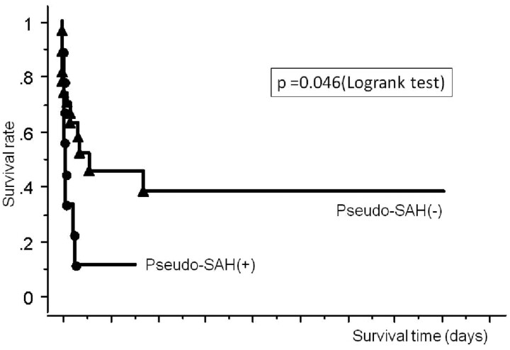
The modified Rankin Scale scores of the pseudo-SAH(+) group (mean score, 5.89) were significantly lower than those of the pseudo-SAH(−) group (mean score, 4.50) (Mann-Whitney U test, P = .02). With the positive pseudo-SAH finding as a predictor of a poor prognosis (modified Rankin Scale score, 4–6), all patients in the pseudo-SAH(+) group and 20 of 28 patients in the pseudo-SAH(−) group showed the poor prognosis, yielding the sensitivity, specificity, and accuracy of 31%, 100%, and 46%, respectively. The pseudo-SAH(+) group had a significantly lower survival rate than the pseudo-SAH(−) group (Fig 7; logrank test, P = .046).
Fig 7.
Survival curves comparing the pseudo-SAH(+) and the pseudo-SAH(−) groups.
Discussion
A pseudo-SAH is a brain CT finding that is seen as HDAs along the basal cisterns, the Sylvian vallecula/fissure, the tentorium cerebelli, or the cortical sulci in patients with severe brain edema, though no SAH is seen at autopsy or lumbar puncture. Pseudo-SAH has been considered rare, though its incidence has not been investigated. We reviewed consecutive cases of patients resuscitated from nontraumatic CPA and found that “pseudo” is not so rare a finding, with an incidence of 20%.
Chronologic Changes in the Pseudo-SAH Findings
In the pseudo-SAH(+) group, pseudo-SAH findings were observed in all CT studies performed on the third day or later. In 1 patient, the findings persisted for 268 days. In the 2 patients in whom the CT values of the HDAs were evaluated sequentially, no obvious change in the CT values was noted. As far as we know, no report has described changes in pseudo-SAH findings with time. In patients with typical SAH, the CT attenuation of a SAH decreases with time: The detection rate on CT is 100% until 2 days after onset, approximately 50% at 1 week, 30% at 2 weeks, and 0% after 3 weeks.13 In view of such temporal changes for a hemorrhage, the relative constancy of the pseudo-SAH findings suggests that a pseudo-SAH does not represent a true hemorrhage.
Presumed Mechanism of Pseudo-SAH Based on Consideration of the CT Values of High-Attenuation Areas and Brain Parenchyma
The mechanism for the development of a pseudo-SAH has not been elucidated fully. Avrahami et al2 suggested that severe brain edema compresses the dural sinuses, compromising the venous drainage from the brain and resulting in engorgement of the superficial veins, which stand out against the edematous low-attenuated brain parenchyma, mimicking an SAH. Given et al3 proposed a similar mechanism and included narrowing or disappearance of hypoattenuated CSF space as an additional factor generating a pseudo-SAH.
In this study, the CT values of HDAs in the pseudo-SAH(+) group ranged from 30 to 42.0 HU and were significantly lower than those of the SAH group; the ranges barely overlapped. The CT value of blood is linearly positively correlated with the hematocrit and is approximately 42 HU in a healthy individual with a hematocrit of approximately 45%.14 However, blood that has leaked from the vasculature or formed a hematoma has a much higher attenuation because of the rapid absorption of plasma. Therefore, the CT values measured in the pseudo-SAH(+) group support the speculation that the finding reflects dilated vessels.
The CT values of the brain parenchyma in the pseudo-SAH(+) and pseudo-SAH(−) groups were significantly lower than those of the other groups. In addition, the mean CT value of the pseudo-SAH(+) group was significantly lower than that of the pseudo-SAH(−) group, though their ranges overlapped. Therefore, a decrease in brain attenuation may not be the only cause of the pseudo-SAH finding. However, the discrepancy regarding the presence of findings of pseudo-SAH might be attributable to differences in the degree of brain edema between the pseudo-SAH(+) and pseudo-SAH(−) groups. All the CT images showing pseudo-SAH findings also showed severe brain edema, whereas most of the images without it showed only mild edema, if any. Therefore, our results support the hypothesis that congested dilated superficial veins and severe brain edema have a synergistic effect that results in the finding of pseudo-SAH (ie, blood in the superficial vessels stands out as HDAs relative to the hypoattenuation of severely edematous brain). A reduction in the CSF space due to the swollen brain may also be contributory.
The fact that the CT attenuation of pseudo-SAH lesions is lower than that of true SAH should distinguish SAH-like CT findings in patients resuscitated from CPA. If an SAH-like finding with modest HDAs is found along the cisterns, cerebral sulci, and tentorium associated with severe brain edema, pseudo-SAH should be suggested rather than true SAH. Measuring the CT value should help with the differentiation, because the value for pseudo-SAH is <43 HU and does not overlap the values seen in acute SAH, at least on the day of onset. Another differentiating point may be the absence of intraventricular high-attenuation lesions. All the patients in the SAH-CPA group had intraventricular hematomas as well as severe SAH extending diffusely along the subarachnoid spaces. Indeed, patients with SAH do not always have intraventricular hematoma in general, but SAH causing CPA must be severe enough to reflux into the ventricular systems.
Pseudo-SAH Findings versus Prognosis
Patients in the pseudo-SAH(+) group had a significantly poorer prognosis than those in the pseudo-SAH(−) group. Although no study has analyzed the relationship between pseudo-SAH and prognosis statistically, several reports on pseudo-SAH have stated that patients with pseudo-SAH caused by various diseases, including postresuscitation, encephalitis, brain tumor, and cerebral infarction, had a poor prognosis.1,4–7 In our series, which focused on patients after resuscitation of CPA, more marked brain edema with CT hypoattenuation was seen in the patients who were pseudo-SAH-positive than in those who were negative. This suggests that more severe brain damage likely develops from an early stage in patients with pseudo-SAH. Once severe brain edema increases the intracranial pressure, the resulting reduction in brain perfusion would worsen any cerebral hypoxia, which would, in turn, exaggerate the brain edema, triggering a vicious cycle.
In this study, the findings of pseudo-SAH appeared by the third day in all patients in the pseudo-SAH(+) group. Therefore, this finding serves as an early indicator of a poor prognosis. The sensitivity of this indicator was not high (31%) because not all patients with a poor prognosis were pseudo-SAH positive, but the specificity was extremely high (100%). In 7 of the 20 patients with a poor prognosis in the pseudo-SAH(−) group, the last CT examination was performed before the third day after the onset. If CT examinations had been repeated on the fourth day or later, these patients might have eventually shown the findings of pseudo-SAH. Consequently, the sensitivity of the finding as an indicator of a poor prognosis could have been higher.
Limitations
The major problems of this study are that the absence of SAH was confirmed in only 1 patient in the pseudo-SAH(+) group at autopsy. Two patients in the pseudo-SAH(+) group had no intracranial HDA on the initial CT, and the SAH-like HDA emerged on follow-up studies obtained a few days later and was interpreted as pseudo-SAH. In these patients, delayed development of an SAH was considered unlikely because they were nontrauma patients. The other 2 patients in whom the CT values were measured repeatedly and who showed no reduction during 1 month were considered to be in the pseudo-SAH group. In the remaining 4 patients, true SAH could not be completely ruled out. In all patients in pseudo-SAH(+) group, however, the causes of CPA were identified such as suffocation, ventricle fibrillation, carbon monoxide intoxication, anaphylactic shock, and hypovolemic shock. None of them presented with clinical symptoms suggesting SAH, so it is less likely that SAH was associated or developed later. Because the CT values of the SAH-like HDAs in the pseudo-SAH(+) group were significantly lower than those in the SAH group with no overlap, our interpretation of the pseudo-SAH(+) group appears to have been reasonable.
Although MR imaging is thought to be useful to determine whether the HDAs on CT are hemorrhage or not, none of the pseudo-SAH(+) group underwent MR imaging in this study. Because all patients were in very poor general status with a ventilator, it would be quite difficult and dangerous to perform MR imaging studies. CSF examination by lumbar puncture is another way to determine whether SAH exists or not, but this procedure is contraindicated for patients with severe brain edema in principle. From these points of view, radiologists should know the phenomenon of the pseudo-SAH found in patients with hypoxic brain damage and be familiar with its imaging characteristics to avoid these additional dangerous procedures.
Conclusion
A pseudo-SAH finding is a CT pseudolesion that shows SAH-like findings, in which the cisterns and cerebral sulci appear hyperattenuated relative to the brain parenchyma. This is a synergistic result of distention of the superficial vessels arising from elevated intracranial pressure and severe brain edema manifesting as hypoattenuated parenchyma. Following resuscitation, pseudo-SAH may develop within 3 days after the onset of CPA in approximately 20% of patients. This indicates severe brain damage and suggests a poor prognosis, providing important information for deciding treatment strategies.
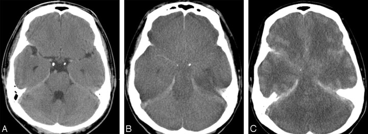
Fig 3.
A 34-year-old man of the pseudo-SAH(+) group. He had CPA due to suffocation. He was resuscitated immediately but did not recover from coma. A, On the first day, no abnormal finding is seen. B, On the eighth day, the brain shows diffuse low attenuation with obliteration of cisterns and cerebral sulci and narrowed ventricles. HDAs mimicking SAH are noted along the bilateral Sylvian valleculae and tentorium cerebelli. CT values of the HDA of the Sylvian vallecula and the brain parenchyma are 36 and 23.5 HU, respectively. C, On the 129th day, brain edema becomes more severe. The SAH-like HDAs become more prominent.
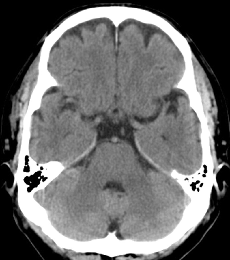
Fig 4.
A 60-year-old man of the pseudo-SAH(−) group. He had CPA due to ventricular fibrillation followed by immediate resuscitation and resultant full recovery. On the thirteenth day, CT shows no demonstrable abnormality.
References
- 1.Spiegel SM, Fox AJ, Vinuela F, et al. Increased density of tentorium and falx: a false positive CT sign of subarachnoid hemorrhage. Can Assoc Radiol J 1986;37:243–47 [PubMed] [Google Scholar]
- 2.Avrahami E, Katz R, Rabin A, et al. CT diagnosis of non-traumatic subarachnoid haemorrhage in patients with brain edema. Eur J Radiol 1998;28:222–25 [DOI] [PubMed] [Google Scholar]
- 3.Given CA 2nd, Burdette JH, Elster AD, et al. Pseudo-subarachnoid hemorrhage: a potential imaging pitfall associated with diffuse cerebral edema. AJNR Am J Neuroradiol 2003;24:254–56 [PMC free article] [PubMed] [Google Scholar]
- 4.Chute DJ, Smialek JE. Pseudo-subarachnoid hemorrhage of the head diagnosed by computerized axial tomography: a postmortem study of ten medical examiner cases. J Forensic Sci 2002;47:360–65 [PubMed] [Google Scholar]
- 5.Mahmoud al-Yamany, Deck J, Bernstein M. Pseudo-subarachnoid hemorrhage: a rare neuroimaging pitfall. Can J Neurol Sci 1999;26:57–59 [PubMed] [Google Scholar]
- 6.Thomas GL, Stachowski ER. Pseudosubarachnoid haemorrhage on CT brain scan: an unusual presentation of diffuse hypoxic brain injury. Intensive Care Med 2007;33:2038–40 [DOI] [PubMed] [Google Scholar]
- 7.Phan TG, Wijdicks EF, Worrell GA, et al. False subarachnoid hemorrhage in anoxic encephalopathy with brain swelling. J Neuroimaging 2000;10:236–38 [DOI] [PubMed] [Google Scholar]
- 8.Rabinstein AA, Pittock SJ, Miller GM, et al. Pseudosubarachnoid haemorrhage in subdural haematoma. J Neurol Neurosurg Psychiatry 2003;74:1131–32 [DOI] [PMC free article] [PubMed] [Google Scholar]
- 9.Huang D, Abe T, Ochiai S, et al. False positive appearance of subarachnoid hemorrhage on CT with bilateral subdural hematomas. Radiat Med 1999;17:439–42 [PubMed] [Google Scholar]
- 10.Mendelsohn DB, Moss ML, Chason DP, et al. Acute purulent leptomeningitis mimicking subarachnoid hemorrhage on CT. J Comput Assist Tomogr 1994;18:126–28 [DOI] [PubMed] [Google Scholar]
- 11.Belsare G, Lee AG, Maley J, et al. Pseudo-subarachnoid hemorrhage and cortical visual impairment as the presenting sign of gliomatosis cerebri. Semin Ophthalmol 2004;19:78–80 [DOI] [PubMed] [Google Scholar]
- 12.Schievink WI, Maya MM, Tourje J, et al. Pseudo-subarachnoid hemorrhage: a CT-finding in spontaneous intracranial hypotension. Neurology 2005;65:135–37 [DOI] [PubMed] [Google Scholar]
- 13.van Gijn J, van Dongen KJ. The time course of aneurysmal haemorrhage on computed tomograms. Neuroradiology 1982;23:153–56 [DOI] [PubMed] [Google Scholar]
- 14.PF New, Aronow S. Attenuation measurements of whole blood and blood fractions in computed tomography. Radiology 1976;121:635–40 [DOI] [PubMed] [Google Scholar]


