Abstract
SUMMARY: Fractionated stereotactic radiation has become the standard treatment of meningioma of the optic nerve sheath. The mechanism responsible for improvement in visual function is unclear, because neuroimaging after treatment usually shows no discernable change in tumor appearance. We report immediate regression of optociliary shunt vessels in a patient after radiation treatment of an optic nerve sheath meningioma. This observation indicates that radiation treatment can cause rapid reduction of optic nerve compression, even without appreciable reduction in the size of the meningioma.
Collateral retinochoroidal vessels straddling the optic disc border, known as optociliary shunt vessels, are a classic finding in patients with optic nerve sheath meningioma.1 In healthy patients, retinal blood leaves the eye via the central retinal vein, which runs down the center of the optic nerve. The growth of a sheath meningioma causes gradual compression of the central retinal vein, forcing blood to find alternative drainage routes. Retinochoroidal vessels, which are normally microscopic, become engorged by retinal blood that is shunted from the optic disc (opto) to the choroid (ciliary). The retinal blood is then able to exit the eye via the choroidal vortex veins, bypassing the obstruction created by the optic nerve sheath tumor.
It has been established that fractionated stereotactic radiation is the optimal treatment of primary optic nerve sheath meningioma.2-5 It is unknown why radiation is effective, given that optic nerve sheath meningiomas are benign tumors. Usually the tumor does not appear smaller on orbital scans obtained after radiation treatment.
We describe a patient with immediate recovery of visual function, resolution of optic nerve swelling, and shrinkage of optociliary shunts after treatment of an optic nerve sheath meningioma with stereotactic 3D conformal radiation therapy. Shrinkage of the optociliary shunt vessels suggests that radiation treatment causes visual improvement by partially relieving compression of the optic nerve.
Case Report
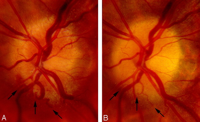
A 50-year-old woman reported a 3-year history of decreased visual acuity and transient visual obscurations. The right eye was normal. The left eye had a best corrected visual acuity of 20/30 and a relative afferent pupillary defect. The optic disc was swollen, with blurred margins and congestion of the surface capillaries. Dilated optociliary shunt vessels were present at the inferior pole (Fig 1A).
Fig 1.
A, Pretreatment view of the edematous left optic disc, showing congestion of surface capillaries and dilated optociliary shunt vessels (arrows). B, Optic disc appearance just before the end of radiation treatment. The optic disc edema has nearly resolved and the optociliary shunt vessels have shrunk (arrows).
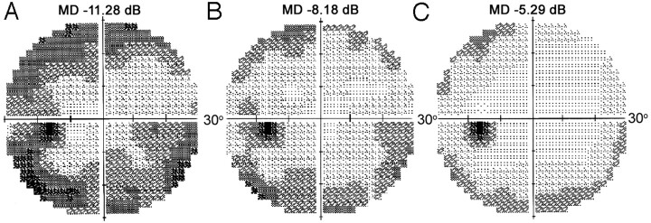
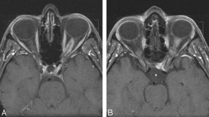
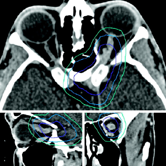
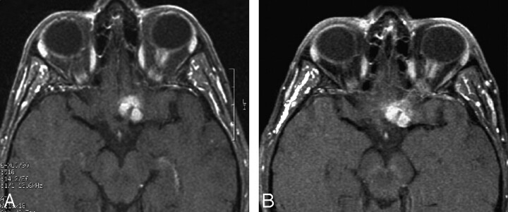
Automated threshold perimetry in the left eye showed peripheral visual field constriction, with a mean deviation of −11.28 dB (Fig 2A). MR imaging revealed tubular enlargement and enhancement of the optic nerve sheath, consistent with a meningioma (Fig 3A). The mass extended through the optic canal, ending abruptly 3 mm anterior to the optic chiasm (Fig 3B). The patient underwent fractionated stereotactic external beam radiation treatment to prevent invasion of the optic chiasm and progression of visual loss in the left eye. The total dose was 5400 cGy, given in 30 sessions of 180 cGy for 42 days (Fig 4). After 15 treatment sessions, automated perimetry showed improvement, registering a mean deviation of −8.18 dB (Fig 2B). Two days before completion of treatment, the visual acuity had improved to 20/25, and the visual field deficit was only −5.29 dB (Fig 2C). It is remarkable that the edema in the optic disc had nearly resolved, along with the optociliary shunt vessels (Fig 1B). Despite this improvement in the appearance of the optic disc, an MR performed 3 months later showed no change in the size or extent of the tumor (Fig 5).
Fig 2.
A, Humphrey visual field of the left eye before treatment shows generalized constriction with a mean deviation −11.28 dB. B, After 3 weeks of treatment, the visual field shows improvement, with a mean deviation of −8.18 dB. C, Two days before completing radiation treatment, the visual field mean deviation is only −5.29 dB.
Fig 3.
A, Axial T1-weighted MR image with fat suppression and gadolinium demonstrates enhancement of the left optic nerve sheath. B, The mass extends into the optic nerve canal, terminating just proximal to the optic chiasm.
Fig 4.
Axial, sagittal, and coronal contrast CT images showing the isodose distribution map. A maximum radiation dose of 5400 cGy was planned to encompass the tumor at 90% isoattenuated line (purple, 5200 cGy; blue, 4500 cGy; turquoise, 3600 cGy).
Fig 5.
A, Axial T1-weighted MR image with fat suppression and gadolinium showing the meningioma before treatment. B, Three months after treatment, there is no change in the size or enhancement of the tumor.
Discussion
Previous reports have described the regression of optociliary shunt vessels after radiation treatment of meningioma of the optic nerve sheath.6,7 The noteworthy feature of the case of our patient was that the shrinkage of the optociliary shunt vessels was immediate and occurred without any apparent change in the size of the tumor. Radiation treatment seems to work by relaxing the compression of the optic nerve, even without obvious radiologic evidence of tumor shrinkage. Reduction in tumor compression presumably allows increased blood flow through the central retinal vein, lessening the driving force for blood flow through the optociliary collateral vessels. It also accounts for the rapid improvement in optic disc edema and partial recovery of optic nerve function.
Advances in radiation therapy have permitted more accurate localization of meningioma of the optic nerve sheath, minimizing radiation exposure to adjacent radiosensitive structures, such as the retina. Visual function can improve rapidly, even before the full course of treatment sessions has been administered. It has been suggested that this rapid response to treatment could be used to titrate the radiation dosage, rather than using a full dose of 5400 cGy for all patients.8
Footnotes
This work was supported by an unrestricted grant from Research to Prevent Blindness, New York, NY.
References
- 1.Newman NJ. Topical diagnosis of tumors. In: Miller NR, Newman NJ, Biousse V, et al, eds. Walsh and Hoyt's Clinical Neuro-ophthalmology, 6th ed. Philadelphia: Lippincott, Williams & Wilkins;2005. :1346
- 2.Turbin RE, Thompson CR, Kennerdell JS, et al. A long-term visual outcome comparison in patients with optic nerve sheath meningioma managed with observation, surgery, radiotherapy, or surgery and radiotherapy. Ophthalmology 2002;109:890–99 [DOI] [PubMed] [Google Scholar]
- 3.Liu JK, Forman S, Hershewe GL, et al. Optic nerve sheath meningiomas: visual improvement after stereotactic radiotherapy. Neurosurgery 2002;50:950–55 [DOI] [PubMed] [Google Scholar]
- 4.Andrews DW, Faroozan R, Yang BP, et al. Fractionated stereotactic radiotherapy for the treatment of optic nerve sheath meningiomas: preliminary observations of 33 optic nerves in 30 patients with historical comparison to observation with or without prior surgery. Neurosurgery 2002;51:890–902 [DOI] [PubMed] [Google Scholar]
- 5.Narayan S, Cornblath WT, Sandler HM, et al. Preliminary visual outcomes after three-dimensional conformal radiation therapy for optic nerve sheath meningioma. Int J Radiat Oncol Biol Phys 2003;56:537–43 [DOI] [PubMed] [Google Scholar]
- 6.Smith JL, Vuksanovic MM, Yates BM, et al. Radiation therapy for primary optic nerve meningiomas. J Clin Neuroophthalmol 1981;1:85–99 [PubMed] [Google Scholar]
- 7.Mashayekhi A, Shields JA, Shields CL. Involution of retinochoroidal shunt vessel after radiotherapy for optic nerve sheath meningioma. Eur J Ophthalmol 2004;14:61–64 [DOI] [PubMed] [Google Scholar]
- 8.Vagefi MR, Larson DA, Horton JC. Optic nerve sheath meningioma: visual improvement during radiation treatment. Am J Ophthalmol 2006;142:343–44 [DOI] [PubMed] [Google Scholar]


