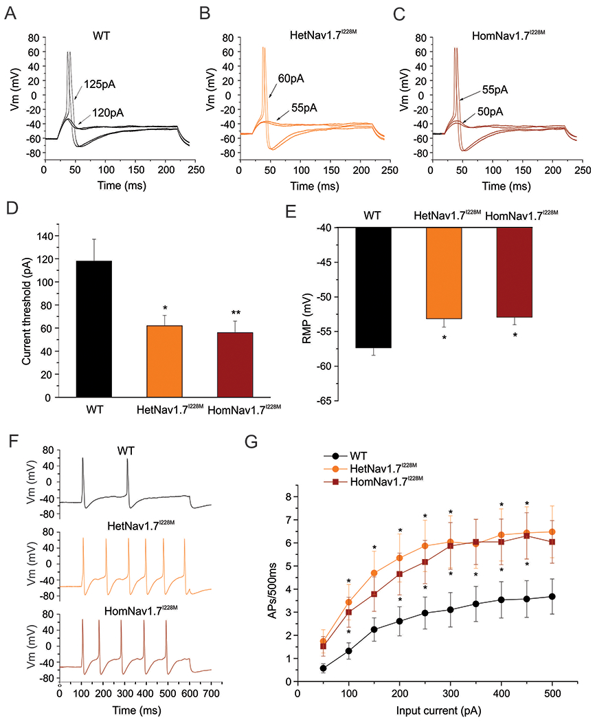
Figure 1. Small-diameter DRG neurons from HetNav1.7I228M and HomNav1.7I228M mutant mice are hyper-excitable compared to WT control DRG neurons.

(A) Representative traces of a small-diameter DRG neuron from WT mouse, showing subthreshold responses to current injections up to 120 pA and subsequent action potentials evoked by current injections above 125pA (current threshold for this neuron). (B) Representative traces of a small-diameter DRG neuron from HetNav1.7I228M mouse, showing a lower current threshold (60 pA) for action potential generation. (C) Representative traces of a small-diameter DRG neuron from HomNav1.7I228M mouse, showing a lower current threshold (55 pA) for action potential generation. (D) Small-diameter DRG neurons from HetNav1.7I228M and HomNav1.7I228M mice display significantly reduced current threshold compared to WT control neurons (* p<0.05; ** p<0.01, one-way ANOVA followed by Tukey post hoc test). (E) Small-diameter DRG neurons from HetNav1.7I228M and HomNav1.7I228M mice display significantly depolarized RMP compared to WT control neurons (* p<0.05, one-way ANOVA followed by Tukey post hoc test). (F) Representative responses of small-diameter DRG neurons from WT, HetNav1.7I228M and HomNav1.7I228M mice, respectively, to 500 ms depolarizing current 3x the threshold for action potential generation. (G) Comparison of firing frequency of small-diameter DRG neurons from WT, HetNav1.7I228M and HomNav1.7I228M mutant mice, in response to graded 500 ms depolarising current stimuli from 50 to 500 pA in 50 pA increments. Nav1.7I228M mutant groups show significantly higher firing frequencies compared to WT group (* p<0.05, one-way ANOVA followed by Tukey post hoc test).
