Figure 2.
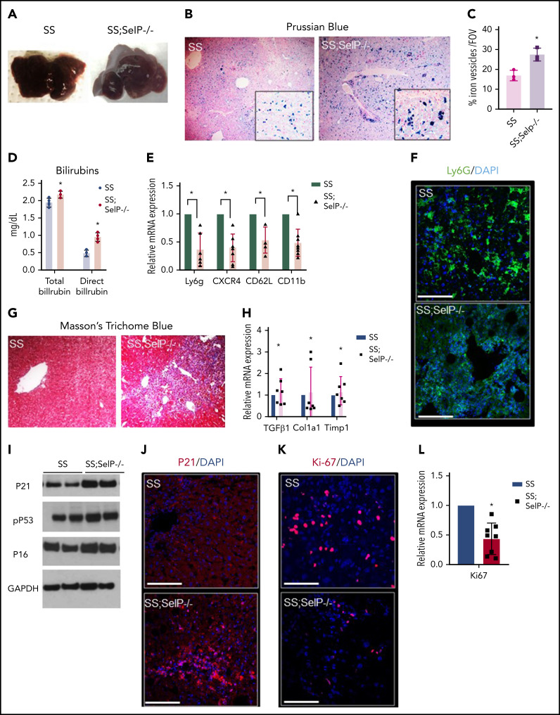
SS-Selp−/− mice exhibit chronic organ injury and exacerbated senescence. (A) Gross specimen of livers of SS and SS-Selp−/− mice. Both the SS and the SS-Selp−/− mice livers were dark and manifested white spots suggestive of progressive injury. (B) Prussian blue staining for iron showed increased iron deposition with a mixed distribution in hepatocytes and Kupffer cells in SS-Selp−/− liver as compared with SS liver. (C) Quantification of iron staining/FOV. (D) Serum direct and total bilirubin levels in SS and SS-Selp−/− mice. (E) Quantitative reverse transcription polymerase chain reaction (qRT-PCR) analysis exhibit reduced mRNA expression of markers of inflammatory cells (including CXCR4, Ly6G, CD11b, and Cd62L) in SS-Selp−/− liver as compared with SS liver. (F) Immunofluorescence for Ly6G showed an increased accumulation in SS mouse liver, which was reduced in SS-Selp−/− liver. Scale bars, 20 µM. (G) Trichome staining of SS and SS-Selp−/− liver sections revealed increased perisinusoidal and periductular fibrosis in SS-Selp−/− liver. Original magnification, ×10. (H) Analysis of mRNA expression by qRT-PCR showed an increase in mRNA expression of TGFβ, Col1A1, and TIMP1 in SS-Selp−/− liver as compared with SS liver. (I) Western blot for P21, P16INK4a, and phosphor-P53 antibodies exhibits increased expression in the liver of SS-Selp−/− as compared with SS. (J) IF of P21 exhibits significant enrichment in SS-Selp−/− liver as compared with SS liver. DAPI, 4′,6-diamidino-2-phenylindole. (K) IF of Ki-67 exhibits reduced hepatocyte proliferation in SS-Selp−/− liver as compared with SS liver. Scale bars, 20 μm (J-K). (L) qRT-PCR analysis of SS and SS-Selp−/− liver exhibits reduced expression of Ki-67 in SS-Selp−/− liver as compared with SS. *P < .05.

