

Abstract
A part of every science is its history of development. Many individuals and events around the world, as have been attested on the stamps issued from different countries, have contributed to evolve the science of orthopedic surgery; however, some may have been ignored. The current brief history tries to present an insight to major milestones in the history of orthopedic science and also the involved people. Philatelic version of orthopedic history by no mean is a comprehensive history. Orthopedic surgery and science does continue to progress. Certain peaks and memorable events in orthopedic science may appear on postal stamps in future.
Key Words: History of orthopedics, rthopedic science, rthopedic surgery, hilately, hilatelic history
Introduction
A part of every science is its history of development. To look for new researches and achievements we have to know what had been happened before and what experiences have been tried. History of orthopedics recognizes many events and individuals; however, some may have been ignored. This fact is particularly correct on the postage stamps. We present a very concise history of orthopedic science and its milestones on postal stamps. Philatelic version of orthopedic history by no mean is a comprehensive history. It is a journey through the past to present. The stamps emphasize on certain peaks and memorable events in the evolvement of orthopedic surgery.
Concepts of orthopedics in the ancient period
On the occasion of Cairo international orthopedic conference in 1988, Egypt issued a stamp that demonstrated the emblem of Egyptian Orthopedic Association [Figure 1]. The stamp showing a bonesetter manipulating and reducing a shoulder dislocation is an example of antiquity of manipulation in history of orthopedics.
Figure 1.

the emblem of Egyptian Orthopedic Association a bonesetter manipulating and reducing a shoulder dislocation
In the ancient world it was believed that some diseases were derived from excess blood; therefore, phlebotomy was a common practice of surgery on the limbs. The surgeon on the Greek stamp is about to incise an antecubital vein of a patient [Figure 2].
Figure 2.

A surgeon is about to incise an antecubital vein
Advances of orthopedic science in the medieval period
Razi (854-925) and Avicenna (980-1035) in the medieval period presented early concepts of musculoskeletal disorders in their written works and established a foundation for modern orthopedic science [Figure 3] (1-3). Razi wrote monographs about foot, artheralgia, gout, pain in the lower limb, and fracture treatment in his books. He established a hospital in Baghdad and devoted a special department to fracture treatment. Razi wrote a book about arthralgia and gout. Avicenna presented numerous concepts of musculoskeletal disorders in his masterpiece of medical books, “The Canon of Medicine” [Figure 3]. In Cannon, he wrote about anatomy and differentiated between nerves and tendons. Avicenna described nerve and tendon injuries, compression neuropathy, neuroma formation, and advocated nerve repair for the first time. He described the treatments for fractures, dislocations, osteoclasis for malunions, osteomyelitis, and compartment syndrome. Razi and Avicenna applied casts consisting of lime (calcium oxide) and egg white to immobilize fractured limbs. In that period, bone setters first made a wooden frame and put the fractured limb in it. Next, the space between the limb and frame was filled with a mixture of lime and egg whites. The wooden frame was removed after the cast stiffened (3).
Figure 3.

Commemorating Razi’s 1100th birth anniversary and Ibn-Sina’ (Avicenna) “The Cannon” writing anniversary on stamps issued by Iran
During the Renaissance period Leonardo Da Vinci (1452- 1519) and Andreas Vesalius (1514-1564) advanced anatomy to previously unknown levels. They recognized each part of the body including bones, muscles, nerves, ligaments, central nervous system, and internal organs. Da Vinci and Vesalius presented detail descriptions and extraordinary anatomic drawings that had been discovered up to then [Figures 4; 5]. Advances in dissection of the human body made human anatomy an evolving science that paved the path of surgery to become a major branch of medicine (4).
Figure 4.

Anatomic drawings of Leonardo Da Vinci
Figure 5.

Anatomic drawings of Andreas Vesalius
Advances in orthopedic science from the nineteenth century onwards
In the middle of the 19th century, advent of anesthetic substances, development of anesthesiology, and introducing of antiseptic principles in the practice of surgery helped surgeons to perform more successful surgeries.
Antonius Mathijsen (1805-1875), a Dutch military, introduced a technique for application of gypsum cast [Figure 6]. He used gypsum powder between the layers of unbleached cotton and linen bandages that when soaked in water, could be easily used immediately. The cast would stiffen in a few minutes, while allowing the surgeon to mold the cast to the limb in its suitable position. Casting in this way was relatively light and easy and could be removed by wetting and unwinding the bandages (3).
Figure 6.

Antonius Mathijsen, pioneer of plaster casting technique
Nikolay Pirogov (1810-1881) was a pioneer surgeon and anatomist who introduced topographical and cross-sectional anatomy as the new branches of anatomy [Figure 7]. Topographical anatomy helps surgeons in their surgical approaches. Pirogov invented several new surgical techniques. The best known is Pirogov amputation. In 1847 Pirogov visited France and Germany and saw patients with Syme amputation. On returning to Russia Pirogov modified Syme amputation technique to preserve the posterior part of the calcaneous by attaching the Achilles tendon in the posterior flap. This modification, now known as Pirogov amputation, is the first osteoplastic foot amputation. Pirogov was a pioneer of battlefield surgery. Because of the huge number of wounded soldiers Pirogov classified the patients based on the severity of their injuries into four groups. This was the first use of triage in the management of mass casualties. In 1864 Pirogov published a classic book on “principles of battlefield surgery” based on his experiences in the Crimean and Caucasian wars in Heidelberg, Germany. The textbook became a standard textbook on the battle field surgery until the Second World War.
Figure 7.

Nikolay Pirogov stamps issued by Russia and Ukraine. He was a pioneer anatomist, surgeon and battlefield surgeon
In 1895 Wilhelm Conrad Roentgen (1845-1923) obtained the first radiograph from his wife’s hand in Germany [Figure 8]. Radiography became an integral part of skeletal surgery and the most intimate friend of orthopedic surgeons as it demonstrates what have been done, what surgeons are doing , and what they are going to do.
Figure 8.

Stamps for the 100th anniversary of Wilhelm Conrad Roentgen
Due to infection as the lethal complication of open reduction during the 19th surgery, non-surgical treatment was the preferred method for musculoskeletal disorders. A pioneer surgeon of non-surgical treatment, was the Austrian surgeon Adolf Lorenz (1856- 1946). He had severe contact dermatitis with carbolic acid which was used as antiseptic hand wash in that time. So, he tried dry surgeries. The term “dry surgery” (bloodless surgery) was used to describe procedures that did not require incision, such as manipulation, using pulleys and traction, and casting techniques for injured or diseased limbs and spine. Lorenz developed a number of non-surgical treatments of children with musculoskeletal problems such as clubfoot, developmental hip dysplasia, musculoskeletal tuberculosis and spinal deformities. The main concept of nonsurgical treatment of pediatric orthopedic problems involved correction of deformities by patient promotion of re-modeling of the growing tissues and structures by means of successive stretching and manipulation to break tendons, ligaments and physeal plates, then, plaster casting until it healed into the proper form. A stamp issued by Austria shows Adolf Lorenz and a crooked growing tree splinted with a straight stake, a symbol of the type of “dry surgery” that Lorenz used to correct skeletal disorders in children [Figure 9] (5).
Figure 9.
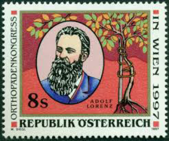
Adolf Lorenz and a crooked growing tree splinted with a straight stake, a symbol of the type of “dry surgery”.
Until the early 20th century, there was no discrimination between surgeons. Surgeons performed operations on various part of the body that they could do. So, it was not strange that Felix Guyon (1831-1920) who described “Loge de Guyon” at the wrist subsequently turned his interest to urology and became a founder and president of Urology Society [Figure 10].
Figure 10.

Felix Guyon stamp issued by France
In the late of 19th surgery many surgeons tried internal fixation to treat fractures; however, because of unsuitable alloy materials, corrosion of the implant occurred regularly. Also, lack of antibiotics led many experiences to catastrophic consequences. Finding a suitable alloy, improvement in the design of implants and evolution in surgical techniques took more than a century. Albin Lambotte (1866- 1955), a Belgium surgeon, revolutionized the fracture treatment with internal fixation. Lambotte endeavors were considered as a role model by the Swiss-German Association for the Study of Internal Fixation (AO). In 2017, Belgium issued a stamp in commemoration of the breakthrough of fracture treatment with internal fixation and to honor Albin Lambotte [Figure 11].
Figure 11.

Commemoration stamp for Albin Lambotte, the pioneer of internal fixation
During the great World War I, Hermann von Krukenberg, a German military surgeon introduced an innovative surgery for below-elbow amputees. Regarding the concept of chopsticks he separated the radius and ulnar bones to make two chopsticks and turned the forearm stump to a pincer. Each bone was supplied by muscles and skin coverage [Figure 12]. The main advantage of the operation was to reconstruct two sensate functional stumps. Krukenberg operation is considered primarily for blind patients with bilateral below-elbow amputations. This kind of mutilation happens when a mine explodes in the hands of an individual.
Figure 12.

A stamp issued by Bangladesh demonstrates bilateral Krukenberg operations
In 1915 brothers William (1861–1939) and Charles Mayo (1865–1939) established The Mayo Graduate School of Medicine. Mayo brothers believed that surgeon who performs surgeries on only one field of surgery gain more experiences and have an integrated patient care. Mayo brothers encouraged the concept of professionalism and specialty in different branches of surgery including orthopedics [Figure 13].
Figure 13.

The stamp pictures James Earle Fraser’s statues of Dr. William and Dr. Charles Mayo in surgical gowns
In the 20th century further advances in anti-septic techniques such as using surgical gloves, discovery of antibiotics, blood banking and transfusion, improved surgical techniques, and improved orthopedic implants provided better measures that orthopedic science could improve a better quality of life for patients. A British surgeon, Sir John Charnley (1911-1982), was the pioneer of the theories and practice of low friction arthroplasty. For 15 years Charnley with a team of engineers and biologist concentrated on low friction arthroplasties. Their investigations on mechanical, material, and surgical techniques led to benefit patients from hip arthroplasties from 1962 onward and other arthroplasties for the other joints. A stamp issued by United Kingdom commemorates Charnley’s achievement [Figure 14].
Figure 14.

Stamp for total hip arthroplasty as breakthrough in orthopedic science by Sir John Charnley
A stamp issued by Japan in 1978 on the occasion of 50th anniversary and 14th congress of the International Society of Orthopedic Surgeons which was held in Kyoto demonstrates hip arthroplasty as the symbol of congress. The symbol of orthopedic surgery which is a splinted growing tree is also demonstrated in the background [Figure 15]
Figure 15.

Memorial Stamp issued by Japan in 1978 on the occasion of 50th anniversary and 14th congress of the International Society of Orthopedic Surgeons depicting total hip arthroplasty and a splinted growing tree
There are many instances in the history of orthopedic surgery that an individual has made a significant contribution to a special field of orthopedic surgery. Emile Letournal (1927-1992) was a contemporary icon of acetabular and pelvic surgery. His extensive experiences and innovative techniques in acetabular and pelvic surgery are unrivaled in modern trauma and orthopedic surgery. His extraordinary understanding of complex and difficult acetabular and pelvic fracture has been acknowledged worldwide. Letournal’s diagnosis, description, classification, and surgical techniques for acetabular and pelvic fractures have been stood the test of time and became a standard in the recent decades. Letournal has been honored on a stamp issued by France in 1999 [Figure 16].
Figure 16.

The stamp issued by France in memorial of Emile Letournal (1927-1992) the iconic pioneer of acetabular and pelvic surgery
Josep Trueta (1897-1977) was a Spanish orthopedic surgeon who worked in the University of Oxford between 1935 and 1966. Trueta’s investigations and fundamental research on vascular contribution to osteogenesis, vascular anatomy of bone, growth, repair, and decay of cartilage, and osteoarthritis placed him in the front rank of contributors of orthopedic science. Trueta has been honored on a stamp issued by Spain in 1997 [Figure 17].
Figure 17.

Josep Trueta (1897-1977) a scholar of basic science research in orthopedics
Microsurgery or surgery with an operating microscope allows successful anastomosis of small blood vessels, typically 1 mm or less in diameter, and nerves. Microsurgery allows free transfer of tissue from one part of the body to another and replantation of severed parts. The advances in techniques and technology of microsurgery have been popularized since the early 1960s. This breakthrough in orthopedic surgery has been demonstrated on a stamp issued by China in 1975. The stamp demonstrates replantation of a forearm amputation in a farmer with microsurgery technique [Figure 18].
Figure 18.

Replantation of a forearm in a farmer with microsurgery technique
Gravil Abramovich Illizarov (1921-1992) was a Soviet Union surgeon who developed a technique that is best known for complex, infected fractures and to lengthen the bones. His hypothesis that bone grows if gradually distracted produced remarkable results that were not seen before in orthopedic science. Illizarov apparatus is an external fixator consisting of circular steel holes connected with rods and bone transfixing wires [Figure 19]. Illizarov technique developed a new field in orthopedic surgery that conserves and exploits the unlimited natural plasticity of bone. In 1987, on the occasion of Portuguese and Spanish Speaking Countries Orthopedists Meeting, Cuba commemorated this achievement by demonstrating an Illizarov apparatus on a stamp.
Figure 19.
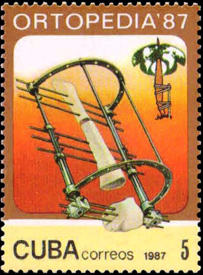
Illizarov apparatus in commemoration of Gravil Abramovich Illizarov on the occasion of Portuguese and Spanish Speaking Countries Orthopedists Meeting
One of the glories in the history of orthopedics is discovery of insulin by an orthopedic surgeon. Sir Frederick G. Banting (1891-1941) who discovered insulin and won the Noble Prize in medicine in 1923 at age 32 was an orthopedic surgeon [Figure 20]. He remains the youngest Noble laureate in physiology and medicine.
Figure 20.

Stamp issued by Canada in commemoration of Sir Frederick G. Banting
Rehabilitation
Rehabilitation is an essential component of orthopedic surgery. According to the World Health Organization “rehabilitation is a set of interventions needed when a person is experiencing or likely to experience limitations in everyday functioning due to ageing or a health condition, including chronic diseases or disorders, injuries or traumas”. The range of rehabilitation interventions is broad. Some examples of rehabilitation in orthopedic surgery include exercises to regain coordination, dexterity and movement of an affected limb following an injury, interventions that improve safety and independence at home and reduce the risk of falls and using prosthesis and orthotics [Figure 21].
Figure 21.

Stamps for rehabilitation to regain coordination, dexterity and movement of the affected limbs, to improve safety and independence at home and reduce the risk of falls, and rehabilitation using prosthesis and orthotics
Prosthesis and orthotics
Losing a part of the limb is not an uncommon injury; however, using prosthesis can improve the quality of life and working of the amputee. Myoelectric functional prostheses were introduced in 1960s; however, the science and technology of the sophisticated prosthesis has been evolutionized and robotic prosthetic hands with individual articulated digits have been produced by the advances in various technology fields. A stamp issued by Belgium in 1987 shows the developed technology of the prosthetic hand. A stamp issued by Great Britain in 2015 shows the development of i-hand. A stamp issued by Italy in 1999 shows the Michelangelo prosthetic robotic hand which is a new state of the art generation of myoelectric prosthetic hands with independent powered opposable thumb and fingers [Figure 22] (6).
Figure 22.

Stamps issued for advanced technology of the hand prosthesis, development of i-hand, and Michelangelo prosthetic robotic hand
Summary
Orthopedic surgery is an evolving science. The current article has tried to present an insight to some of the major milestones in the history of orthopedics and also the involved people. Orthopedic surgery and science continues to progress. Certain peaks and memorable events in orthopedic science such as computer assisted, robotic surgeries, and introduction of new orthopedic implants may find their place on postal stamps in future [Figure 23].
Figure 23.

Synthetic bone-graft materials encourages new bone growth.
References
- 1.Afshar A. Concepts of orthopedic disorders in Avicenna’s Canon of Medicine. Arch Iran Med. 2011;14(2):157–159. [PubMed] [Google Scholar]
- 2.Afshar A. A brief report about the concepts of hand disorders in the Canon of Medicine of Avicenna. J Hand Surg Am. 2011;36(9):1509–1514. doi: 10.1016/j.jhsa.2011.06.018. [DOI] [PubMed] [Google Scholar]
- 3.Afshar A, Kyle RA, Steensma DP. Stamp vignette on medical science: Antonius Mathijsen and plaster casts. Mayo Clin Proceed. 2017;92(5) doi: 10.1016/j.mayocp.2017.01.028. [DOI] [PubMed] [Google Scholar]
- 4.Afshar A, Kyle RA, Steensma DP. Stamp vignette on medical science: Andreas Vesalius and De Fabrica. Mayo Clin Proceed. 2019;94(5):e67–68. doi: 10.1016/j.mayocp.2019.03.018. [DOI] [PubMed] [Google Scholar]
- 5.Afshar A, Kyle RA, Steensma DP. Stamp vignette on medical science: Adolf Lorenz: The Bloodless Surgeon of Vienna. Mayo Clin Proceed. 2017;92(7):e105–e106. doi: 10.1016/j.mayocp.2017.02.025. [DOI] [PubMed] [Google Scholar]
- 6.Afshar A. Evolution of functional hand prostheses on the postal stamps. Journal of Hand and Microsurgery. 2017;9(3):172–174. doi: 10.1055/s-0037-1608744. [DOI] [PMC free article] [PubMed] [Google Scholar]