Int J Mol Med 41: 51-60, 2018; DOI: 10.3892/ijmm.2017.3226
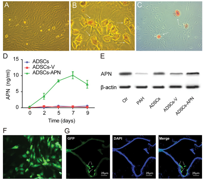
Following the publication of the above article, an interested reader drew the authors' attention that the data featured in Fig. 1B (for adipogenic differentiation of adipose-derived stem cells) and Fig. 1F (for expression of green fluorescent protein of adipose-derived stem cells) of the above article appeared to have already been published as Fig. 1A (for adipogenic differentiation of adipose-derived stem cells) and Fig. 2D (for expression of green fluorescent protein of adipose-derived stem cells) in the following article: Luo L, Lin T, Zheng S, Xie Z, Chen M, Lian G, Xu C, Wang H and Xie L: Adipose-derived stem cells attenuate pulmonary arterial hypertension and ameliorate pulmonary arterial remodeling in monocrotaline-induced pulmonary hypertensive rats. Clin Exp Hypertens 37: 241-248, 2015.
Figure 1.

Adipose-derived stem cell (ADSC) characterization and adiponectin (APN) expression. (A) ADSCs isolated from rats had spindle-shaped and fibroblast-like morphological features. (B) After adipogenic induction, intracellular lipid accumulation was observed by Oil Red O staining. (C) After osteogenic induction, extracellular matrix calcification was detected by Alizarin red staining. (D) APN expression in the various groups. (E) APN protein expression in the lung was evaluated by western blot analysis. (F) Representative fluorescence microscopy image of green fluorescent protein (GFP)-expressing ADSCs in vitro (green). (G) Distribution of intravenously administered ADSCs in lung tissue. GFP-expressing rat ADSCs (green) were incorporated into walls of pulmonary vascular beds. Nuclei were stained by DAPI. Scale bars, 25 µm.
The authors consulted their original data and were able to determine that the duplication of these figure parts had arisen inadvertently during the process of compiling the figure. The revised version of Fig. 1, featuring the corrected data panels for the above-mentioned experiments in Fig. 1B and F, is shown on the next page. The authors confirm that the errors associated with this figure did not have any significant impact on either the results or the conclusions reported in this study, and are grateful to the Editor of International Journal of Molecular Medicine for allowing them the opportunity to publish this Corrigendum. Furthermore, they apologize to the readership of the Journal for any inconvenience caused.


