

Abstract
Objective
To describe a surgical technique using suture tape for reconstruction of the medial patellofemoral ligament (MPFL). This technique restores the stability of the reconstructed ligament and has excellent postoperative outcomes.
Method
This is a retrospective analysis. From January 2016 to June 2018, 17 patients underwent MPFL reconstruction using high‐strength suture (FiberTape; Arthrex) augmentation, with at least 12 months of follow up. There were 11 female and 6 male patients. The mean age at the time of MPFL reconstruction was 22.1 years (range 13–34 years). Clinical outcomes included pain level, knee range of motion, passive patellar hypermobility, and maltracking at follow‐up. The lateral patellofemoral angles, congruence angles, and patellar tilt angles were measured in a skyline view by CT at 30° of knee flexion at 12 months. Functional outcomes were assessed using the Lysholm knee scoring scale, the SF‐12 score, the Tegner score, and the Crosby and Insall grading system at yearly follow‐up.
Result
No patients were lost at the last follow up. One patient had recurrence of patellar dislocation and none of the others had serious complications. The success rate of MPFL repair for preventing recurrent dislocations was 94.1% (16 of 17 knees). Fifteen knees had full range of motion of more than 130°. At follow‐up, 2 knees were judged to have mild hypermobility and none had severe hypermobility or maltracking. Using the Crosby and Insall grading system, 12 knees (70.6%) were graded as excellent, 4 knees (23.5%) as good, 1 knee (5.9%) as fair to poor, and none as worse at the last follow‐up assessment. In all patients, the Lysholm knee score (55.12 ± 13.52 vs 79.88 ± 7.50, P < 0.01), the SF‐12 score (47 ± 9.53 vs 65.24 ± 12.82, P < 0.01), and the Tegner score (2.76 ± 1.39 vs 6.53 ± 1.70, P < 0.01) had improved at their 12‐month follow up. Compared with preoperative radiological findings, there was a significant improvement in lateral patellofemoral angle (−10.24 ± 7.10 vs 6 ± 5.43, P < 0.01), patellar tilt angle (26.53 ± 7.23 vs 9.88 ± 4.24, P < 0.01), and congruence angle (29.59 ± 11.95 vs −8.65 ± 4.86, P < 0.01).
Conclusion
The use of FiberTape in MPFL reconstruction can improve the stability of the knee following surgery and has good midterm clinical results and low complication rates.
Keywords: Medial patellofemoral ligament, Patellar tilt, Patellofemoral joint, Reconstruction, Suture tape
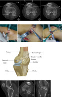
Fig. 1 Measuring methods on a bradiograph. (a) 30° skyline view. (A) Lateral patellofemoral angle was defined as an angle with line (a) to (b). (B) Congruence angle was defined as an angle with line (c) to (d). (C) Patellar tilt angle was defined as an angle with line (e) to (f).Fig. 2 Intraoperative photograph of a left knee as viewed from the medial side.Fig. 3. Two suture tapes were adopted for double‐bundle medial patellofemoral ligament reconstruction and fixed by knotless anchors at the femoral side.Fig. 4. CT arthrogram postoperatively showing the position of the patella tunnel, the knotless anchor, and the patella at the patellofemoral joint.
Introduction
The medial patellofemoral ligament (MPFL) is the main stabilizer against lateral patella translation. Imaging studies and surgical exploration have identified that MPFL injury is associated with patella dislocations. After the first dislocation, the MPFL is torn in up to 90% of patients. Recurrent dislocation occurs in 17% to 69% of patients after the primary dislocation event 1 . Even if there is no further dislocation, it can lead to patellar tilt and abnormal patellar track, eventually resulting in patellofemoral osteoarthritis 2 . There are a variety of causes of patellar dislocation or subluxation, including trochlear dysplasia, patella alta, femoral antetorsion, and an increased tibial tubercle‐trochlear groove distance 3 , 4 . Due to the abnormal physiological bone structure, patella dislocation is more likely to occur in patients participating in sports activities, where the knee is slightly flexed, rotated inwards, and in valgus position.
Most surgical techniques include MPFL reconstructions with tendon allografts or autografts, repair of the MPFL using an anchor‐based reattachment to the adductor tubercle, and medial and lateral patellar retinaculum plasty. Surgical treatment of patellar dislocation has always been a challenge for orthopaedic surgeons due to the complexity of the physical structure and unsatisfactory results, such as recurrent dislocation, persistent anterior patellar pain, and patellar fracture. Thus, conservative treatment has also been a long‐term treatment option in the past. However, the rate of patella redislocation and other complications with this approach has increased significantly in comparison to surgical treatment 5 . Over the past two decades, increasing attention has been paid to MPFL reconstruction for the treatment of recurrent patellar dislocations/subluxations 6 . MPFL reconstruction has become the first choice for treating recurrent patellar dislocation. Good midterm clinical results with up to 97% patient satisfaction and up to 10 years of follow up have been reported 7 . There are many devices (e.g. interference screws and suture anchors and techniques (e.g. via single tunnel or double tunnel technique) available for fixation of a graft to the anatomic insertion site of the MPFL.
However, despite MPFL reconstruction having a high rate of success for patients, the complication rate of 26.1% associated with this procedure is not trivial 8 . The complications of MPFL reconstruction include graft laxity, abnormal patellar track, recurrent dislocations, allogeneic tendon reaction, and donor pain. Inappropriate graft tensioning during MPFL reconstruction may cause altered patellofemoral joint kinematics and contact mechanics, potentially resulting in pain and joint degeneration.
We need a new material with high initial strength, that does not easily relax, with adjustable tension, and with no rejection reaction to replace the current graft reconstruction. In the last few decades, increasing research on materials has been carried out, and it is hoped that synthetic materials can replace autogenous tendons. One study describes a minimally invasive technique to reconstruct the MPFL using an artificial ligament and provides descriptive information about clinical benefits and safety in a broad population of patients with all grades of patellar instability 9 . Reports on FiberTape in MPFL reconstruction demonstrated that it is safe and effective and provides higher initial strength than the hamstring tendon 10 , 11 . The major advantage of using synthetic materials for MPFL reconstruction is that the autologous tendon is retained and, thus, donor complications are reduced.
The purpose of the present paper is outlined as follows. First, we describe a new surgical technique for patellar dislocations using high‐strength suture reconstruction of the MPFL. Second, we evaluate preoperative and postoperative clinical outcomes, including pain level, knee range of motion, passive patellar hypermobility, maltracking and apprehension, and functional outcomes. Third, we evaluate preoperative and postoperative positions of the patella by CT, such as lateral patellofemoral angles, congruence angles, and patellar tilt angles. We expect that the application of this technique will improve the stability of the knee following surgery and will result in good midterm clinical outcomes and low complication rates.
Materials and Methods
Patients
We retrospectively analyzed patients with patellar instability at the Third Hospital of Hebei Medical University. From January 2016 to June 2018, 21 patients underwent MPFL reconstruction using high‐strength suture (FiberTape; Arthrex) augmentation, with at least 12 months of follow up. All patients were diagnosed with patellar instability (patellar tilt, subluxation, or dislocation) by physical examination. Four patients with a history of prior knee surgery (medial tubercle transfer in two and distal femur osteotomy in two) were excluded from the present investigation.
Inclusion and Exclusion Criteria
The inclusion criteria were: (i) primary patellar dislocation; (ii) failure of non‐operative treatments; (iii) without multiple ligament injury; and (iv) body mass index (BMI) <30 kg/m2. The exclusion criteria were: (i) recurrent patellar dislocation; (ii) less than 16 years old and with open epiphyseal plates; (iii) trochlear dysplasia; (iv) femoral anteversion angle >25°; and (iv) tibial tuberosity–trochlear groove (TT–TG) distance ≥20 mm.
Surgical Technique
Anesthesia and Position
All surgical procedures were performed by the same surgical team. The patients received general anesthesia and were placed in the supine position. A tourniquet was tied to the proximal thigh and set to 280 mmHg. The aseptic surgical area was prepared using iodine (2%) and medical alcohol (70%).
Approach
The knee was first examined and the tightness of the lateral structures was assessed. Following this, a knee arthroscopy was performed using standard anteromedial and anterolateral portals to visualize the knee. The lateral retinaculum was released arthroscopically using thermal ablation in all patients. A 4‐cm longitudinal incision was made medial to the patella.
Bone Tunnel
Two guidewires were transversely inserted, one from the proximal one‐third of the medial edge of the patella and the other from the center of the patella. Two polyester suture tapes were pulled through the bone tunnel using a guide pin.
Reconstruction
The suture tape came out from the tunnel and was turned back through the upper surface of the patella to the femur side together with the other end. With the knee at 30° of flexion, the suture tape was shuttled through layer 2 using a 1–2‐cm incision centered at the region of the medial epicondyle of the knee. Then, the two free ends of the polyester suture tape were fixed using a 4.75‐mm knotless anchor (SwiveLock; Arthrex) on the femoral side, while the patella was kept in the center of the patellar groove. The femoral fixation point was determined by the insertion of the intact MPFL between the adductor tubercle and the medial epicondyle (Fig. 1). To prevent overtightening the suture, a curved hemostat was placed under the tape before final fixation and the patella was held with its lateral border aligned to the lateral aspect of the femoral trochlea. The repair was then visualized directly and arthroscopically to confirm the extra‐articular position of the suture tape. After these procedures, the subcutaneous tissue and skin were stitched up.
Fig. 1.

Measuring methods on a bradiograph. (a) 30° skyline view. (A) A patellar tilt angle was defined as an angle with the extension line of the maximum transverse diameter of the cut position of the patella (a) to the posterior line of the internal and external condyle of the femur (b). (B) A congruence angle was defined as an angle with the line drawn through the lower pole of the patella and the deepest point of the chute (c) to the line on the side of the bisector defines the tackle angle (d). (C) A lateral patellofemoral angle was defined as an angle with the line of the tangent of the lateral articular surface of the patella the (e) to highest point of the femoral and external condyle (f).
Postoperative Management
Postoperatively, patients were placed into a range‐of motion brace and allowed to weight‐bear as tolerated with the brace locked in full knee extension. For 2 weeks after MPFL reconstruction, the knee was immobilized with a brace at 45° of knee flexion. After the brace was removed, passive‐assisted and active‐assisted range of knee motion was commenced. Weight‐bearing was gradually increased to full weight‐bearing at 4 weeks postoperatively. Running was allowed at 3 months, followed by a return to previous sporting activity at 6 months.
Clinical Assessments
For assessment of the results, clinical outcomes included pain level, knee range of motion, passive patellar hypermobility, maltracking and apprehension at follow up. The congruence angles, lateral patellofemoral angles, and patellar tilt angles were measured in a skyline view at 30° of knee flexion at 12 months (Fig. 2). The Lysholm knee scoring score SF‐12 score and the Tegner scale were administered to assess the functional impairment due to clinical instability and to evaluate the outcomes of knee ligament surgery. The Crosby and Insall grading system was used to assess outcomes following ligament reconstruction. Using this system, outcomes were classified into four categories (excellent, good, fair to poor, and worse) 12 .
Fig. 2.

Intraoperative photograph of a left knee as viewed from the medial side. (A) Two bone tunnels are established within the patella, one in the upper third of the patella, and the other in the middle. (B) Two suture tapes come out from the double tunnel and turn back through the upper surface of the patella to the femur side together with the other end. (C) The free ends of two polyester suture tapes were fixed using a knotless anchor on the femoral side.
Radiological Assessments
Lateral Patellofemoral Angle
The angle between the line of the highest point of the femoral and external condyle and the tangent of the lateral articular surface of the patella is the lateral patellofemoral angle, which normally opens outward. If the opening is inward or the two lines are parallel, then there is lateral inclination of the patella. When the lines are parallel or converge, it is suggestive of excessive lateral tilt, often found in patients with recurrent patella dislocations (Fig. 2A).
Congruence Angle
A line is drawn through the lower pole of the patella and the deepest point of the chute. The angle of the line on the side of the bisector defines the tackle angle (Fig. 2B). This angle is negative on the inside of the bisector and positive on the outside.
Patellar Tilt Angle
The angle formed by the posterior line of the internal and external condyle of the femur and the extension line of the maximum transverse diameter of the cut position of the patella. A patellar tilt angle of more than 20° indicated abnormal patellar tracking in patients with patellar dislocation (Figs 3 and 4).
Fig. 3.

Two suture tapes were adopted for double‐bundle medial patellofemoral ligament reconstruction and fixed by knotless anchors at the femoral side.
Fig. 4.

CT arthrogram postoperatively showing the position of the patella tunnel, the knotless anchor, and the patella at the patellofemoral joint in the sagittal (A), axial (B), and coronal (C) plane, respectively.
CT Measure
The lateral patellofemoral angles, congruence angles, and patellar tilt angles were measured in a skyline view by CT at 30°of knee flexion. X‐rays were also performed to assess the integrity of the fixation (alignment and positioning) and other complications (arthritis and fracture). All outcomes were measured by a single investigator and confirmed by a senior surgeon. Patellar tracking was evaluated at 0° to 60° of knee flexion through a lateral suprapatellar portal. A median ridge of the patella located above the middle third of the femoral groove was defined as “centrally located,” while a ridge located lateral to the middle third of femoral groove was defined as “laterally shifted.”
Statistical Analysis
The frequency of clinical outcome events was summarized using percentages. Functional outcomes were summarized by calculating the group mean and standard deviation for baseline and follow‐up measurements. The differences between follow‐up and baseline values for each knee were calculated in measurement units. A two‐tailed paired t‐test was used to test for a statistical difference in baseline versus follow‐up measures. If the data had been found to not be normally distributed, the Mann–Whitney U non‐parametric test would have been used. A P‐value of <0.05 was considered statistically significant. Analyses were conducted using SPSS software (Version 21.0, IL, USA).
Results
Demographics
There were 11 female and 6 male patients. The mean age at the time of MPFL reconstruction was 22.1 years (range 13–34 years). The average follow‐up time was 14 months (Table 1). The mean duration of time between MPFL reconstruction and postoperative CT evaluation was 11 months (range 9–15 months). The average BMI was 25.24 ± 4.20 kg/m2.
TABLE 1.
Demographics of the patient characteristics
| Sex (M:F) | Age (years) | Follow‐up time (months) | BMI (kg/m2) | Side (L/R) | |
|---|---|---|---|---|---|
| Patients | 6/11 | 22.06 ± 6.61 | 14.29 ± 4.21 | 24.04 ± 2.45 | 10/7 |
Data are expressed as mean values ± standard deviation. BMI, body mass index.
Subjective Function Score
Using the Crosby and Insall grading system, 12 knees (70.6%) were graded as excellent, 4 knees (23.5%) were good, 1 knee (5.9%) as fair to poor, and none as worse at the last follow‐up assessment. In most patients, the patella trajectory was basically back to normal at their last follow up. In one case, the medial patellar support band remained loose due to a partial release of the femoral fixation. However, the patient did not experience significant pain during knee flexion and light exercise. In all patients, the Lysholm knee score (55.12 ± 13.52 vs 79.88 ± 7.50, P < 0.01), the SF‐12 score (47 ± 9.53 vs 65.24 ± 12.82, P < 0.01) and the Tegner score (2.76 ± 1.39 vs 6.53 ± 1.70, P < 0.01) had improved at their 12‐month follow up. Postoperative functional scores improved significantly in all patients (Table 2).
TABLE 2.
Preoperative and postoperative clinical outcomes of patients
| Preoperation | Postoperation | P‐value | |
|---|---|---|---|
| Lysholm score | 55.12 ± 13.52 | 79.88 ± 7.50 | <0.01 |
| SF‐12 score | 47 ± 9.53 | 65.24 ± 12.82 | <0.01 |
| Tegner | 2.76 ± 1.39 | 6.53 ± 1.70 | <0.01 |
| Crosby and Insall grading system | |||
| Excellent | 0 | 12 (70.6%) | — |
| Good | 0 | 4 (23.5%) | — |
| Poor | 6 (37.5%) | 1 (5.9%) | — |
| Worse | 10 (62.5%) | 0 | — |
Data are expressed as mean values ± standard deviation.
Objective Functional Evaluation
At 2 weeks, all patients had started knee bending and could achieve 70°–90° of flexion. Fifteen knees had full range of motion (more than 130°), while 2 knees had a slight loss of flexion (20°) at their last follow up. In both patients, the quadriceps strength of the affected limb was significantly lower than that of the healthy side due to inactivity. Passive patellar hypermobility with the knee extended and flexed at 20° and maltracking were present in all knees preoperatively. At the follow up, 2 knees were judged to have mild hypermobility and none had severe hypermobility or maltracking. The apprehension test was positive in 8 knees preoperatively and was positive at the follow up in only 1 knee. Generalized ligamentous laxity was observed in 3 patients preoperation and postoperation, which might be related to individuals’ physical condition.
Radiographic Findings
Immediately after MPFL reconstruction, the three indices had improved (Table 3). Although patellar maltracking was reduced in all cases immediately after surgery, the position had shifted laterally to some extent at follow‐up.
TABLE 3.
CT evaluations of comparison between preoperation and postoperation(°)
| Preoperation | Postoperation | P‐value | |
|---|---|---|---|
| Lateral patellofemoral angle | −10.24 ± 7.10 | 6 ± 5.43 | <0.01 |
| Patellar tilt angle | 26.53 ± 7.23 | 9.88 ± 4.24 | <0.01 |
| Congruence angle | 29.59 ± 11.95 | −8.65 ± 4.86 | <0.01 |
Data are expressed as mean values ± standard deviation.
Lateral Patellofemoral Angle
Normally, the opening of the lateral patellofemoral angle is positive to the lateral and negative to the medial when the patella is unstable. Compared with preoperative radiological findings, a significant improvement was found for postoperative lateral patellofemoral angles (−10.24° ± 7.10° vs 6° ± 5.43°, P < 0.01).
Patellar Tilt Angle
The average patellar tilt angle was decreased from 26.53° ± 7.23° preoperatively to 9.88° ± 4.24° postoperatively, with an average improvement of 16.6° (P < 0.01).
Congruence Angle
The preoperatively average congruence angle in 17 patients with 29.59° ± 11.95° was significantly higher than postoperative date with −8.65 ± 4.86 (P < 0.01).
Surgery and Complications
The average operative time was 60 min (range, 50–75 min) and blood loss was 50 mL (range, 40–65 mL). There were no fractures, no damage to nerves or blood vessels, no closure of the wound, or any other errors during the operation. The success rate of MPFL repair for preventing recurrent dislocations was 94.1% (16 of 17 knees). One patient experienced a recurrent lateral patellar dislocation. The femoral side knotless anchor of 1 patient had become loose at the final follow‐up, but graft fixation was stable in the other patients at 12‐month follow up. One patient had a redislocation at 6 months after surgery. Four patients had prominence of the ligament over the medial femoral condyle. Three patients presented with anterior knee pain at their 6‐month follow‐up, with only one of them experiencing knee pain while walking normally at the final follow‐up. Fourteen knees had no complications, and no other major complications were reported (Table 4).
TABLE 4.
Postoperative physical examination outcomes and complications of the patient
| Physical evaluation | Recurrent patellar dislocation | Knee pain | Loss of fixation | Loss of knee flexion | Apprehension test | Passive patellar hypermobility | No complications |
|---|---|---|---|---|---|---|---|
| Frequency | 1 | 1 | 1 | 2 | 1 | 2 | 14 |
| Incidence (%) | 5.9 | 11.8 | 5.9 | 11.8 | 5.9 | 11.8 | 82.4 |
Knee pain in walking normally.
Loss of knee flexion >10°.
Passive patellar Hypermobility was in the knee extended and flexed at 20°.
Discussion
The main findings of this study are that medial patellofemoral ligament reconstruction using the suture tape for patellofemoral joint instability increases the initial stability and achieves a good clinical effect.
Significance of Medial Patellofemoral Ligament Reconstruction
Patellar dislocation affects approximately 30% of individuals, and up to 75% of those with instability; it eventually leads to the rupture or dysfunction of MPFL. Isolated MPFL repairs do not provide adequate functional outcomes and have been associated with a high failure rate 13 , 14 . MPFL reconstruction has become an acceptable treatment option that demonstrates excellent clinical results and has a low repeat dislocation rate 15 . Various graft options exist for MPFL reconstruction, including semitendinosus, gracilis, quadriceps or adductor magnus tendons, part of the patellar tendon, allografts, and artificial ligaments 15 , 16 . These grafts require early biological healing, and failure of biological remodeling can lead to graft elongation, which eventually causes irreversible relaxation and the failure of the operation.
The present study revealed that not all improved patellar trackings after MPFL reconstruction remained intact at follow up. Ostermeier et al. concluded that reconstruction techniques created sufficient stabilization of the patella but that the patellar position was slightly overmedialized following MPFL reconstruction with a semitendinosus autograft, which could lead to overload on the medial retropatellar cartilage 17 . Sandmeier et al. concluded that patellar tracking changed significantly with loss of the medial restraints and improved after MPFL reconstruction using a gracilis tendon. They also noted that patellar tracking was not completely restored 18 . Nomura et al. concluded that the association of definite knee osteoarthritis in MPFL reconstruction was minimal at long‐term follow up and revealed that MPFL reconstruction shows no or only slight progression of knee osteoarthritis 7 .
Emergence of Synthetic Materials
Proper tension and graft length applied in MPFL reconstruction may be necessary to prevent further dislocation after surgery 19 . Conversely, redislocation and return of excessive lateral pressure may occur with reductions in tension. In many knee ligament reconstruction surgeries, biomechanical properties of implanted grafts are known to change with the effects of stress relaxation and graft remodeling. Autogenous tendon grafting has always been the gold standard for reconstruction, but in recent years, with the progress of materials science, artificial ligaments have been reported as an alternative option 9 , 20 . Using suture augmentation as an internal brace against lateral patellar translation, the MPFL may have a better chance of healing and repair of concomitant cartilage lesions is protected from additional instability episodes after surgery. MPFL reconstruction with suture tape augmentation acts as a secondary stabilizer 21 .
Advantages of Synthetic Materials
Polyester suture tape is an ultra‐high strength tape consisting of long chains of ultra‐high molecular weight polyethylene. The beneficial characteristics of an artificial ligament are its initial stiffness and resistance to elongation. Tsushima et al. conducted a biomechanical analysis of MPFL reconstruction, comparing suture tape (FiberTape; Arthrex) with knotless anchors to a semitendinosus tendon autograft with soft anchors. The mean ultimate load of the native MPFL was 130.6 ± 28.7 N and all native MPFL failed at the femoral insertion site. The mean ultimate loads for the reconstructed MPFL using a modern synthetic material with knotless anchors and a semitendinosus tendon autograft with soft anchors were 178.4 ± 4.4 N and 102.8 ± 21.5 N, respectively. FiberTape with knotless anchors was stronger than all native MPFL and a semitendinosus tendon with soft tissue anchors for MPFL reconstruction at time zero 10 . Mehl confirmed that suture tape augmentation of the MPFL resulted in similar primary contact pressures and joint kinematics in comparison with MPFL reconstruction using a tendon graft 22 .
Hopper et al. proposed reconstructing the MPFL using artificial ligaments to allow early mobilization without bracing, and revealed that the procedure was safe, with a low risk of redislocation in all grades of instability 23 . In another study, 20 27 MPFL reconstructions using a mesh‐type artificial ligament and medial retinaculum slip coverage were reviewed using the Crosby and Insall grading system, with an average follow up of 5.9 years. A total of 15 knees (55%) were classified as excellent, 11 knees (41%) as good, 1 knee (4%) as fair/poor, and none as worse. Lee et al. reported that MPFL reconstruction using polyester suture tape and knotless anchors significantly improves patients’ quality of life and results in better postoperative outcomes 11 . The use of FiberTape in MPFL reconstruction is safe and effective, and it significantly improves patients’ quality of life and related postoperative outcome measures. There were no significant differences in all knee scores compared to GT autografts.
Using this technique for MPFL reconstruction, tendon harvesting is unnecessary and, hence, donor site morbidity‐associated complications are eliminated. The semitendinosus and gracilis muscles play an important role in flexing the knee, regulating rotation of the tibia, limiting anterior translation of the tibia, and sharing the stress with the ACL 24 , 25 . Therefore, hamstring tendon harvesting causes significant weakness of knee flexor strength at high knee flexion angles, but such weakness is minimized because the gracilis tendon is preserved in the all‐inside ACL reconstruction 24 .
Femoral tunnels positioned too proximal or distal resulted in significantly elevated medial joint contact pressures and increased medial patellar tilting. This may lead to adverse outcomes, such as early degenerative joint changes or pain in a clinical population. According to Stephen et al., the correct femoral tunnel position restored normal joint kinematics and patellofemoral joint contact pressure 26 . Anatomical double‐bundle reconstruction was performed to reduce the influence on patellofemoral joint contact pressure. Our technique involves the use of FiberTape in MPFL reconstruction; the insertion of the femur should be anatomically located to prevent positioning that is too proximal or distal. An adjustable loop is used on the femur side, which is convenient for adjusting the tension of the FiberTape when it is fixed. On the patella side, we used a double tunnel reconstruction technique, which reduced the risk of patella fracture due to the smaller tunnel diameter. The double tunnel technique is more in line with the biomechanics of the patellofemoral joint, and it can better correct the inclination of the patella and effectively prevent redislocation of the patella 27 , 28 .
The MPFL augmentation repair can be performed quickly in a minimally invasive manner and without the need for tendon autograft or allograft, ultimately reducing the morbidly and cost compared with an MPFL reconstruction. The suture tape has stronger biomechanical properties and resistance to elongation, and the angulation of the suture tape augmentation provides a very secure fixation that can mimic the strength of the native MPFL. Patients are allowed to participate in sports if the neuromuscular function has recovered. Through clinical observation, no local tissue and delayed rejection reaction were found in the reconstruction with suture tape.
Disadvantages of Synthetic Materials
One potential complication of MPFL reconstruction with an artificial ligament is postoperative medial patellofemoral pain caused by an overly constrained patella. This is affected by the fixation angle of the artificial ligament 29 , 30 . Hopper et al. described a method using FiberTape in MPFL reconstruction and stated the importance of avoiding excessive constraint when fixing the suture tape because excess tension leads to irritation and can result in quadriceps inhibition 23 . The MPFL is the main restraining force in the first 20° of flexion against lateral patellar displacement and is disrupted after patellar subluxation or dislocation. Because this tape does not elongate over time, as compared to autografts or allografts, the polyester suture tape fixation angle may need to be equal to or greater than that used for autografts or allografts. Nomura et al. recommended fixation at 60° when using polyester tape (Leeds–Keio artificial ligament); they also used a tension spacer between the polyester tape and the femur during fixation to avoid excessive tensioning 7 . In our study, to avoid excessive patellofemoral joint contact pressure after MPFL reconstruction, polyester suture tape was fixed between 60° to 90° of knee flexion. To prevent overtightening the suture, a curved hemostat was placed under the tape before final fixation and the patella was positioned with its lateral border aligned to the lateral aspect of the femoral trochlea.
There were several limitations in the present study. First, different doctors may have varying results when they perform the same surgery, depending on the surgeon's experience. In addition, a slight difference in femoral and patellar tunnel placement can potentially occur during reconstruction. Second, several advantages are associated with this technique, as outlined earlier, and we have observed excellent clinical results, which could lead to subjective bias. Finally, the short follow‐up time in this study made it impossible to assess patients’ long‐term outcomes. Therefore, further clinical studies are necessary to determine the overall outcomes of this procedure.
Conclusion
The use of FiberTape in MPFL reconstruction can improve the stability of the knee following surgery and has good midterm clinical results and low complication rates.
Disclosure: All authors declare no conflict of interest. No funding was received for this paper.
References
- 1. Gravesen KS, Kallemose T, Blond L, et al. High incidence of acute and recurrent patellar dislocations: a retrospective nationwide epidemiological study involving 24.154 primary dislocations. Knee Surg Sports Traumatol Arthrosc, 2018, 26: 1204–1209. [DOI] [PubMed] [Google Scholar]
- 2. Dandy DJ et al. Chronic patellofemoral instability. J Bone Joint Surg Br, 1996, 78: 328–335. [PubMed] [Google Scholar]
- 3. Balcarek P, Oberthür S, Hopfensitz S, et al. Which patellae are likely to redislocate? Knee Surg Sport Traumatol Arthrosc, 2014, 22: 2308–2314. [DOI] [PubMed] [Google Scholar]
- 4. Köhlitz T, Scheffer S, Jung T, et al. Prevalence and patterns of anatomical risk factors in patients after patellar dislocation: a case control study using MRI. Eur Radiol, 2013, 23: 1067–1074. [DOI] [PubMed] [Google Scholar]
- 5. Mäenpää H, Lehto MU, Patellar dislocation . The long‐term results of nonoperative management in 100 patients. Am J Sports Med, 1997, 25: 213–217. [DOI] [PubMed] [Google Scholar]
- 6. Smith TO, Walker J, Russell N, et al. Outcomes of medial patellofemoral ligament reconstruction for patellar instability: a systematic review. Knee Surg Sports Traumatol Arthrosc, 2007, 15: 1301–1314. [DOI] [PubMed] [Google Scholar]
- 7. Nomura E, Inoue M, Kobayashi S, et al. Long‐term follow‐up and knee osteoarthritis change after medial patellofemoral ligament reconstruction for recurrent patellar dislocation. Am J Sports Med, 2007, 35: 1851–1858. [DOI] [PubMed] [Google Scholar]
- 8. Shah JN, Howard JS, Flanigan DC, et al. A systematic review of complications and failures associated with medial patellofemoral ligament reconstruction for recurrent patellar dislocation. Am J Sports Med, 2012, 40: 1916–1923. [DOI] [PMC free article] [PubMed] [Google Scholar]
- 9. Khemka A, Lord SJ, Doyle Z, et al. Minimally invasive medial patellofemoral ligament reconstruction for patellar instability using an artificial ligament: a two year follow‐up. Knee, 2016, 23: 261–266. [DOI] [PubMed] [Google Scholar]
- 10. Tsushima T, Tsukada H, Saaki S, et al. Biomechanical analysis of medial patellofemoral ligament reconstruction: FiberTape® with knotless anchors versus a semitendinosus tendon autograft with soft anchors. J Orthop Sci, 2019, 24: 663–667. [DOI] [PubMed] [Google Scholar]
- 11. Lee PYF, Golding D, Rozewicz S, Chandratreya A. Modern synthetic material is a safe and effective alternative for medial patellofemoral ligament reconstruction. Knee Surg Sports Traumatol Arthrosc, 2018, 26: 2716–2721. [DOI] [PubMed] [Google Scholar]
- 12. Crosby EB, Insall J. Recurrent dislocation of the patella. Relation of treatment to osteoarthritis. J Bone Joint Surg Br, 1976, 58: 9–13. [PubMed] [Google Scholar]
- 13. Arendt EA, Moeller A, Agel J, et al. Clinical outcomes of medial patellofemoral ligament repair in recurrent (chronic) lateral patella dislocations. Knee Surg Sports Traumatol Arthrosc, 2011, 19: 1909–1914. [DOI] [PubMed] [Google Scholar]
- 14. Camp CL, Krych AJ, Dahm DL, et al. Medial patellofemoral ligament repair for recurrent patellar dislocation. Am J Sports Med, 2010, 38: 2248–2254. [DOI] [PubMed] [Google Scholar]
- 15. Lippacher S, Dreyhaupt J, Williams SR, et al. Reconstruction of the medial patellofemoral ligament: clinical outcomes and return to sports. Am J Sports Med, 2014, 42: 1661–1668. [DOI] [PubMed] [Google Scholar]
- 16. Krebs C, Tranovich M, Andrews K, Ebraheim N, et al. The medial patellofemoral ligament: review of the literature. J Orthop, 2018, 15: 596–599. [DOI] [PMC free article] [PubMed] [Google Scholar]
- 17. Ostermeier S, Holst M, Bohnsack M, et al. In vitro measurement of patellar kinematics following reconstruction of the medial patellofemoral ligament. Knee Surg Sports Traumatol Arthrosc, 2007, 15: 276–285. [DOI] [PubMed] [Google Scholar]
- 18. Sandmeier RH, Burks RT, Bachus KN, Billings A, et al. The effect of reconstruction of the medial patellofemoral ligament on patellar tracking. Am J Sports Med, 2010, 28: 345–349. [DOI] [PubMed] [Google Scholar]
- 19. Mountney J, Senavongse W, Amis AA, et al. Tensile strength of the medial patellofemoral ligament before and after repair or reconstruction. J Bone Joint Surg Br, 2005, 87: 36–40. [PubMed] [Google Scholar]
- 20. Nomura E, Horiuchi Y, Kihara M, et al. A mid‐term follow‐up of medial patellofemoral ligament reconstruction using an artificial ligament for recurrent patellar dislocation. Knee, 2000, 7: 211–215. [DOI] [PubMed] [Google Scholar]
- 21. Horibe S, Shino K, Nagano J, et al. Replacing the medial collateral ligament with an allogenic tendon graft. An experimental canine study. J Bone Joint Surg Br, 1990, 72: 1044–1049. [DOI] [PubMed] [Google Scholar]
- 22. Mehl J, Otto A, Comer B, et al. Repair of the medial patellofemoral ligament with suture tape augmentation leads to similar primary contact pressures and joint kinematics like reconstruction with a tendon graft: a biomechanical comparison. Knee Surg Sports Traumatol Arthrosc, 2020, 28: 478–488. [DOI] [PubMed] [Google Scholar]
- 23. Hopper GP, Heusdens CHW, Dossche L, et al. Medial patellofemoral ligament repair with suture tape augmentation. Arthrosc Tech, 2019, 8: e1–e5. [DOI] [PMC free article] [PubMed] [Google Scholar]
- 24. Tashiro T, Kurosawa H, Kawakami A, et al. Influence of medial hamstring tendon harvest on knee flexor strength after anterior cruciate ligament reconstruction. A detailed evaluation with comparison of single‐ and double‐tendon harvest. Am J Sports Med, 2003, 31: 522–529. [DOI] [PubMed] [Google Scholar]
- 25. MacWilliams BA, Wilson DR, DesJardins JD, et al. Hamstrings cocontraction reduces internal rotation, anterior translation, and anterior cruciate ligament load in weight‐bearing flexion. J Orthop Res, 1999, 17: 817–822. [DOI] [PubMed] [Google Scholar]
- 26. Stephen JM, Kaider D, Lumpaopong P, et al. The effect of femoral tunnel position and graft tension on patellar contact mechanics and kinematics after medial patellofemoral ligament reconstruction. Am J Sports Med, 2014, 42: 364–372. [DOI] [PubMed] [Google Scholar]
- 27. Kita K, Tanaka Y, Toritsuka Y, et al. Factors affecting the outcomes of double‐bundle medial patellofemoral ligament reconstruction for recurrent patellar dislocations evaluated by multivariate analysis. Am J Sports Med, 2015, 43: 2988–2996. [DOI] [PubMed] [Google Scholar]
- 28. Mae T, Shino K, Matsumoto N, et al. Anatomic double‐bundle anterior cruciate ligament reconstruction using hamstring tendons with minimally required initial tension. Art Ther, 2010, 26: 1289–1295. [DOI] [PubMed] [Google Scholar]
- 29. Kita K, Horibe S, Toritsuka Y, et al. Effects of medial patellofemoral ligament reconstruction on patellar tracking. Knee Surg Sports Traumatol Arthrosc, 2012, 20: 829–837. [DOI] [PubMed] [Google Scholar]
- 30. Panni AS, Alam M, Cerciello S, et al. Medial patellofemoral ligament reconstruction with a divergent patellar transverse 2‐tunnel technique. Am J Sports Med, 2011, 39: 2647–2655. [DOI] [PubMed] [Google Scholar]