PLOS ONE logoLink to PLOS ONE
. 2021 May 17;16(5):e0246221. doi: 10.1371/journal.pone.0246221

Effect of luminal surface structure of decellularized aorta on thrombus formation and cell behavior

Mako Kobayashi 1,, Masako Ohara 2,, Yoshihide Hashimoto 1, Naoko Nakamura 2, Toshiya Fujisato 3, Tsuyoshi Kimura 1, Akio Kishida 1,*
Editor: Michiya Matsusaki4
PMCID: PMC8128234  PMID: 33999919

Abstract

Due to an increasing number of cardiovascular diseases, artificial heart valves and blood vessels have been developed. Although cardiovascular applications using decellularized tissue have been studied, the mechanisms of their functionality remain unknown. To determine the important factors for preparing decellularized cardiovascular prostheses that show good in vivo performance, the effects of the luminal surface structure of the decellularized aorta on thrombus formation and cell behavior were investigated. Various luminal surface structures of a decellularized aorta were prepared by heating, drying, and peeling. The luminal surface structure and collagen denaturation were evaluated by immunohistological staining, collagen hybridizing peptide (CHP) staining, and scanning electron microscopy (SEM) analysis. To evaluate the effects of luminal surface structure of decellularized aorta on thrombus formation and cell behavior, blood clotting tests and recellularization of endothelial cells and smooth muscle cells were performed. The results of the blood clotting test showed that the closer the luminal surface structure is to the native aorta, the higher the anti-coagulant property. The results of the cell seeding test suggest that vascular cells recognize the luminal surface structure and regulate adhesion, proliferation, and functional expression accordingly. These results provide important factors for preparing decellularized cardiovascular prostheses and will lead to future developments in decellularized cardiovascular applications.

Introduction

Cardiovascular disease is one of the leading causes of death worldwide [1,2]. Due to a shortage of available donor organs and limitations of current artificial cardiovascular prostheses, artificial heart valves and vessels with anti-thrombotic and anti-infectious properties, high durability, growth potential, and no need for re-replacement surgery are desired.

Recently, decellularized tissues, which are the extracellular matrix obtained by removing cellular components from living tissues, have been widely developed. They have attracted increasing interest for a variety of applications in regenerative medicine [3]. Although the development of decellularized cardiovascular tissues has been studied [46], few have been clinically applied as of yet [3]. One of the reasons for this is that the mechanisms of decellularized tissues enabling a high biocompatibility and functionality are not yet fully understood.

Previously, we reported that decellularized aorta prepared by high-hydrostatic pressurization (HHP) showed good in vivo performance, including early reendothelialization and anti-thrombogenicity [7]. The HHP method disrupts the cells inside the tissue, and the cell debris can be removed by a series of washing processes without use of any surfactants. To determine the factors related to good in vivo performance, we previously evaluated mechanical properties and protein permeability, and it was found that HHP decellularized aortas have properties and functions similar to those of native aortas [8,9]. Furthermore, observation of the luminal surface structure showed that the luminal surface structure of HHP decellularized aorta was maintained, in contrast to other decellularized aortas prepared by different methods. Therefore, we hypothesized that vascular endothelial cell recruitment and anti-thrombogenicity occur in HHP-treated aortas because their luminal surface structure is maintained. Thus, the purpose of this study was to clarify the effects of the luminal surface structure on thrombus formation and vascular cell behavior using HHP decellularized aortas. To prepare decellularized aortas with various luminal surface structures, the luminal surface of decellularized aortas was disrupted by heating, drying, and peeling. The luminal surface structure was evaluated by immunohistological staining, collagen hybridizing peptide (CHP) staining, and scanning electron microscopy (SEM) analysis. To evaluate the effects of luminal surface structure of decellularized aorta on thrombus formation and cell behavior, blood clotting tests and recellularization of endothelial cells and smooth muscle cells were performed.

Materials and methods

Materials

Fresh porcine aortas and blood were obtained from a local slaughterhouse (Tokyo Shibaura Zouki, Tokyo, Japan). DNase I (10104159001) was purchased from Roche Diagnostics (Tokyo, Japan). Magnesium chloride hexahydrate (MgCl2・6H2O) (135–15055), sodium chloride (NaCl), phosphate-buffered saline (PBS) (164–23551), neutral buffered (pH 7.4) solution of 10% formalin (062–01661), tert-butyl alcohol (028–03386), and proteinase-K (169–21041) were purchased from FUJIFILM Wako Pure Chemical Corp. (Osaka, Japan). Ethanol (99.5%, 14033–80) was purchased from Kanto Chemical Co., Inc. (Tokyo, Japan). Glutaraldehyde (25%, G004) was obtained from TAAB Laboratories Equipment, Ltd. (Berkshire, England) and Paraplast Plus® (P3683-1KG) was purchased from Sigma (St. Louis, MO, USA). Phenol/chloroform (311–90151) and ISOGEN with a spin column (318–07511) were purchased from Nippon Gene (Tokyo, Japan). Anti-collagen IV (1340–01) was obtained from SouthernBiotech (Alabama, USA). The collagen-hybridizing peptide, 5-FAM conjugate (F-CHP) (FLU60) was purchased from 3 Helix, Inc. (Utah, USA). Quant-iT™ PicoGreen™ dsDNA Assay Kit (P7581), Lambda DNA (#SD0011), and ProLong Gold Antifade Mountant with 4’,6-diamidino-2-phenylindole (DAPI) (P36935) were obtained from Thermo Fisher Scientific K.K. (Tokyo, Japan). Human umbilical vein endothelial cells (HUVECs) (C-12208), human aortic smooth muscle cells (HAoSMCs) (C-12533), and smooth muscle cell growth medium 2 kit (C-22162) were purchased from Takara Bio (Shiga, Japan). EGM™-2 BulletKit™ (CC-3162) was obtained from Lonza (Chiba, Japan). Calcein-AM solution (148504-34-1) was purchased from Dojindo Laboratories (Kumamoto, Japan). THUNDERBIRD® SYBR qPCR Mix (QPS-201) was purchased from Toyobo (Osaka, Japan).

Preparation of decellularized porcine aorta

Fresh porcine aortas were purchased from a local slaughterhouse and stored at 4°C until use. The aortas were washed with saline, and the surrounding tissue and fat were trimmed. The trimmed aortas were then cut longitudinally. These aortas were circularly cut out with an inner diameter of 15 mm using a hollow punch. The disked aortas were packed in plastic bags with saline and hermetically sealed. After the cells were destroyed by hydrostatic pressurization at 1000 MPa and 30°C for 10 min using a hydrostatic pressurization system (Dr. Chef, Kobelco, Tokyo, Japan), the samples were washed with DNase (0.2 mg/mL) and MgCl2 (50 mM) in saline at 4°C for 7 d, followed by a change of the washing solution to 80% ethanol in saline at 4°C for 3 d, and then to saline at 4°C for 3 d to remove cell debris in the tissues.

To prepare decellularized aorta with various luminal surface structures, some HHP decellularized aortas were placed in a sterile flask with saline and placed in a heating mantle at 90°C for 1 h (hereinafter referred to as HHP 90°C tunica-intima). Some of the aortas were placed on a sterile drape with the inner surface facing up and air-dried on a clean bench for 30 min (hereinafter called HHP 30 min dried tunica-intima). The inner membrane was peeled off of some decellularized aortas to expose the tunica media (HHP tunica media). To prepare tunica media, the outer membrane of the decellularized HHP aorta was held with tweezers, while the inner membrane was peeled off with another tweezer.

DNA quantification

The decellularized aortas were freeze-dried and dissolved in lysis buffer containing 50 μg/mL proteinase- K, 50 mM Tris-HCl, 1% sodium dodecyl sulfate (SDS), 10 mM sodium chloride (NaCl), and 20 mM ethylenediaminetetraacetic acid (EDTA) at 55°C overnight. DNA extraction and purification were performed using phenol/chloroform and ethanol precipitation. The residual DNA content in the native and decellularized tissues was quantified using Quant-iT PicoGreen dsDNA reagent against a λDNA standard curve (0–1000 ng/mL) using a microplate reader at an excitation of 480 nm and an emission of 525 nm (Cytation 5, BioTek Instruments, Inc., Vermont, USA). The measurements were normalized to a tissue dry weight of 20 mg.

Histological evaluation of decellularized aortas

The native aorta and decellularized aortas were fixed by immersion in a neutral-buffered (pH 7.4) solution of 10% formalin in PBS for 24 h at 25°C and dehydrated in graded ethanol. The samples were then immersed in xylene and embedded in paraffin. The paraffin samples were cut into 4 μm-thick sections for H-E and 5 μm-thick sections for anti-type IV collagen staining.

F-CHP staining

After paraffin was removed by rinsing with xylene and graded ethanol, all slides were rinsed twice with deionized water and three times with 1 × PBS to remove detergents. A solution containing 15 μm of F-CHP diluted with PBS was heated to 80°C for 5 min to dissociate trimeric CHP into monomers. The heated CHP solution was cooled immediately in an ice bath to avoid thermal damage to the tissue sections. CHP solution was dropped onto each section, and the samples were incubated in a humidity chamber at 4°C for 2 h. The slides were then washed three times with PBS before mounting.

Scanning electron microscope (SEM) observation

A scanning electron microscope (S-4500/EMAX-700, Hitachi, Ltd., Tokyo, Japan) was used. The aortas were fixed with 2.5% glutaraldehyde in PBS and gradually dehydrated in ethanol. The dehydrated aortas were placed in tert-butyl alcohol and then vacuum-dried. Before observation, the surfaces of the decellularized aortas were coated with gold.

Evaluation of thrombogenicity of decellularized aortas

Fresh porcine blood was obtained from a local slaughterhouse and stored at 4°C until use. Blood coagulation tests were performed according to a previously described protocol [10]. Briefly, whole blood containing 0.324% citric acid was prepared with a tenth of calcium chloride (CaCl2) for the coagulation of blood for 15 min on glass (C022221, Matsunami Glass Ind.,Ltd.). A stainless-steel tray covered with a moistened paper towel was floated in a 37°C water bath. Whole blood was dropped onto the cover glass lined up on the tray. Each cover glass was picked up at 2, 4, 6, 8, 10, and 15 min, washed with saline, and then checked for whether the whole blood had coagulated at 15 min. When the concentration of CaCl2 was determined, the same amounts of CaCl2 and whole blood were mixed. Glass, polytetrafluoroethylene (PTFE) (7-359-01, Flonchemical Co., Ltd., Oosaka, Japan), and disked decellularized aortas were placed on the tray in the same way. Fifty μl of whole blood containing CaCl2 were dropped onto the samples, which were washed with saline at the same time intervals as described above, and photographs were taken. Glass, PTFE, and all decellularized aortas were then placed into a 24-well plate filled with deionized water, resulting in hemolysis. After 24 h, the absorbance at 576 nm of the hemolyzed blood was measured using an absorbance meter. The obtained absorbance measurements were substituted into the following equation for calculating the percentage of the hemoglobin content in each sample.

Hemoglobincontent(%)=AbsorbanceofeachsampleAbsorbanceofdeioinizedwaterAbsorbanceofwholeblood×100(%)

Cell seeding

The disked aortas (inner diameter 15 mm) were placed onto a 24-well tissue culture plate, and a stainless-steel ring with a 13 mm inner and 15 mm outer diameter was placed onto them to avoid curling. HUVECs were seeded with 2 × 10⁴ cells/cm2 and HAoSMCs were seeded with 1 × 10⁴ cells/cm2 on the surface of the decellularized aortas and incubated at 37°C under 5% CO2 conditions for 7 d.

Measurement of cell proliferation

HUVECs and HAoSMCs were stained with calcein-AM and then incubated for 30 min at 37°C. The cells were observed under a fluorescence microscope (BZ-X710, Keyence Corp., Osaka, Japan). The number of cells was counted in sections from all samples, and the cell density was calculated using the counted cell numbers.

Quantitative reverse-transcription polymerase chain reaction (qPCR)

Total RNA was extracted from HAoSMCs on TCPS and decellularized aortas using ISOGEN with a spin column according to the manufacturer’s instructions. GAPDH was used to normalize the gene expression. Quantitative PCR was performed using Power SYBR Green PCR Master Mix on a StepOnePlus system (Thermo Fisher Scientific K.K., Tokyo, Japan) with the Delta Delta Ct method. The forward and reverse primer sequences are shown in Table 1.

Table 1. List of forward and reverse primer for reverse transcription polymerase chain reaction.

Gene sequence Forward Reverse
human GAPDH GGAGCGAGATCCCTCCAAAAT GGCTGTTGTCATACTTCTCATGG
human SMA ATGAAGGATGGCTGGAACAG GCGTGGCTATTCCTTCGTTA
human CNN CATCTGCAGGCTGACATTGA AGCTAAGAGAAGGGCGGAAC
human SM22 CGGTAGTGCCCATCATTCTT AACAGCCTGTACCCTGATGG

Statistical analysis

The quantitative analysis of residual DNA (Fig 1), F-CHP intensity (Fig 3(F)), and cell density (Figs 6 and 7) are expressed using the mean ± standard deviation (SD). The quantitative analysis of the blood coagulation rate data (Fig 5) and qRT-PCR (Fig 8) were expressed using the mean ± standard error of the mean (SE). The Student’s t-test was used to determine significant differences (Fig 1). Tukey’s multiple-comparison test was used to test for statistical significances in Figs 3(F), 5(B), 6(K), 7(K), 8(A) and 8(B).

Fig 1. Quantitative analysis of residual double stranded DNA (dsDNA) in untreated and decellularized aortas.
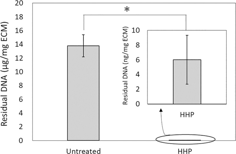
Fig 1

*p < 0.001.

Fig 3. Fluorescence images showing F-CHP staining on sections.
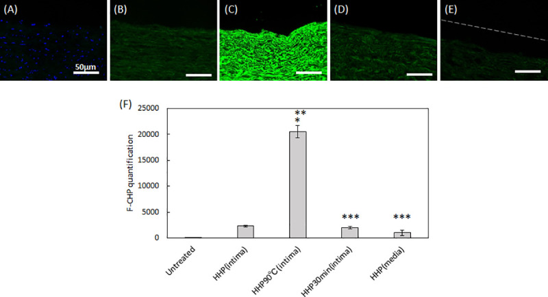
Fig 3

(A) untreated aorta, (B) HHP tunica-intima, (C) HHP 90°C tunica-intima, (D) HHP 30 min dried tunica-intima, (E) HHP tunica-media. Scale bar: 50 μm. (F) integrated F-CHP signals quantified from images of the tissue sections. The asterisk (*) indicates statistical significance in comparison with the untreated aorta (p < 0.01). Two asterisks (**) indicate statistical significance in comparison with HHP tunica-intima (p < 0.01). Three asterisks (***) indicate statistical significance in comparison with HHP 90°C tunica-intima (p < 0.01).

Fig 6.

Fig 6

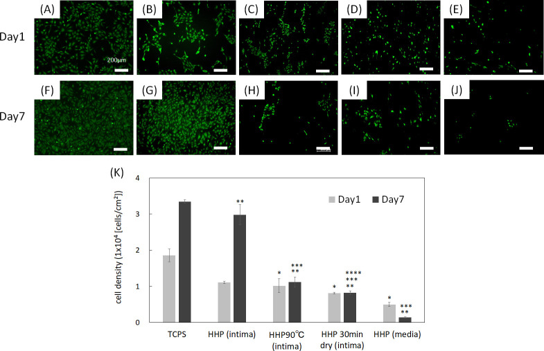
Fluorescence images of HUVECs at day 1 and day 7 on (A), (F) TCPS, (B), (G) HHP tunica-intima, (C), (H) HHP 90°C tunica-intima, (D), (I) HHP 30 min dried tunica-intima, (E), (J) HHP tunica-media. Scale bar: 200 μm. (K) Cell density. The cell density of each sample was compared each day. Asterisk (*) indicates statistical significance in comparison with TCPS on day 1 (p < 0.05). Two asterisks (**) indicate statistical significance in comparison with TCPS on day 7 (p < 0.05). Three asterisks (***) indicate statistical significance in comparison with HHP tunica-intima on day 7 (p < 0.05). Four asterisks (****) indicate statistical significance in comparison with HHP 90°C tunica-intima on day 7 (p < 0.05).

Fig 7.

Fig 7

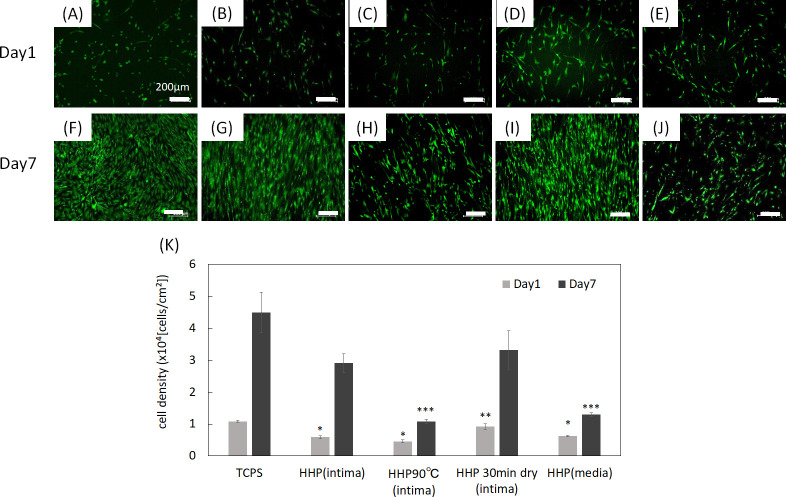
Fluorescence images of HAoSMCs on day 1 and day 7 on (A), (F) TCPS, (B), (G) HHP tunica-intima, (C), (H) HHP 90°C tunica-intima, (D), (I) HHP 30 min dried tunica-intima, (E), (J) HHP tunica-media. Scale bar: 200 μm. (K) Cell density. The cell density of each sample was compared each day. Asterisk (*) indicates statistical significance in comparison with TCPS on day 1 (p < 0.05). Two asterisks (**) indicate statistical significance in comparison with HHP 90°C tunica-intima on day 1 (p < 0.05). Three asterisks (***) indicate statistical significance in comparison with TCPS on day 7 (p < 0.05).

Fig 5.

Fig 5

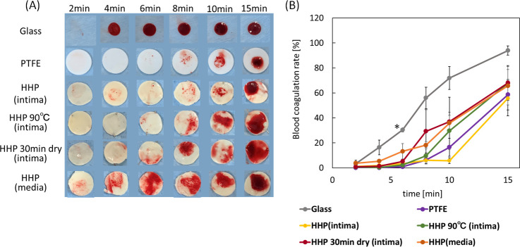
(A) Photograph of blood coagulation times of decellularized aortas with various basement membrane structures. (B) Blood coagulation rate of decellularized aortas. Error bars represent S.E. Asterisk (*) shows a significant difference (p < 0.05) between glass and PTFE, HHP (intima), HHP 90°C (intima), HHP 30 min dry (intima) at 6 min.

Fig 8.

Fig 8

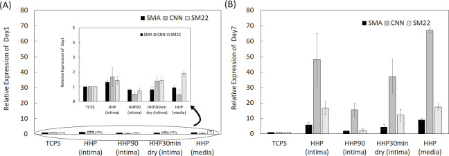
mRNA expression of SMA, CNN and SM22 in HAoSMCs on TCPS and decellularized aortas, (A) on day 1 and (B) on day 7. The relative expression of each gene was compared with all samples for statistical analysis.

Results

Porcine aortas were decellularized, and the amount of residual DNA was measured (Fig 1). The amount of DNA remaining in the HHP-treated aorta was significantly lower than that in the untreated aorta.

Fig 2(A)–2(J) show H-E and Type IV collagen-stained native aortas and decellularized aortas. The efficacy of decellularization was verified using H&E staining. No nuclei were detected in sections with decellularized aortas after HHP treatment (Fig 2(B)–2(E)). The tunica intima of the HHP-treated aorta and the HHP 30 min dried tunica-intima were observed to be similar to that of the untreated aorta (Fig 2(B) and 2(D)). HHP 90°C tunica intima had larger wave-like shapes and a flatter fibril structure than the native aorta (Fig 2(C)). In the tunica media of HHP-treated aorta, the gaps between collagen fibrils were enlarged due to the peeling of the tunica intima (Fig 2(E)). Immunostaining for type IV collagen was also performed. Type IV collagen is the main component of the basement membrane and forms networks that provide structural support for endothelial cells. The type IV collagen layer was preserved in HHP tunica intima, even after drying (Fig 2(G) and 2(I)). HHP 90°C tunica intima was weakly stained, so it is assumed that type IV collagen was denatured by the heating treatment (Fig 2(H)).

Fig 2.
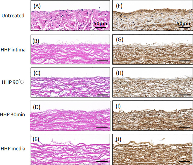
Fig 2

H-E staining (A-E), type IV collagen staining (F-J) of untreated and decellularized aortas. (A)(F) Untreated aorta, (B)(G) HHP tunica-intima, (C)(H) HHP 90°C tunica-intima, (D)(I) HHP 30 min dried tunica-intima, (E)(J) HHP tunica-media. Scale bar: 50 μm.

CHP staining, which specifically targets denatured collagen chains, was performed (Fig 3(A)–3(E)), and the fluorescence signals in the scanned images were quantified (Fig 3(F)). As expected, no fluorescence intensity except for DAPI was detected in the untreated aorta (Fig 3(A) and 3(F)). HHP tunica intima exhibited a low intensity signal, suggesting slightly denatured collagen (Fig 3(B) and 3(F)). In HHP 90°C tunica intima, a strong fluorescence signal was detected, indicating that the collagen was completely destroyed by the heating process (Fig 3(C) and 3(F)). No significant differences were observed between the fluorescence image and the intensity signal of the HHP tunica intima and that of the HHP 30 min dried tunica intima and HHP tunica media (Fig 3(D)–3(F)).

SEM was used to analyze the fiber structures of the decellularized aortas (Fig 4). The fibers were observed in magnified 3.0k SEM images (Fig 4(A)–4(E)), while the surfaces were observed in 100 SEM images (Fig 4(F)–4(J)). For HHP-treated tunica intima, the fiber bundles were oriented longitudinally and similar to in the untreated aortas (Fig 4(B) and 4(G)). As for the HHP 90°C tunica-intima, shrunken and wavy fibers were observed in the magnified images compared with HHP tunica-intima (Fig 4(C)), and smooth plane surfaces without ruggedness are shown in Fig 4(H). As for the HHP 30 min dried tunica-intima, there was not much difference in fiber structure compared to HHP tunica intima; however, numerous fine cracks were observed on the surface of HHP 30 min dried tunica-intima (Fig 4(I)). The HHP-treated tunica media exhibited a rough surface due to the peeling of the tunica intima (Fig 4(E) and 4(J)). Based on the above observations, various luminal surface structures of decellularized aortas were prepared.

Fig 4. SEM observation of surface.

Fig 4

(A)(F) Untreated aorta, (B)(G) HHP tunica-intima, (C)(H) HHP 90°C tunica-intima, (D)(I) HHP 30 min dried tunica-intima, (E)(J) HHP tunica-media. Scale bar: (A)–(E) 10 μm, (F)–(J) 500 μm.

In this study, the Lee-White test was used to examine the effects of luminal surface structure on the thrombogenicity of decellularized aortas. Fig 5(A) shows an in vitro evaluation of a blood clotting test for decellularized aortas. Glass and PTFE were used as the negative and positive controls, respectively. On glass, a blood clot formed at 4 min, while no clot formation occurred until 15 min on PTFE. As for the HHP-treated tunica-intima, no clot formation was observed until 15 min, which is almost the same result as for PTFE. As for HHP 90°C tunica intima, HHP 30 min dried tunica intima, and HHP treated tunica media, thrombus formation occurred within 10 min. The result in Fig 5(B) shows the measurement of the absorbance of the hemolyzed blood clot. The difference between the decellularized samples and PTFE was not statistically significant; in short, all HHP decellularized aortas showed a high anti-coagulability. As shown in Fig 5(B), however, HHP tunica intima showed the highest anti-coagulability among all samples at each time point.

It is widely known that endothelial cell coverage on the luminal surface of cardiovascular grafts immediately after transplantation is required for preventing thrombus formation [11,12]. Endothelial cells also regulate vessel tone, platelet and leukocyte activation, and SMC migration and proliferation [13]. In this study, HUVECs were chosen for evaluating the recellularization efficacy and cell behavior of decellularized aortas with various luminal surface structures. The attachment and proliferation of HUVECs on decellularized aortas were observed using a fluorescence microscope. The number of cells was then counted in sections from all samples, and the cell density was calculated using the counted cell numbers (Fig 6(K)). As can be seen in the results, HUVECs adhered on all samples. HUVECs proliferated well on HHP-treated tunica intima (Fig 6(B), 6(G) and 6(K)). The attachment of HUVECs on HHP 90°C tunica-intima and HHP 30 min dried tunica-intima were nearly equal to that of the HHP treated tunica intima, but the morphology of the cells was different. HUVECs on HHP tunica-intima extended, while HUVECs on HHP 90°C and HHP 30 min dried tunica-intima did not extend and showed a round shape (Fig 6(C), 6(D), 6(H) and 6(I)). It is considered that HUVECs were shed during cell culture, resulting in little cell attachment on HHP 90°C and HHP 30 min dried tunica-intima on day 7 (Fig 6(K)). As for the HHP treated tunica media, the initial adhesion of HUVECs was low, and the number of HUVECs decreased during the 7 days of cultivation (Fig 6(E), 6(J) and 6(K)).

Vascular smooth muscle cells (VSMCs) are present in the tunica media (middle layer) and have secretory ability of the major extracellular proteins, including collagen, elastin, and proteoglycans, all of which influence the mechanical properties [14]. Furthermore, VSMCs can switch between contractile and synthetic phenotypes in response to changes in the local environment [15,16]. They usually exhibit a contractile phenotype and express contractile proteins such as α-smooth muscle actin (SMA), smooth muscle calponin (CNN) and SM22 α (SM22). In the contractile phenotype, they have a low proliferative rate to maintain the ECM of the tunica media [15]. In response to vascular injury, including tissue damage, they alter their phenotype to a synthetic state and reduce the expression of contractile proteins, with increased proliferation and remodeling of the ECM to facilitate migration [1719]. In this study, the effects of the luminal surface structure of the decellularized aorta on VSMC proliferation and phenotype were investigated. Human aorta smooth muscle cells (HAoSMCs) were seeded on TCPS and decellularized aortas. The cell density was calculated by counting the number of cells on each sample (Fig 7(K)). As shown in Fig 7(A)–7(J), HAoSMCs adhered on all samples. HAoSMCs proliferated on HHP-treated tunica-intima and HHP 30 min dried tunica intima (Fig 7(B), 7(D), 7(G), 7(I) and 7(K)). The initial attachment of HAoSMCs onto HHP 90°C tunica-intima and HHP tunica media was nearly equal to that of HHP tunica intima, but the cells did not proliferate (Fig 7(C), 7(E), 7(H), 7(J) and 7(K)).

The expression levels of SMA, CNN, and SM22 from HAoSMCs on the samples, which were expressed in the contractile phenotype, were examined by qRT-PCR analysis. On day 1, HAoSMCs on all samples showed a low gene expression (Fig 8(A)). In contrast, on day 7, the expression ratio of the contractile phenotype genes was high, except for tunica intima heated at 90°C (Fig 8(B)).

Discussion

To identify the effect of the luminal surface structure of the decellularized aorta on thrombus formation and cell behavior, porcine aortas were decellularized using the HHP method, which has been previously reported to have good in vivo performance [7]. Their luminal surfaces were then processed by heating, drying, and peeling. The luminal surface structure and components were evaluated using type IV collagen immunostaining, CHP staining, and SEM analysis. Type IV collagen immunostaining was performed to evaluate the maintenance of the basement membrane of the decellularized aorta. HHP decellularization did not affect type IV collagen, but type IV collagen was not stained after the heating process. This result indicates that type IV collagen was denatured and gelatinized during the heating process to 90°C. CHP staining is a method used to examine the unfolding of collagen molecules in decellularized tissues [20]. Compared with the untreated aorta, slightly unfolded collagen was observed when aortas were decellularized using the HHP method. The image and quantification analysis of HHP 90°C heated tunica intima clearly showed the denaturation of collagen molecules. SEM observation was performed to observe the fiber structure of the decellularized aorta. In the HHP 90°C heated tunica intima, the fibers appeared shrunken and wavy because the collagen fibers had been denatured by heat treatment. This result correlates with a previous report which showed that collagen fibers shrink, and that their fiber diameters increase when they are treated with temperatures at or above 50°C [21,22].

The effect of the luminal surface of the decellularized aorta on thrombus formation was assessed by a blood clotting test. The results showed that the anti-coagulability was higher for HHP-treated tunica-intima and lower for decellularized aortas with disordered luminal surface structures (HHP 90°C tunica-intima, HHP 30 min dried tunica-intima, HHP tunica-media). Previously, it was reported that damaging the luminal surface of grafts by drying decreased graft patency after transplantation [23]. Therefore, this in vitro result correlates with the in vivo results and indicates that the more similar the luminal surface structure is to the native aorta, the higher the anti-coagulant property.

The effect of the luminal surface of the decellularized aorta on cell behavior and gene expression was evaluated. Endothelial cells and smooth muscle cells are the main cellular components of the aorta and interact with each other to maintain the function and mechanical properties of the vessel [24]. It is important to evaluate whether these vascular cells, which play essential roles in vivo, recognize the place where they should exist and proliferate and express their functions properly. HUVECs and HAoSMCs were seeded on decellularized aortas, and the phenotype of HAoSMCs was evaluated by qRT-PCR. Regarding the qRT-PCR results on day 1, it was assumed that the gene expression ratio was low in all samples because the phenotype of the adhered HAoSMCs could not have switched their phenotype in such a short period. HUVECs adhered and proliferated on HHP decellularized tunica-intima because the luminal surface structure and basement membrane were well maintained. HAoSMCs showed high proliferation as they recognized the luminal surface as tunica intima. This cell behavior seems to be correlated with the event of neointima hyperplasia occurring, because the population of SMCs migrates from the media and proliferates within the intima during neointima formation [25,26]. However, the expression of HAoSMC contractile phenotype marker genes was detected in the results of qRT-PCR on day 7. It is assumed that the cell density exceeded the capacity of the luminal surface, so that HAoSMCs shifted the synthetic phenotype to the contractile phenotype. As for the HHP 90°C heated tunica intima, in which luminal surface structure and collagen were denatured, initial adhesion of both HUVECs and HAoSMCs was low and did not proliferate. This may be because the luminal surface structure was disordered, and type IV collagen was denatured by the heating process. As for the HHP 30 min dried tunica intima, in which the luminal surface structure was cracked by drying, the number of attached HUVECs during cell culturing was low, while HAoSMCs adhered well and proliferated. Based on this result, it was suggested that drying out the luminal surface of the decellularized aorta may lead to neointima hyperplasia. This in vitro result correlates with a previous in vivo study that showed vessel occlusion when the intima were dried before implantation [23]. As for the HHP tunica media, initial adhesion of HUVECs was observed, but they were shed during cell culturing. This may be because the basement membrane, which supports endothelial cells, was removed. HAoSMCs cultured on HHP tunica media did not proliferate. This phenomenon is similar to the behavior of vascular smooth cells in vivo for which it has been reported that smooth muscle cells within media exhibit the stable contractile type with low proliferative capacity [15]. Thus, HAoSMCs recognized the luminal surface structure and component of tunica media and exhibited a stable contractile type with low proliferative capacity in vitro. These results suggest that HUVECs and HAoSMCs recognize the luminal surface and component of the decellularized aorta and proliferate and/or express their functions properly. Therefore, good maintenance of the luminal surface structure of the decellularized aorta so that it is similar to that of the native aorta is one of the key factors in preparing decellularized cardiovascular applications.

Conclusion

In the present study, decellularized aortas with different luminal surface structures were prepared to investigate the effects of their structures on thrombus formation and cell behavior. The results of the blood clotting test showed that the more similar the luminal surface structure is to that of the native aorta, the higher the anti-coagulant property. From the results of cell seeding and qRT-PCR, it was found that endothelial cells and smooth muscle cells recognized the luminal surface structure and components of the decellularized aorta and regulated adhesion, proliferation, and functional expression. These results provide useful insights into future developments of decellularized cardiovascular applications.

Supporting information

S1 File. Quantitative analysis of residual dsDNA.

(XLSX)

S2 File. F-CHP intensity.

(XLSX)

S3 File. Blood coagulation rate.

(XLSX)

S4 File. HUVEC cell density.

(XLSX)

S5 File. HAoSMC cell density.

(XLSX)

S6 File. qRT-PCR_HAoSMC day 1.

(XLSX)

S7 File. qRT-PCR_HAoSMC day7.

(XLSX)

Data Availability

All relevant data are within the manuscript and its Supporting Information files.

Funding Statement

This work was supported in part by a Grant-Aid for Scientific Research (B) (16H03180) from JSPS, the Creative Scientific Research of the Viable Material via Integration of Biology and Engineering from MEXT and the Cooperative Research Project of Research Center for Biomedical Engineering from MEXT.

References

  • 1.Virani SS, Alonso A, Benjamin EJ, Bittencourt MS, Callaway CW, Carson AP, et al. Heart Disease and Stroke Statistics-2020 Update: A Report From the American Heart Association. Circulation. 2020;141(9):E139–E596. 10.1161/CIR.0000000000000757 [DOI] [PubMed] [Google Scholar]
  • 2.Mendis S, Davis S, Norrving B. Organizational Update The World Health Organization Global Status Report on Noncommunicable Diseases 2014; One More Landmark Step in the Combat Against Stroke and Vascular Disease. Stroke. 2015;46(5):E121–E2. 10.1161/STROKEAHA.115.008097 [DOI] [PubMed] [Google Scholar]
  • 3.Nakamura N, Kimura T, Kishida A. Overview of the Development, Applications, and Future Perspectives of Decellularized Tissues and Organs. Acs Biomaterials Science & Engineering. 2017;3(7):1236–44. [DOI] [PubMed] [Google Scholar]
  • 4.Pashneh-Tala S, MacNeil S, Claeyssens F. The Tissue-Engineered Vascular Graft-Past, Present, and Future. Tissue Engineering Part B-Reviews. 2016;22(1):68–100. 10.1089/ten.teb.2015.0100 [DOI] [PMC free article] [PubMed] [Google Scholar]
  • 5.Naso F, Gandaglia A. Different approaches to heart valve decellularization: A comprehensive overview of the past 30years. Xenotransplantation. 2018;25(1). [DOI] [PubMed] [Google Scholar]
  • 6.Moroni F, Mirabella T. Decellularized matrices for cardiovascular tissue engineering. Am J Stem Cells. 2014;3(1):1–20. [PMC free article] [PubMed] [Google Scholar]
  • 7.Funamoto S, Nam K, Kimura T, Murakoshi A, Hashimoto Y, Niwaya K, et al. The use of high-hydrostatic pressure treatment to decellularize blood vessels. Biomaterials. 2010;31(13):3590–5. 10.1016/j.biomaterials.2010.01.073 [DOI] [PubMed] [Google Scholar]
  • 8.Wu PL, Kimura T, Tadokoro H, Nam K, Fujisato T, Kishida A. Relation between the tissue structure and protein permeability of decellularized porcine aorta. Materials Science & Engineering C-Materials for Biological Applications. 2014;43:465–71. 10.1016/j.msec.2014.06.041 [DOI] [PubMed] [Google Scholar]
  • 9.Wu PL, Nakamura N, Kimura T, Nam K, Fujisato T, Funamoto S, et al. Decellularized porcine aortic intima-media as a potential cardiovascular biomaterial. Interactive Cardiovascular and Thoracic Surgery. 2015;21(2):189–94. 10.1093/icvts/ivv113 [DOI] [PubMed] [Google Scholar]
  • 10.Kobayashi M, Ohara M, Hashimoto Y, Nakamura N, Fujisato T, Kimura T, et al. In vitro evaluation of surface biological properties of decellularized aorta for cardiovascular use. Journal of Materials Chemistry B; 2020. p. 10977–89. 10.1039/d0tb01830a [DOI] [PubMed] [Google Scholar]
  • 11.Pearson JD. ENDOTHELIAL-CELL FUNCTION AND THROMBOSIS. Baillieres Clinical Haematology. 1994;7(3):441–52. 10.1016/s0950-3536(05)80092-7 [DOI] [PubMed] [Google Scholar]
  • 12.van Hinsbergh VWM. Endothelium-role in regulation of coagulation and inflammation. Seminars in Immunopathology. 2012;34(1):93–106. 10.1007/s00281-011-0285-5 [DOI] [PMC free article] [PubMed] [Google Scholar]
  • 13.Yau JW, Teoh H, Verma S. Endothelial cell control of thrombosis. Bmc Cardiovascular Disorders. 2015;15. 10.1186/s12872-015-0124-z [DOI] [PMC free article] [PubMed] [Google Scholar]
  • 14.Matsumoto T, Nagayama K. Tensile properties of vascular smooth muscle cells: Bridging vascular and cellular biomechanics. Journal of Biomechanics. 2012;45(5):745–55. 10.1016/j.jbiomech.2011.11.014 [DOI] [PubMed] [Google Scholar]
  • 15.Owens GK, Kumar MS, Wamhoff BR. Molecular regulation of vascular smooth muscle cell differentiation in development and disease. Physiological Reviews. 2004;84(3):767–801. 10.1152/physrev.00041.2003 [DOI] [PubMed] [Google Scholar]
  • 16.Davis-Dusenbery Brandi N., Wu C, Hata A. Micromanaging vascular smoothe muscle cell differentiation and phenotypic modulation. ARTERIOSCLEROSIS THROMBOSIS AND VASCULAR BIOLOGY. 2011;31(11):2370–2377. [DOI] [PMC free article] [PubMed] [Google Scholar]
  • 17.Davis BN, Hilyard AC, Nguyen PH, Lagna G, Hata A. Induction of MicroRNA-221 by Platelet-derived Growth Factor Signaling Is Critical for Modulation of Vascular Smooth Muscle Phenotype. Journal of Biological Chemistry. 2009;284(6):3728–38. 10.1074/jbc.M808788200 [DOI] [PMC free article] [PubMed] [Google Scholar]
  • 18.Durham AL, Speer MY, Scatena M, Giachelli CM, Shanahan CM. Role of smooth muscle cells in vascular calcification: implications in atherosclerosis and arterial stiffness. Cardiovascular Research. 2018;114(4):590–600. 10.1093/cvr/cvy010 [DOI] [PMC free article] [PubMed] [Google Scholar]
  • 19.Milutinovic A, Suput D, Zorc-Pleskovic R. Pathogenesis of atherosclerosis in the tunica intima, media, and adventitia of coronary arteries: An updated review. Bosnian Journal of Basic Medical Sciences. 2020;20(1):21–30. 10.17305/bjbms.2019.4320 [DOI] [PMC free article] [PubMed] [Google Scholar]
  • 20.Zitnay JL, Li Y, Qin Z, San BH, Depalle B, Reese SP, et al. Molecular level detection and localization of mechanical damage in collagen enabled by collagen hybridizing peptides. Nature Communications. 2017;8. 10.1038/ncomms14913 [DOI] [PMC free article] [PubMed] [Google Scholar]
  • 21.Hayashi K, Thabit G, Massa KL, Bogdanske JJ, Cooley AJ, Orwin JF, et al. The effect of thermal heating on the length and histologic properties of the glenohumeral joint capsule. American Journal of Sports Medicine. 1997;25(1):107–12. 10.1177/036354659702500121 [DOI] [PubMed] [Google Scholar]
  • 22.Kirsch KM, Zelickson BD, Zachary CB, Tope WD. Ultrastructure of collagen thermally denatured by microsecond domain pulsed carbon dioxide laser. Archives of Dermatology. 1998;134(10):1255–9. 10.1001/archderm.134.10.1255 [DOI] [PubMed] [Google Scholar]
  • 23.Kurokawa S, Hashimoto Y, Funamoto S, Yamashita A, Yamazaki K, Ikeda T, et al. In vivo recellularization of xenogeneic vascular grafts decellularized with high hydrostatic pressure method in a porcine carotid arterial interpose model. bioRxiv. [DOI] [PMC free article] [PubMed] [Google Scholar]
  • 24.Truskey GA. Endothelial Cell Vascular Smooth Muscle Cell Co-Culture Assay For High Throughput Screening Assays For Discovery of Anti-Angiogenesis Agents and Other Therapeutic Molecules. Int J High Throughput Screen. 2010;2010(1):171–181. 10.2147/IJHTS.S13459 [DOI] [PMC free article] [PubMed] [Google Scholar]
  • 25.Christie DB, Kang J, Ashley DW, Mix W, Lochner FK, Solis MM, et al. Accelerated migration and proliferation of smooth muscle cells cultured from neointima induced by a vena cava filter. American Surgeon. 2006;72(6):491–6. 10.1177/000313480607200606 [DOI] [PubMed] [Google Scholar]
  • 26.Hoglund VJ, Dong XR, Majesky MW. Neointima Formation A Local Affair. Arteriosclerosis Thrombosis and Vascular Biology. 2010;30(10):1877–9. 10.1161/ATVBAHA.110.211433 [DOI] [PMC free article] [PubMed] [Google Scholar]

Decision Letter 0

Michiya Matsusaki

10 Feb 2021

PONE-D-21-01350

Effect of luminal surface structure of decellularized aorta on thrombus formation and cell behavior

PLOS ONE

Dear Dr. Kishida,

Thank you for submitting your manuscript to PLOS ONE. After careful consideration, we feel that it has merit but does not fully meet PLOS ONE’s publication criteria as it currently stands. Therefore, we invite you to submit a revised version of the manuscript that addresses the points raised during the review process.

Please carefully check the comments from reviewers and make a revision to satisfy it. I will add an additional reviewer to correctly judge your revision.

Please submit your revised manuscript by Mar 25 2021 11:59PM. If you will need more time than this to complete your revisions, please reply to this message or contact the journal office at plosone@plos.org. When you're ready to submit your revision, log on to https://www.editorialmanager.com/pone/ and select the 'Submissions Needing Revision' folder to locate your manuscript file.

Please include the following items when submitting your revised manuscript:

  • A rebuttal letter that responds to each point raised by the academic editor and reviewer(s). You should upload this letter as a separate file labeled 'Response to Reviewers'.

  • A marked-up copy of your manuscript that highlights changes made to the original version. You should upload this as a separate file labeled 'Revised Manuscript with Track Changes'.

  • An unmarked version of your revised paper without tracked changes. You should upload this as a separate file labeled 'Manuscript'.

If you would like to make changes to your financial disclosure, please include your updated statement in your cover letter. Guidelines for resubmitting your figure files are available below the reviewer comments at the end of this letter.

If applicable, we recommend that you deposit your laboratory protocols in protocols.io to enhance the reproducibility of your results. Protocols.io assigns your protocol its own identifier (DOI) so that it can be cited independently in the future. For instructions see: http://journals.plos.org/plosone/s/submission-guidelines#loc-laboratory-protocols

We look forward to receiving your revised manuscript.

Kind regards,

Michiya Matsusaki

Academic Editor

PLOS ONE

Journal Requirements:

When submitting your revision, we need you to address these additional requirements.

1. Please ensure that your manuscript meets PLOS ONE's style requirements, including those for file naming. The PLOS ONE style templates can be found at

https://journals.plos.org/plosone/s/file?id=wjVg/PLOSOne_formatting_sample_main_body.pdf and

https://journals.plos.org/plosone/s/file?id=ba62/PLOSOne_formatting_sample_title_authors_affiliations.pdf

[Note: HTML markup is below. Please do not edit.]

Reviewers' comments:

Reviewer's Responses to Questions

Comments to the Author

1. Is the manuscript technically sound, and do the data support the conclusions?

The manuscript must describe a technically sound piece of scientific research with data that supports the conclusions. Experiments must have been conducted rigorously, with appropriate controls, replication, and sample sizes. The conclusions must be drawn appropriately based on the data presented.

Reviewer #1: Yes

Reviewer #2: No

**********

2. Has the statistical analysis been performed appropriately and rigorously?

Reviewer #1: Yes

Reviewer #2: N/A

**********

3. Have the authors made all data underlying the findings in their manuscript fully available?

The PLOS Data policy requires authors to make all data underlying the findings described in their manuscript fully available without restriction, with rare exception (please refer to the Data Availability Statement in the manuscript PDF file). The data should be provided as part of the manuscript or its supporting information, or deposited to a public repository. For example, in addition to summary statistics, the data points behind means, medians and variance measures should be available. If there are restrictions on publicly sharing data—e.g. participant privacy or use of data from a third party—those must be specified.

Reviewer #1: Yes

Reviewer #2: Yes

**********

4. Is the manuscript presented in an intelligible fashion and written in standard English?

PLOS ONE does not copyedit accepted manuscripts, so the language in submitted articles must be clear, correct, and unambiguous. Any typographical or grammatical errors should be corrected at revision, so please note any specific errors here.

Reviewer #1: Yes

Reviewer #2: No

**********

5. Review Comments to the Author

Please use the space provided to explain your answers to the questions above. You may also include additional comments for the author, including concerns about dual publication, research ethics, or publication ethics. (Please upload your review as an attachment if it exceeds 20,000 characters)

Reviewer #1: Dear Authors,

(Ms. No.; PONE-D-21-01350)

In this study, the authors investigate the thrombus formation and the in vitro behaviors of vascular cells on decellularized aorta focusing on the surface structure. The in vitro results of your study are indicating that vascular events should be affected by the surface structure of decellularized aorta. This finding will give us useful knowledge for designing suitable decellularized tissue for clinical applications

I would like to suggest the authors to clarify the following criticisms.

1) Introduction, line 53 - 56

Could you please add the details of why you hit upon your hypothesis focusing on the luminal surface structure of the HHP treated aorta. Many conceivable factors might affect the biological responses to HHP treated aortas.

I recommend making the distinction between molecular structure and tissue structure.

2) 2.1. Preparation of decellularized porcine aorta

Please describe more details about the treatments after decellularization. Do you have any reason why you decided 90 degrees for heating treatment? Did you dry decellularized aorta in the air for 30 min? How did you peel off the inner membrane from the decellularized aorta with?

3) 2.3. Histological evaluation of decellularized aortas

My concern is that the reagents used in the process to make the section for collagen staining might affect the structure of the collagen molecule. Is the collagen molecule not denatured by xylene, ethanol, and paraffin? Is the frozen section not suitable for this experiment?

4) 2.6. Evaluation of thrombogenicity of decellularized aortas

What from did you obtain the whole blood for this evaluation? Please add the information about approvals if you obtained the whole blood from human donors or animals.

5) 2.7 Cell seeding

Please indicate the size of decellularized aortas. I think that TAKARA Bio Inc. is located in Shiga prefecture, not Tokyo.

6) 2.9. qPCR

The authors forgot to indicate the Table of primers. There is no Table in the manuscript. I previously make an apology if I overlooked it.

7) 2.10. Statistical analysis

As you know, the one-way ANOVA is suitable for evaluating the significant differences among more than three groups.

8) 3. Results and Discussion, line 154

Please remove “Discussion” because you subsequently prepared “4. Discussion”.

9) 3. Results, line 210-211

“Glass and PTFE were used as negative and positive controls, respectively.”

Is glass a positive control, right? I think the position between Glass and PTFE should be placed opposite. Please add the further details (manufacture and name) of the Glass and PTFE you used.

10) 3. Results, line 226 – 227

“It is widely known that endothelial cell seeding on luminal surface of cardiovascular grafts are required to prevent thrombus formation (9, 10).”

This sentence is complicated for me. What did you mean by “cell seeding”? Is “cell seeding” meaning that cells are seeded on cardiovascular grafts ex vivo?

Also, “,,,,,grafts are” should be corrected to “,,,,,,grafts is” in this sentence.

11) Figure 3

Were the parameters of CLSM fixed for the observation of all samples? How did you adjust and calibrate the fluorescence intensity for all observations? The fluorescence intensity should be calibrated because the tissue has self-fluorescence and you can control the fluorescence intensity by changing the parameter for CLSM observation.

I recommend doing the quantitative evaluation of collagen denaturation using the fluorescence intensity of these pictures.

12) Fig. 6

I agree that both types of cells recognized “something” on decellularized aortas and the “something” is relating to the denaturation of the collagen molecule. But, I cannot believe that cells directly recognize the denatured collagen because the amino acid sequence of collagen did not change by the denaturation process. The initial adhesion of HUVECs is similar between HHPi and HHPi-90 degrees, but the proliferation behavior of adherent HUVECs was completely different. This means that HUVECs recognize the adhesive sites in collagen molecule on both aortas as well. Then, HUVECs was affected by “something” and was not able to proliferate on HHPi-90 degrees. Do you have any speculation about “something”? Was the tissue structure of HHPi-90 degrees maintained during cell culture for 7 days?

13) Fig. 8

Please add the enlarged view in Fig. 8 (A). It was so difficult to recognize a difference in the gene expression level of all samples. Please perform the statistical analysis. In my opinion, you can remove this figure if you do not use it for discussion.

14) 4. Discussion, line 314-315

“Cell seeding on decellularized aortas and the evaluation of mRNA expression of those cells by RT-PCR were performed.”

Does “those cells” mean both HUVECs and AOSMCs? If so, the gene expression analysis of HUVECs should be added to this manuscript.

15)

There are several incomprehensible sentences and grammatical errors in this manuscript. [For example, line 304-308 and 310-314]

I recommend that this manuscript should be checked carefully or get the English proofreading.

Reviewer #2: The manuscript studies the effect of luminal surface structure on thrombus formation and cell behavior using HHP decellularized aortas. The luminal surface structure was disrupted by heating, drying and peeling. The resultant surfaces were analyzed by immunohistological staining, collagen hybridizing peptide (CHP) staining and scanning electron microscopy (SEM) analysis. Overall, the manuscript has a poor-written language and requires major revision in several parts. It lacks novelty, and the data is not interesting. Here are some points that may improve the quality of the manuscript:

1) The lines from 59 to 66 are copied from the abstract.

2) Information (company-cat number for antibody) for chemicals reagents and instruments need to be mentioned in the materials and methods.

3) How does the inner membrane peel off? explain more on methods.

4) Figures legends should not be in the manuscript.

5) Why did the Author investigate the binding and proliferation of SMC in the lumen? These cells reside in medial layer.

6) Scale bar in figure 2 is absent.

7) CF-CHP staining images in Figure 3 have different contrast, limiting meaningful comparison between images.

8) In line 197, the authors mentioned that SEM analysis showed thicker fibers of HHP 90 ºC tunica-intima. Quantification of fiber diameters from SEM images among the samples should be provided with statistical analysis.

9) In line 199, from Fig. 4D and I, the authors indicated more gap between fibers on HHP 30 min dried tunica intima. The gap distance among the samples has to be quantified and analyzed statistically.

10) In line 226, the paragraph has only one sentence.

11) The images in figure 4G-I have different contrast compared with other images in the same figure.

12) Statistical analysis in Figures 6K, 7K, and 8 is absent.

13) The manuscript has grammatical errors, several too-short sentences, and the language needs revision.

[NOTE: If reviewer comments were submitted as an attachment file, they will be attached to this email and accessible via the submission site. Please log into your account, locate the manuscript record, and check for the action link "View Attachments". If this link does not appear, there are no attachment files.]

While revising your submission, please upload your figure files to the Preflight Analysis and Conversion Engine (PACE) digital diagnostic tool, https://pacev2.apexcovantage.com/. PACE helps ensure that figures meet PLOS requirements. To use PACE, you must first register as a user. Registration is free. Then, login and navigate to the UPLOAD tab, where you will find detailed instructions on how to use the tool. If you encounter any issues or have any questions when using PACE, please email PLOS at figures@plos.org. Please note that Supporting Information files do not need this step.

PLoS One. 2021 May 17;16(5):e0246221. doi: 10.1371/journal.pone.0246221.r002

Author response to Decision Letter 0


30 Mar 2021

Response to reviewers

Title: Effect of luminal surface structure of decellularized aorta on thrombus formation and cell behavior.

Thank you for your information together with feedback from the reviewers of the above paper. We have read reviewer’s comments very carefully. We have revised the manuscript according to the comments and suggestions of reviewers.

Reviewer #1: Dear Authors,

(Ms. No.; PONE-D-21-01350)

In this study, the authors investigate the thrombus formation and the in vitro behaviors of vascular cells on decellularized aorta focusing on the surface structure. The in vitro results of your study are indicating that vascular events should be affected by the surface structure of decellularized aorta. This finding will give us useful knowledge for designing suitable decellularized tissue for clinical applications

I would like to suggest the authors to clarify the following criticisms.

Answer:

We appreciate your kind comment. We response the reviewer’s questions point by point as below.

1) Introduction, line 53 - 56

Could you please add the details of why you hit upon your hypothesis focusing on the luminal surface structure of the HHP treated aorta. Many conceivable factors might affect the biological responses to HHP treated aortas.

I recommend making the distinction between molecular structure and tissue structure.

Answer:

We have previously reported that reendothelialization and high patency were observed in HHP decellularized aorta, when decellularized aortas prepared by different decellularization methods were implanted in vivo (1). To find the factors related to good in vivo performance, we previously evaluated mechanical properties and protein permeability, and it was found that HHP decellularized aortas have similar properties and functions similar to those of native aortas (2), (3). Furthermore, observation of the luminal surface structure showed that structure of HHP decellularized aorta was maintained, in contrast to other decellularized aortas prepared by different methods. Therefore, we proposed hypothesis that vascular endothelial cell recruitment and anti-thrombogenicity occur on the HHP treated aorta since its luminal surface structure on aorta is well maintained. Thus, we prepared HHP decellularized aortas with different surface structure and evaluated the effects on vascular cell behavior.

We added the reason why we hit upon the hypothesis in P.4, line 57-62.

Reference

1) Funamoto S, Nam K, Kimura T, Murakoshi A, Hashimoto Y, Niwaya K, et al. The use of high-hydrostatic pressure treatment to decellularize blood vessels. Biomaterials. 2010;31(13): 3590-3595. DOI: 10.1016/j.biomaterials.2010.01.073.

2) Wu PL, Kimura T, Tadokoro H, Nam K, Fujisato T, Kishida A. Relation between the tissue structure and protein permeability of decellularized porcine aorta. Mater Sci Eng C Mater Biol Appl. 2014;43: 465-471. DOI: 10.1016/j.msec.2014.06.041.

3) Wu PL, Nakamura N, Kimura T, Nam K, Fujisato T, Funamoto S, et al. Decellularized porcine aortic intima-media as a potential cardiovascular biomaterial. Interact Cardiovasc Thorac Surg. 2015;21(2): 189-194. DOI: 10.1093/icvts/ivv113.

2) 2.1. Preparation of decellularized porcine aorta

Please describe more details about the treatments after decellularization. Do you have any reason why you decided 90 degrees for heating treatment? Did you dry decellularized aorta in the air for 30 min? How did you peel off the inner membrane from the decellularized aorta with?

Answer:

Since it is known that collagen denatures at temperatures above 50°C, we firstly tried to heat samples

at 60°C which was insufficient for collagen denaturation at fiber structure and CHP staining. So, we

set the temperature at 90°C to completely damage collagen fibers. As for the 30 min dried tunica-intima, disked HHP decellularized tunica-intima were air-dried in a clean bench for 30 min with the lumen surfaces upward. To prepare tunica media, the outer membrane of the decellularized HHP aorta was held with tweezer while inner membrane was peeled off with another tweezer.

We added the detailed methods of preparing HHP 30 min dried decellularized aorta and HHP tunica-media in P.7, line 111-112 and 114-116.

3) 2.3. Histological evaluation of decellularized aortas

My concern is that the reagents used in the process to make the section for collagen staining might affect the structure of the collagen molecule. Is the collagen molecule not denatured by xylene, ethanol, and paraffin? Is the frozen section not suitable for this experiment?

Answer:

Since no fluorescence intensity except DAPI was detected in untreated aorta from the results of F-CHP staining, we believe that collagen molecule was not denatured by xylene, ethanol and paraffin. We consider that this is because both untreated aorta and decellularized aortas were fixed using glutaraldehyde before staining.

4) 2.6. Evaluation of thrombogenicity of decellularized aortas

What from did you obtain the whole blood for this evaluation? Please add the information about approvals if you obtained the whole blood from human donors or animals.

Answer:

 We apologize for not specifying it in the manuscript. Porcine whole blood was purchased from local slaughterhouse. This product is for research experiment and no need approvals. We added “Materials” in the “Materials and Methods” section in P.5-6, line 75-96.

5) 2.7 Cell seeding

Please indicate the size of decellularized aortas. I think that TAKARA Bio Inc. is located in Shiga prefecture, not Tokyo.

Answer:

As written in P.6, line 100-101, the trimmed aortas were cut along the longitudinal direction and were fabricated with an inner diameter of 15 mm with a hollow punch. Therefore, “the disked aorta” which is mentioned in Section 2.7 Cell seeding indicates 15 mm diameter punched aortas. We added the word “inner diameter 15 mm” in P.10, line 167.

As you have pointed out, we revised the location of TAKARA Bio Inc. in P.6, line 93. Thank you for your correction.

6) 2.9. qPCR

The authors forgot to indicate the Table of primers. There is no Table in the manuscript. I previously make an apology if I overlooked it.

Answer:

We apologize for not indicating the Table of primers in the manuscript. We added the list of primers used in this experiment as Table 1 in P11, line 186.

7) 2.10. Statistical analysis

As you know, the one-way ANOVA is suitable for evaluating the significant differences among more than three groups.

Answer:

We performed one-way ANOVA, Tukey’s multiple comparison test in Figure 3(F), 6(K), 7(K) and 8(A)(B). In Figure 3(F), the F-CHP intensity was compared among different groups. In Figure 6(K) and 7(K), the cell density of each sample was compared by day. We added the symbols in those figures and the meaning of symbols were written in the legend of each figure. For figure 8(A) and (B), the relative expression of each gene was compared by samples, but the difference was not statistically significant. According to the above statistical analysis, we revised the sentence in the section of “Statistical Analysis” in P.12, line 189-195.

8) 3. Results and Discussion, line 154

Please remove “Discussion” because you subsequently prepared “4. Discussion”.

Answer:

Noted. We deleted the “Discussion” in P.12, line 197. Thank you for your correction.

9) 3. Results, line 210-211

“Glass and PTFE were used as negative and positive controls, respectively.”

Is glass a positive control, right? I think the position between Glass and PTFE should be placed opposite. Please add the further details (manufacture and name) of the Glass and PTFE you used.

Answer:

For blood clotting test, glass and polytetrafluoroethylene (PTFE) were used as a negative and positive control, respectively, since the absence of blood clot is considered as positive. These materials are often used as reference materials in studies of blood coagulation (4), (5). The exposure of blood to glass surface which has negatively charged surface greatly shortens the clotting time, since contact of blood with glass surface initiate contact activation of the platelets and Factor XII. On the other hand, PTFE is widely used as a biomaterial for blood-contacting medical devices because of its properties, such as good biocompatibility, chemical stability and anti-cellular adhesiveness.

We added the details (manufacture and name) of Glass and PTFE in section “Evaluation of thrombogenicity of decellularized aortas.” in P.9-10, line 150-151 and 156-157.

Reference

4) Sperling C, Fischer M, Maitz MF, Werner C. Blood coagulation on biomaterials requires the combination of distinct activation processes. Biomaterials. 2009;30(27):4447-56.

5) Li JM, Singh MJ, Nelson PR, Hendricks GM, Itani M, Rohrer MJ, et al. Immobilization of human thrombomodulin to expanded polytetrafluoroethylene. Journal of Surgical Research. 2002;105(2):200-8.

10) 3. Results, line 226 – 227

“It is widely known that endothelial cell seeding on luminal surface of cardiovascular grafts are required to prevent thrombus formation (9, 10).”

This sentence is complicated for me. What did you mean by “cell seeding”? Is “cell seeding” meaning that cells are seeded on cardiovascular grafts ex vivo?

Also, “,,,,,grafts are” should be corrected to “,,,,,,grafts is” in this sentence.

Answer:

We apologize for confusing you. We revised above sentence as “It is widely known that endothelial cell coverage on luminal surface of cardiovascular grafts immediately after transplantation are required to prevent thrombus formation” in P.17, line 272-274.

As you mentioned, we revised the “are” to “is” in P.17, line 273. Thank you for your correction.

11) Figure 3

Were the parameters of CLSM fixed for the observation of all samples? How did you adjust and calibrate the fluorescence intensity for all observations? The fluorescence intensity should be calibrated because the tissue has self-fluorescence and you can control the fluorescence intensity by changing the parameter for CLSM observation.

I recommend doing the quantitative evaluation of collagen denaturation using the fluorescence intensity of these pictures.

Answer:

We used fluorescence microscope (BZ-X710, Keyence Corp., Osaka, Japan)), not CLSM. The photos of Figure 3 were taken at same exposure time, 1/40 sec. 

According to your suggestions, F-CHP staining was analyzed in ImageJ software using measurements of the mean intensity and area of all remaining pixels after a background subtraction. The signal quantification was shown as integrated intensity (the mean intensity times area). We added this graph as Figure 3(F) in the manuscript.

Figure. The integrated F-CHP signals quantifies from images of tissue section. Numbers are presented as mean + standard deviation. The * sign indicates statistical significance in comparison to the untreated aorta (p < 0.01). The ** sign indicates statistical significance in comparison to the HHP (intima) (p < 0.01). The *** sign indicates statistical significance in comparison to the HHP 90°C (intima) (p < 0.01).

12) Fig. 6

I agree that both types of cells recognized “something” on decellularized aortas and the “something” is relating to the denaturation of the collagen molecule. But, I cannot believe that cells directly recognize the denatured collagen because the amino acid sequence of collagen did not change by the denaturation process. The initial adhesion of HUVECs is similar between HHPi and HHPi-90 degrees, but the proliferation behavior of adherent HUVECs was completely different. This means that HUVECs recognize the adhesive sites in collagen molecule on both aortas as well. Then, HUVECs was affected by “something” and was not able to proliferate on HHPi-90 degrees. Do you have any speculation about “something”? Was the tissue structure of HHPi-90 degrees maintained during cell culture for 7 days?

Answer:

We consider that vascular cells seeded on each sample recognized the structure and component of

luminal surface, and regulated adhesion, proliferation and functional expression. As for HHP intima and HHP 90°C intima, the number of adhered HUVECs was almost the same, but the morphology of them was different. HUVECs on HHP intima extended, while HUVECs on HHP 90°C intima didn’t extend and showed round shape. This phenomenon occurred on HHP 30min dried intima and tunica-media. Therefore, we assumed that HUVECs on HHP 90°C sloughed off during cell culture, so that the total number of HUVECs on day7 was few. We added above sentences in the manuscript in P.17, line 281-287.

Also, the tissue structure of HHP90°C remained same during cell culture.

13) Fig. 8

Please add the enlarged view in Fig. 8 (A). It was so difficult to recognize a difference in the gene expression level of all samples. Please perform the statistical analysis. In my opinion, you can remove this figure if you do not use it for discussion.

Answer:

We added the enlarged view in Figure 8(A). The sentences about Fig 8(A) were mentioned in P.23, line 375-377.

14) 4. Discussion, line 314-315

“Cell seeding on decellularized aortas and the evaluation of mRNA expression of those cells by RT-PCR were performed.”

Does “those cells” mean both HUVECs and AOSMCs? If so, the gene expression analysis of HUVECs should be added to this manuscript.

Answer:

We apologize again for confusing you. We did not perform the gene expression analysis of HUVECs. Therefore, we revised the sentence as “HUVEC and HAoSMCs were seeded on decellularized aortas, and the phenotype of HAoSMCs was evaluated by qRT-PCR.” in P.22-23, line 373-374.

15)

There are several incomprehensible sentences and grammatical errors in this manuscript. [For example, line 304-308 and 310-314]

I recommend that this manuscript should be checked carefully or get the English proofreading.

Answer: 

I requested native speakers of English to proofread our English writing after revising manuscript. I have attached the certificate of English editing.

Reviewer #2: The manuscript studies the effect of luminal surface structure on thrombus formation and cell behavior using HHP decellularized aortas. The luminal surface structure was disrupted by heating, drying and peeling. The resultant surfaces were analyzed by immunohistological staining, collagen hybridizing peptide (CHP) staining and scanning electron microscopy (SEM) analysis. Overall, the manuscript has a poor-written language and requires major revision in several parts. It lacks novelty, and the data is not interesting. Here are some points that may improve the quality of the manuscript:

Answer: We appreciate your valuable suggestions on our manuscript. We response the reviewer’s questions point by point as below.

1) The lines from 59 to 66 are copied from the abstract.

Answer:

We deleted the sentence which you mentioned.

2) Information (company-cat number for antibody) for chemicals reagents and instruments need to be mentioned in the materials and methods.

Answer:

We added detailed information of the chemical reagents in “Materials” in the Materials and Methods section in P.5-6, line 74-96. Detailed information of instruments was mentioned at each experiment method section.

3) How does the inner membrane peel off? explain more on methods.

Answer:

We apologize for not specifying the detailed method. To prepare tunica media, the outer membrane of the decellularized HHP aorta was held with tweezer while inner membrane was peeled off with another tweezer. We added the detailed methods in P.7, line 114-116.

4) Figures legends should not be in the manuscript.

Answer: 

We followed the PLOS ONE guidelines for figure caption which mentions “Each figure caption should appear directly after the paragraph in which they are first cited.” Please let us know if we misunderstood, we will revise them.

5) Why did the Author investigate the binding and proliferation of SMC in the lumen? These cells reside in medial layer.

Answer:

We would like to evaluate the effect of luminal surface structure of decellularized aorta on vascular cell attachment and proliferation. Therefore, we seeded endothelial cell and smooth muscle on the lumen of decellularized aorta and compared their behavior.

6) Scale bar in figure 2 is absent.

Answer:

We apologize for not mentioning the scale. Scale bar indicates 50μm. We added the scale in Figure 2 and also noted in figure legend.

7) CF-CHP staining images in Figure 3 have different contrast, limiting meaningful comparison between images.

Answer:

We used fluorescence microscope (BZ-X710, Keyence Corp., Osaka, Japan)), and the photos of Figure 3 were taken at same exposure time, 1/40 sec. 

F-CHP staining was analyzed in ImageJ software using measurements of the mean intensity and area of all remaining pixels after a background subtraction. The signal quantification was shown as integrated intensity (the mean intensity times area). We added this graph as Figure 3(F) in the manuscript.

Figure. The integrated F-CHP signals quantifies from images of tissue section. Numbers are presented as mean + standard deviation. The * sign indicates statistical significance in comparison to the untreated aorta (p < 0.01). The ** sign indicates statistical significance in comparison to the HHP (intima) (p < 0.01). The *** sign indicates statistical significance in comparison to the HHP 90°C (intima) (p < 0.01).

8) and 9) In line 197, the authors mentioned that SEM analysis showed thicker fibers of HHP 90 ºC tunica-intima. Quantification of fiber diameters from SEM images among the samples should be provided with statistical analysis.

In line 199, from Fig. 4D and I, the authors indicated more gap between fibers on HHP 30 min dried tunica intima. The gap distance among the samples has to be quantified and analyzed statistically.

Answer:

As you mentioned, it has to be quantified and analyzed to assess the fiber thickness and gaps between fibers. We are very interested in your suggestion, but we consider that the difference won’t be statistically significant. Therefore, we revised the sentence in P. 15, line 242-247, not including fiber thickness and gaps.

10) In line 226, the paragraph has only one sentence.

Answer:

We inserted a line break by mistake. Thank you for your correction.

11) The images in figure 4G-I have different contrast compared with other images in the same figure.

Answer:

We adjusted the contrast of SEM images and changed Figure 4.

12) Statistical analysis in Figures 6K, 7K, and 8 is absent.

Answer:

Statistical analysis was performed with one-way ANOVA, Tukey’s multiple comparison test in Figure 6(K), 7(K) and 8(A)(B). In Figure 6(K) and 7(K), the cell density of each sample was compared by day. We added the symbols in those figures. For figure 8(A) and (B), the relative expression of each gene was compared by samples, but the difference was not statistically significant.

13) The manuscript has grammatical errors, several too-short sentences, and the language needs revision.

Answer: 

I requested native speakers of English to proofread our English writing after revising manuscript. I have attached the certificate of English editing.

Attachment

Submitted filename: Response to Reviewers.docx

Decision Letter 1

Michiya Matsusaki

4 May 2021

Effect of luminal surface structure of decellularized aorta on thrombus formation and cell behavior

PONE-D-21-01350R1

Dear Dr. Kishida,

We’re pleased to inform you that your manuscript has been judged scientifically suitable for publication and will be formally accepted for publication once it meets all outstanding technical requirements.

Within one week, you’ll receive an e-mail detailing the required amendments. When these have been addressed, you’ll receive a formal acceptance letter and your manuscript will be scheduled for publication.

An invoice for payment will follow shortly after the formal acceptance. To ensure an efficient process, please log into Editorial Manager at http://www.editorialmanager.com/pone/, click the 'Update My Information' link at the top of the page, and double check that your user information is up-to-date. If you have any billing related questions, please contact our Author Billing department directly at authorbilling@plos.org.

If your institution or institutions have a press office, please notify them about your upcoming paper to help maximize its impact. If they’ll be preparing press materials, please inform our press team as soon as possible -- no later than 48 hours after receiving the formal acceptance. Your manuscript will remain under strict press embargo until 2 pm Eastern Time on the date of publication. For more information, please contact onepress@plos.org.

Kind regards,

Michiya Matsusaki

Academic Editor

PLOS ONE

Additional Editor Comments (optional):

Reviewers' comments:

Reviewer's Responses to Questions

Comments to the Author

1. If the authors have adequately addressed your comments raised in a previous round of review and you feel that this manuscript is now acceptable for publication, you may indicate that here to bypass the “Comments to the Author” section, enter your conflict of interest statement in the “Confidential to Editor” section, and submit your "Accept" recommendation.

Reviewer #1: All comments have been addressed

**********

2. Is the manuscript technically sound, and do the data support the conclusions?

The manuscript must describe a technically sound piece of scientific research with data that supports the conclusions. Experiments must have been conducted rigorously, with appropriate controls, replication, and sample sizes. The conclusions must be drawn appropriately based on the data presented.

Reviewer #1: Yes

**********

3. Has the statistical analysis been performed appropriately and rigorously?

Reviewer #1: Yes

**********

4. Have the authors made all data underlying the findings in their manuscript fully available?

The PLOS Data policy requires authors to make all data underlying the findings described in their manuscript fully available without restriction, with rare exception (please refer to the Data Availability Statement in the manuscript PDF file). The data should be provided as part of the manuscript or its supporting information, or deposited to a public repository. For example, in addition to summary statistics, the data points behind means, medians and variance measures should be available. If there are restrictions on publicly sharing data—e.g. participant privacy or use of data from a third party—those must be specified.

Reviewer #1: Yes

**********

5. Is the manuscript presented in an intelligible fashion and written in standard English?

PLOS ONE does not copyedit accepted manuscripts, so the language in submitted articles must be clear, correct, and unambiguous. Any typographical or grammatical errors should be corrected at revision, so please note any specific errors here.

Reviewer #1: Yes

**********

6. Review Comments to the Author

Please use the space provided to explain your answers to the questions above. You may also include additional comments for the author, including concerns about dual publication, research ethics, or publication ethics. (Please upload your review as an attachment if it exceeds 20,000 characters)

Reviewer #1: Dear Authors,

(PONE-D-21-01350R1)

Thank you for your earnest responses to my comments. I have confirmed the revised manuscript is addressing all comments. I recommend accepting the revised manuscript.

**********

7. PLOS authors have the option to publish the peer review history of their article (what does this mean?). If published, this will include your full peer review and any attached files.

If you choose “no”, your identity will remain anonymous but your review may still be made public.

Do you want your identity to be public for this peer review? For information about this choice, including consent withdrawal, please see our Privacy Policy.

Reviewer #1: No

Acceptance letter

Michiya Matsusaki

7 May 2021

PONE-D-21-01350R1

Effect of luminal surface structure of decellularized aorta on thrombus formation and cell behavior

Dear Dr. Kishida:

I'm pleased to inform you that your manuscript has been deemed suitable for publication in PLOS ONE. Congratulations! Your manuscript is now with our production department.

If your institution or institutions have a press office, please let them know about your upcoming paper now to help maximize its impact. If they'll be preparing press materials, please inform our press team within the next 48 hours. Your manuscript will remain under strict press embargo until 2 pm Eastern Time on the date of publication. For more information please contact onepress@plos.org.

If we can help with anything else, please email us at plosone@plos.org.

Thank you for submitting your work to PLOS ONE and supporting open access.

Kind regards,

PLOS ONE Editorial Office Staff

on behalf of

Dr. Michiya Matsusaki

Academic Editor

PLOS ONE

Associated Data

    This section collects any data citations, data availability statements, or supplementary materials included in this article.

    Supplementary Materials

    S1 File. Quantitative analysis of residual dsDNA.

    (XLSX)

    S2 File. F-CHP intensity.

    (XLSX)

    S3 File. Blood coagulation rate.

    (XLSX)

    S4 File. HUVEC cell density.

    (XLSX)

    S5 File. HAoSMC cell density.

    (XLSX)

    S6 File. qRT-PCR_HAoSMC day 1.

    (XLSX)

    S7 File. qRT-PCR_HAoSMC day7.

    (XLSX)

    Attachment

    Submitted filename: Response to Reviewers.docx

    Data Availability Statement

    All relevant data are within the manuscript and its Supporting Information files.


    Articles from PLoS ONE are provided here courtesy of PLOS

    RESOURCES