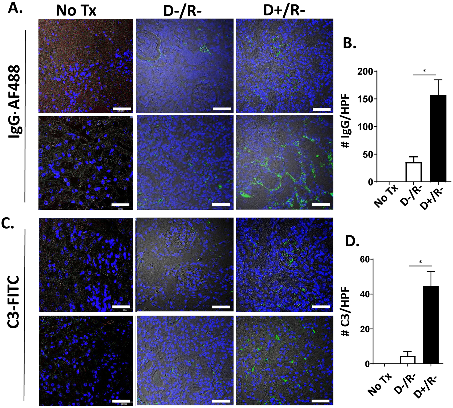
FIGURE 1.

IgG and C3 staining of CMV infected and uninfected renal allografts.
(A, C) Allografts of MCMV D−/R− and D+/R− transplants, without immunosuppression, were fixed at post-transplant day 7 and stained with IgG-AF488 (A) or C3-FITC (C). Kidneys from non-transplant C57BL/6 mice (“No Tx”) served as staining controls (left panels). Representative images of cortex (upper panels) and medulla (lower panels) are shown, with brightfield overlay to orient tissue morphology (60x; white bar=50 μm). (B, D) Fluorescence immunostaining for IgG (B) or C3 (D) was quantitated for 10 high power fields (HPF) per kidney using Image J software. Average positive staining was calculated for each graft and compared among the groups (n=3/group).
* p<0.05.
